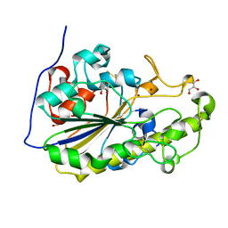
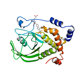
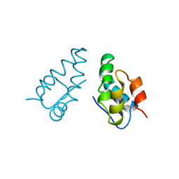
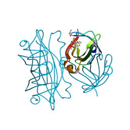
1DFF
 
 | | PEPTIDE DEFORMYLASE | | 分子名称: | PEPTIDE DEFORMYLASE, ZINC ION | | 著者 | Chan, M.K, Gong, W, Rajagopalan, P.T.R, Hao, B, Tsai, C.M, Pei, D. | | 登録日 | 1997-08-19 | | 公開日 | 1998-09-02 | | 最終更新日 | 2024-02-07 | | 実験手法 | X-RAY DIFFRACTION (2.88 Å) | | 主引用文献 | Crystal structure of the Escherichia coli peptide deformylase.
Biochemistry, 36, 1997
|
|
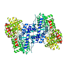
5LEZ
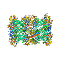 | | Human 20S proteasome complex with Oprozomib in Mg-Acetate at 2.2 Angstrom | | 分子名称: | ACETATE ION, MAGNESIUM ION, PENTAETHYLENE GLYCOL, ... | | 著者 | Schrader, J, Henneberg, F, Mata, R, Tittmann, K, Schneider, T.R, Stark, H, Bourenkov, G, Chari, A. | | 登録日 | 2016-06-30 | | 公開日 | 2016-08-17 | | 最終更新日 | 2024-01-10 | | 実験手法 | X-RAY DIFFRACTION (2.19 Å) | | 主引用文献 | The inhibition mechanism of human 20S proteasomes enables next-generation inhibitor design.
Science, 353, 2016
|
|
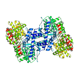
5LF1
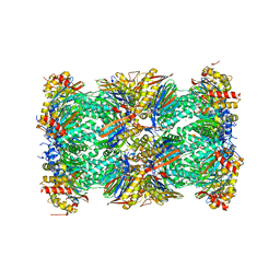 | | Human 20S proteasome complex with Dihydroeponemycin at 2.0 Angstrom | | 分子名称: | CHLORIDE ION, MAGNESIUM ION, PENTAETHYLENE GLYCOL, ... | | 著者 | Schrader, J, Henneberg, F, Mata, R, Tittmann, K, Schneider, T.R, Stark, H, Bourenkov, G, Chari, A. | | 登録日 | 2016-06-30 | | 公開日 | 2016-08-17 | | 最終更新日 | 2024-01-10 | | 実験手法 | X-RAY DIFFRACTION (2 Å) | | 主引用文献 | The inhibition mechanism of human 20S proteasomes enables next-generation inhibitor design.
Science, 353, 2016
|
|
5MX9
 | | High resolution crystal structure of the MCR-2 catalytic domain | | 分子名称: | GLYCEROL, Phosphatidylethanolamine transferase Mcr-2, ZINC ION | | 著者 | Hinchliffe, P, Coates, K, Walsh, T.R, Spencer, J. | | 登録日 | 2017-01-22 | | 公開日 | 2017-08-09 | | 最終更新日 | 2024-01-17 | | 実験手法 | X-RAY DIFFRACTION (1.12 Å) | | 主引用文献 | 1.12 angstrom resolution crystal structure of the catalytic domain of the plasmid-mediated colistin resistance determinant MCR-2.
Acta Crystallogr F Struct Biol Commun, 73, 2017
|
|
5N41
 | | Archaeal DNA polymerase holoenzyme - SSO6202 at 1.35 Ang resolution | | 分子名称: | 1,2-ETHANEDIOL, PolB1 Binding Protein 2 | | 著者 | Yan, J, Beattie, T.R, Rojas, A.L, Schermerhorn, K, Gristwood, T, Trinidad, J.C, Albers, S.V, Roversi, P, Gardner, A.F, Abrescia, N.G.A, Bell, S.D. | | 登録日 | 2017-02-09 | | 公開日 | 2017-05-17 | | 最終更新日 | 2024-01-17 | | 実験手法 | X-RAY DIFFRACTION (1.351 Å) | | 主引用文献 | Identification and characterization of a heterotrimeric archaeal DNA polymerase holoenzyme.
Nat Commun, 8, 2017
|
|
1BZC
 | | HUMAN PTP1B CATALYTIC DOMAIN COMPLEXED WITH TPI | | 分子名称: | 4-CARBAMOYL-4-{[6-(DIFLUORO-PHOSPHONO-METHYL)-NAPHTHALENE-2-CARBONYL]-AMINO}-BUTYRIC ACID, PROTEIN (PROTEIN-TYROSINE-PHOSPHATASE) | | 著者 | Groves, M.R, Yao, Z.J, Jr Burke, T.R, Barford, D. | | 登録日 | 1998-10-27 | | 公開日 | 1999-02-16 | | 最終更新日 | 2023-08-09 | | 実験手法 | X-RAY DIFFRACTION (2.35 Å) | | 主引用文献 | Structural basis for inhibition of the protein tyrosine phosphatase 1B by phosphotyrosine peptide mimetics.
Biochemistry, 37, 1998
|
|
1BW7
 | |
1BWW
 | | BENCE-JONES IMMUNOGLOBULIN REI VARIABLE PORTION, T39K MUTANT | | 分子名称: | PROTEIN (IG KAPPA CHAIN V-I REGION REI) | | 著者 | Uson, I, Pohl, E, Schneider, T.R, Dauter, Z, Schmidt, A, Fritz, H.J, Sheldrick, G.M. | | 登録日 | 1998-09-29 | | 公開日 | 1998-10-07 | | 最終更新日 | 2023-08-09 | | 実験手法 | X-RAY DIFFRACTION (1.7 Å) | | 主引用文献 | 1.7 A structure of the stabilized REIv mutant T39K. Application of local NCS restraints.
Acta Crystallogr.,Sect.D, 55, 1999
|
|
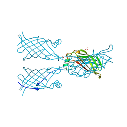
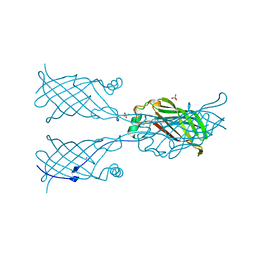
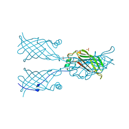
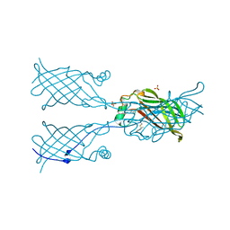
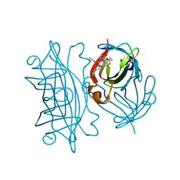
1EGJ
 | | DOMAIN 4 OF THE BETA COMMON CHAIN IN COMPLEX WITH AN ANTIBODY | | 分子名称: | 2-acetamido-2-deoxy-beta-D-glucopyranose, ANTIBODY (HEAVY CHAIN), ANTIBODY (LIGHT CHAIN), ... | | 著者 | Rossjohn, J, McKinstry, W.J, Woodcock, J.M, McClure, B.J, Hercus, T.R, Parker, M.W, Lopez, A.F, Bagley, C.J. | | 登録日 | 2000-02-15 | | 公開日 | 2001-02-15 | | 最終更新日 | 2020-07-29 | | 実験手法 | X-RAY DIFFRACTION (2.8 Å) | | 主引用文献 | Structure of the activation domain of the GM-CSF/IL-3/IL-5 receptor common beta-chain bound to an antagonist.
Blood, 95, 2000
|
|
1CBW
 | | BOVINE CHYMOTRYPSIN COMPLEXED TO BPTI | | 分子名称: | BOVINE CHYMOTRYPSIN, BPTI, SULFATE ION | | 著者 | Hynes, T.R, Scheidig, A.J, Kossiakoff, A.A. | | 登録日 | 1996-12-22 | | 公開日 | 1997-07-23 | | 最終更新日 | 2023-08-09 | | 実験手法 | X-RAY DIFFRACTION (2.6 Å) | | 主引用文献 | Crystal structures of bovine chymotrypsin and trypsin complexed to the inhibitor domain of Alzheimer's amyloid beta-protein precursor (APPI) and basic pancreatic trypsin inhibitor (BPTI): engineering of inhibitors with altered specificities.
Protein Sci., 6, 1997
|
|
1AUM
 | | HIV CAPSID C-TERMINAL DOMAIN (CAC146) | | 分子名称: | HIV CAPSID | | 著者 | Hill, C.P, Gamble, T.R, Yoo, S, Vajdos, F.F, Von Schwedler, U.K, Worthylake, D.K, Wang, H, Mccutcheon, J.P, Sundquist, W.I. | | 登録日 | 1997-08-29 | | 公開日 | 1998-01-14 | | 最終更新日 | 2024-04-03 | | 実験手法 | X-RAY DIFFRACTION (3 Å) | | 主引用文献 | Structure of the carboxyl-terminal dimerization domain of the HIV-1 capsid protein.
Science, 278, 1997
|
|
1EKA
 | | NMR AND MOLECULAR MODELING REVEAL THAT DIFFERENT HYDROGEN BONDING PATTERNS ARE POSSIBLE FOR GU PAIRS: ONE HYDROGEN BOND FOR EACH GU PAIR IN R(GGCGUGCC)2 AND TWO FOR EACH GU PAIR IN R(GAGUGCUC)2 | | 分子名称: | RNA (5'-R(*GP*AP*GP*UP*GP*CP*UP*C)-3') | | 著者 | Chen, X, McDowell, J.A, Kierzek, R, Krugh, T.R, Turner, D.H. | | 登録日 | 2000-03-07 | | 公開日 | 2000-11-13 | | 最終更新日 | 2024-05-22 | | 実験手法 | SOLUTION NMR | | 主引用文献 | Nuclear magnetic resonance spectroscopy and molecular modeling reveal that different hydrogen bonding patterns are possible for G.U pairs: one hydrogen bond for each G.U pair in r(GGCGUGCC)(2) and two for each G.U pair in r(GAGUGCUC)(2).
Biochemistry, 39, 2000
|
|
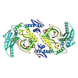
1EM6
 | | HUMAN LIVER GLYCOGEN PHOSPHORYLASE A COMPLEXED WITH GLCNAC AND CP-526,423 | | 分子名称: | (4S)-2-METHYL-2,4-PENTANEDIOL, BIS[5-CHLORO-1H-INDOL-2-YL-CARBONYL-AMINOETHYL]-ETHYLENE GLYCOL, LIVER GLYCOGEN PHOSPHORYLASE, ... | | 著者 | Rath, V.L, Ammirati, M, Danley, D.E, Ekstrom, J.L, Hynes, T.R, Olson, T.V, Hoover, D.J. | | 登録日 | 2000-03-16 | | 公開日 | 2000-11-01 | | 最終更新日 | 2020-07-29 | | 実験手法 | X-RAY DIFFRACTION (2.2 Å) | | 主引用文献 | Human liver glycogen phosphorylase inhibitors bind at a new allosteric site.
Chem.Biol., 7, 2000
|
|
1EEK
 | | SOLUTION STRUCTURE OF A NONPOLAR, NON HYDROGEN BONDED BASE PAIR SURROGATE IN DNA. | | 分子名称: | 5'-D(*CP*GP*CP*AP*TP*(DFT)P*GP*TP*TP*AP*CP*C)-3', 5'-D(*GP*GP*TP*AP*AP*CP*(MBZ)P*AP*TP*GP*CP*G)-3' | | 著者 | Kool, E.T, Krugh, T.R, Guckian, K.M. | | 登録日 | 2000-02-01 | | 公開日 | 2000-02-16 | | 最終更新日 | 2024-05-22 | | 実験手法 | SOLUTION NMR | | 主引用文献 | Solution Structure of a Nonpolar, Non-Hydrogen-Bonded Base Pair Surrogate in DNA
J.Am.Chem.Soc., 122, 2000
|
|
1EKD
 | | NMR AND MOLECULAR MODELING REVEAL THAT DIFFERENT HYDROGEN BONDING PATTERNS ARE POSSIBLE FOR GU PAIRS: ONE HYDROGEN BOND FOR EACH GU PAIR IN R(GGCGUGCC)2 AND TWO FOR EACH GU PAIR IN R(GAGUGCUC)2 | | 分子名称: | RNA (5'-R(*GP*GP*CP*GP*UP*GP*CP*C)-3') | | 著者 | Chen, X, McDowell, J.A, Kierzek, R, Krugh, T.R, Turner, D.H. | | 登録日 | 2000-03-07 | | 公開日 | 2000-11-13 | | 最終更新日 | 2024-05-22 | | 実験手法 | SOLUTION NMR | | 主引用文献 | Nuclear magnetic resonance spectroscopy and molecular modeling reveal that different hydrogen bonding patterns are possible for G.U pairs: one hydrogen bond for each G.U pair in r(GGCGUGCC)(2) and two for each G.U pair in r(GAGUGCUC)(2).
Biochemistry, 39, 2000
|
|
1EXV
 | | HUMAN LIVER GLYCOGEN PHOSPHORYLASE A COMPLEXED WITH GLCNAC AND CP-403,700 | | 分子名称: | (4S)-2-METHYL-2,4-PENTANEDIOL, LIVER GLYCOGEN PHOSPHORYLASE, N-acetyl-beta-D-glucopyranosylamine, ... | | 著者 | Rath, V.L, Ammirati, M, Danley, D.E, Ekstrom, J.L, Hynes, T.R, Olson, T.V, Hoover, D.J. | | 登録日 | 2000-05-04 | | 公開日 | 2000-11-01 | | 最終更新日 | 2020-07-29 | | 実験手法 | X-RAY DIFFRACTION (2.4 Å) | | 主引用文献 | Human liver glycogen phosphorylase inhibitors bind at a new allosteric site.
Chem.Biol., 7, 2000
|
|
5VL8
 | | Coordination Chemistry within a Protein Host: Regulation of the Secondary Coordination Sphere | | 分子名称: | COPPER (II) ION, GLYCEROL, Streptavidin, ... | | 著者 | Mann, S.I, Heinisch, T, Ward, T.R, Borovik, A.S. | | 登録日 | 2017-04-25 | | 公開日 | 2018-04-18 | | 最終更新日 | 2023-10-04 | | 実験手法 | X-RAY DIFFRACTION (1.7 Å) | | 主引用文献 | Coordination chemistry within a protein host: regulation of the secondary coordination sphere.
Chem. Commun. (Camb.), 54, 2018
|
|
6D3F
 | | Crystal Structure of the PTP epsilon D2 domain | | 分子名称: | Receptor-type tyrosine-protein phosphatase epsilon | | 著者 | Lountos, G.T, Raran-Kurussi, S, Zhao, B.M, Dyas, B.K, Austin, B.P, Burke Jr, T.R, Ulrich, R.G, Waugh, D.S. | | 登録日 | 2018-04-16 | | 公開日 | 2018-10-17 | | 最終更新日 | 2023-10-04 | | 実験手法 | X-RAY DIFFRACTION (2.271 Å) | | 主引用文献 | High-resolution crystal structures of the D1 and D2 domains of protein tyrosine phosphatase epsilon for structure-based drug design.
Acta Crystallogr D Struct Biol, 74, 2018
|
|
5W17
 | |
5W2V
 | |
6CGO
 | | Molecular basis for condensation domain-mediated chain release from the enacyloxin polyketide synthase | | 分子名称: | Condensation domain protein, PHOSPHATE ION | | 著者 | Valentic, T.R, Tsai, S.C, Challis, G.L, Lewandowski, J.R, Kosol, S, Gallo, A, Griffiths, D, Masschelein, J.L, Jenner, M, De los Santos, E. | | 登録日 | 2018-02-20 | | 公開日 | 2019-02-27 | | 最終更新日 | 2024-03-13 | | 実験手法 | X-RAY DIFFRACTION (2 Å) | | 主引用文献 | Structural basis for chain release from the enacyloxin polyketide synthase.
Nat.Chem., 11, 2019
|
|
5W2D
 | |
5W30
 | | Crystal structure of mutant CJ YCEI protein (CJ-N48C) with monobromobimane guest structure | | 分子名称: | 3-(bromomethyl)-2,5,6-trimethyl-1H,7H-pyrazolo[1,2-a]pyrazole-1,7-dione, Putative periplasmic protein, SULFATE ION, ... | | 著者 | Huber, T.R, Snow, C.D. | | 登録日 | 2017-06-07 | | 公開日 | 2018-01-03 | | 最終更新日 | 2023-10-04 | | 実験手法 | X-RAY DIFFRACTION (2.75 Å) | | 主引用文献 | Installing Guest Molecules at Specific Sites within Scaffold Protein Crystals.
Bioconjug. Chem., 29, 2018
|
|
5VL5
 | | Coordination Chemistry within a Protein Host: Regulation of the Secondary Coordination Sphere | | 分子名称: | COPPER (II) ION, Streptavidin, [N-(3-{bis[2-(pyridin-2-yl-kappaN)ethyl]amino-kappaN}propyl)-5-(2-oxohexahydro-1H-thieno[3,4-d]imidazol-4-yl)pentanamide](azido)(hydroxy)copper | | 著者 | Mann, S.I, Heinisch, T, Ward, T.R, Borovik, A.S. | | 登録日 | 2017-04-24 | | 公開日 | 2018-04-18 | | 最終更新日 | 2024-03-13 | | 実験手法 | X-RAY DIFFRACTION (1.46 Å) | | 主引用文献 | Coordination chemistry within a protein host: regulation of the secondary coordination sphere.
Chem. Commun. (Camb.), 54, 2018
|
|
5W3A
 | |


