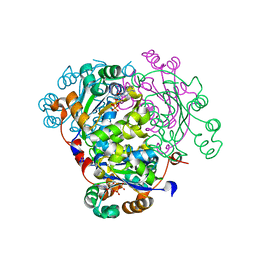
3ALS
 
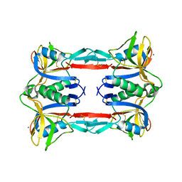 | | Crystal structure of CEL-IV | | 分子名称: | CALCIUM ION, Lectin CEL-IV, C-type | | 著者 | Hatakeyama, T, Hozawa, T, Ishii, K, Kamiya, T, Goda, S, Kusunoki, M, Unno, H. | | 登録日 | 2010-08-07 | | 公開日 | 2011-01-19 | | 最終更新日 | 2023-11-01 | | 実験手法 | X-RAY DIFFRACTION (3 Å) | | 主引用文献 | Galactose recognition by a tetrameric C-type lectin, CEL-IV, containing the EPN carbohydrate recognition motif
J.Biol.Chem., 286, 2011
|
|
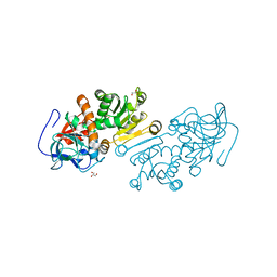
1ZD9
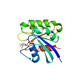 | | Structure of human ADP-ribosylation factor-like 10B | | 分子名称: | ADP-ribosylation factor-like 10B, GUANOSINE-5'-DIPHOSPHATE, MAGNESIUM ION | | 著者 | Choe, J, Atanassova, A, Arrowsmith, C, Edwards, A, Sundstrom, M, Bochkarev, A, Park, H, Structural Genomics Consortium (SGC) | | 登録日 | 2005-04-14 | | 公開日 | 2005-05-17 | | 最終更新日 | 2024-02-14 | | 実験手法 | X-RAY DIFFRACTION (1.7 Å) | | 主引用文献 | Structure of human ADP-ribosylation factor-like 10B
to be published
|
|
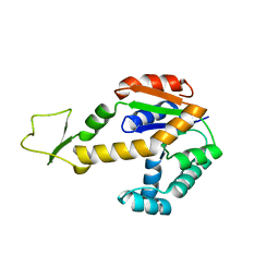
3AW0
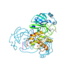 | | Structure of SARS 3CL protease with peptidic aldehyde inhibitor | | 分子名称: | 3C-Like Proteinase, peptide ACE-SER-ALA-VAL-LEU-HIS-H | | 著者 | Akaji, K, Konno, H, Mitsui, H, Teruya, K, Hattori, Y, Ozaki, T, Kusunoki, M, Sanjho, A. | | 登録日 | 2011-03-09 | | 公開日 | 2011-12-14 | | 最終更新日 | 2023-11-15 | | 実験手法 | X-RAY DIFFRACTION (2.3 Å) | | 主引用文献 | Structure-Based Design, Synthesis, and Evaluation of Peptide-Mimetic SARS 3CL Protease Inhibitors.
J.Med.Chem., 54, 2011
|
|
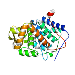
3AW1
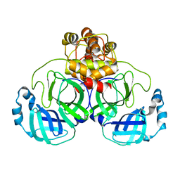 | | Structure of SARS 3CL protease auto-proteolysis resistant mutant in the absent of inhibitor | | 分子名称: | 3C-Like Proteinase | | 著者 | Akaji, K, Konno, H, Mitsui, H, Teruya, K, Hattori, Y, Ozaki, T, Kusunoki, M, Sanjho, A. | | 登録日 | 2011-03-09 | | 公開日 | 2011-12-14 | | 最終更新日 | 2023-11-01 | | 実験手法 | X-RAY DIFFRACTION (2 Å) | | 主引用文献 | Structure-Based Design, Synthesis, and Evaluation of Peptide-Mimetic SARS 3CL Protease Inhibitors.
J.Med.Chem., 54, 2011
|
|
1XWS
 | | Crystal Structure of the human PIM1 kinase domain | | 分子名称: | 3-{1-[3-(DIMETHYLAMINO)PROPYL]-1H-INDOL-3-YL}-4-(1H-INDOL-3-YL)-1H-PYRROLE-2,5-DIONE, Proto-oncogene serine/threonine-protein kinase Pim-1, SULFATE ION | | 著者 | Knapp, S, Debreczeni, J, Bullock, A, von Delft, F, Sundstrom, M, Arrowsmith, C, Edwards, A, Guo, K. | | 登録日 | 2004-11-02 | | 公開日 | 2004-11-16 | | 最終更新日 | 2024-03-13 | | 実験手法 | X-RAY DIFFRACTION (1.8 Å) | | 主引用文献 | Crystal structure of human PIM1 kinase domain
To be Published
|
|
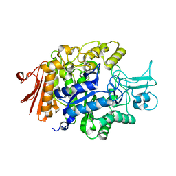
3AJ7
 | | Crystal Structure of isomaltase from Saccharomyces cerevisiae | | 分子名称: | CALCIUM ION, Oligo-1,6-glucosidase | | 著者 | Yamamoto, K, Miyake, H, Kusunoki, M, Osaki, S. | | 登録日 | 2010-05-26 | | 公開日 | 2010-08-11 | | 最終更新日 | 2023-11-01 | | 実験手法 | X-RAY DIFFRACTION (1.3 Å) | | 主引用文献 | Crystal structures of isomaltase from Saccharomyces cerevisiae and in complex with its competitive inhibitor maltose
Febs J., 277, 2010
|
|
1ZBY
 | | High-Resolution Crystal Structure of Native (Resting) Cytochrome c Peroxidase (CcP) | | 分子名称: | Cytochrome c peroxidase, PROTOPORPHYRIN IX CONTAINING FE | | 著者 | Bonagura, C.A, Bhaskar, B, Shimizu, H, Li, H, Sundaramoorthy, M, McRee, D.E, Goodin, D.B, Poulos, T.L. | | 登録日 | 2005-04-09 | | 公開日 | 2005-05-03 | | 最終更新日 | 2023-10-25 | | 実験手法 | X-RAY DIFFRACTION (1.2 Å) | | 主引用文献 | High-resolution crystal structures and spectroscopy of native and compound I cytochrome c peroxidase
Biochemistry, 42, 2003
|
|
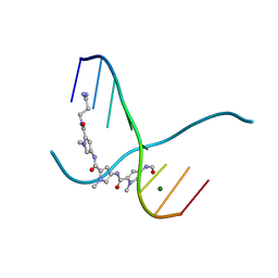
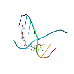
4ITD
 | | Structures of DNA duplexes containing O6-carboxymethylguanine, a lesion associated with gastrointestinal cancer, reveal a mechanism for inducing transition mutation | | 分子名称: | 2'-(4-HYDROXYPHENYL)-5-(4-METHYL-1-PIPERAZINYL)-2,5'-BI-BENZIMIDAZOLE, DNA (5'-D(*CP*GP*CP*GP*(C6G)P*AP*TP*TP*CP*GP*CP*G)-3'), MAGNESIUM ION | | 著者 | Zhang, F, Suzuki, K, Tsunoda, M, Wilkinson, O, Millington, C.L, Williams, D.M, Morishita, E.C, Takenaka, A. | | 登録日 | 2013-01-18 | | 公開日 | 2013-05-08 | | 最終更新日 | 2024-03-20 | | 実験手法 | X-RAY DIFFRACTION (1.94 Å) | | 主引用文献 | Structures of DNA duplexes containing O6-carboxymethylguanine, a lesion associated with gastrointestinal cancer, reveal a mechanism for inducing pyrimidine transition mutations
Nucleic Acids Res., 41, 2013
|
|
3ATW
 | | Structure-Based Design, Synthesis, Evaluation of Peptide-mimetic SARS 3CL Protease Inhibitors | | 分子名称: | 3C-Like Proteinase, peptide ACE-THR-VAL-ALC-HIS-H | | 著者 | Akaji, K, Konno, H, Mitsui, H, Teruya, K, Hattori, Y, Ozaki, T, Kusunoki, M, Sanjho, A. | | 登録日 | 2011-01-20 | | 公開日 | 2011-12-14 | | 最終更新日 | 2023-11-15 | | 実験手法 | X-RAY DIFFRACTION (2.36 Å) | | 主引用文献 | Structure-Based Design, Synthesis, and Evaluation of Peptide-Mimetic SARS 3CL Protease Inhibitors.
J.Med.Chem., 54, 2011
|
|
3AVZ
 | | Structure of SARS 3CL protease with peptidic aldehyde inhibitor containing cyclohexyl side chain | | 分子名称: | 3C-Like Proteinase, peptide ACE-SER-ALA-VAL-ALC-HIS-H | | 著者 | Akaji, K, Konno, H, Mitsui, H, Teruya, K, Hattori, Y, Ozaki, T, Kusunoki, M, Sanjho, A. | | 登録日 | 2011-03-09 | | 公開日 | 2011-12-14 | | 最終更新日 | 2023-11-15 | | 実験手法 | X-RAY DIFFRACTION (2.46 Å) | | 主引用文献 | Structure-Based Design, Synthesis, and Evaluation of Peptide-Mimetic SARS 3CL Protease Inhibitors.
J.Med.Chem., 54, 2011
|
|
1Y6L
 | | Human ubiquitin conjugating enzyme E2E2 | | 分子名称: | Ubiquitin-conjugating enzyme E2E2 | | 著者 | Walker, J.R, Avvakumov, G.V, Newman, E.M, Mackenzie, F, Kozieradzki, I, Bochkarev, A, Sundstrom, M, Arrowsmith, C, Edwards, A, Dhe-Paganon, S, Structural Genomics Consortium (SGC) | | 登録日 | 2004-12-06 | | 公開日 | 2005-01-11 | | 最終更新日 | 2023-08-23 | | 実験手法 | X-RAY DIFFRACTION (1.85 Å) | | 主引用文献 | A human ubiquitin conjugating enzyme (E2)-HECT E3 ligase structure-function screen.
Mol Cell Proteomics, 11, 2012
|
|
1ZDN
 | | Ubiquitin-conjugating enzyme E2S | | 分子名称: | SODIUM ION, Ubiquitin-conjugating enzyme E2S | | 著者 | Walker, J.R, Avvakumov, G.V, Xue, S, Newman, E.M, Mackenzie, F, Sundstrom, M, Arrowsmith, C, Edwards, A, Bochkarev, A, Dhe-Paganon, S, Structural Genomics Consortium (SGC) | | 登録日 | 2005-04-14 | | 公開日 | 2005-05-03 | | 最終更新日 | 2023-08-23 | | 実験手法 | X-RAY DIFFRACTION (1.93 Å) | | 主引用文献 | A human ubiquitin conjugating enzyme (E2)-HECT E3 ligase structure-function screen.
Mol Cell Proteomics, 11, 2012
|
|
1ZKC
 | | Crystal Structure of the cyclophiln_RING domain of human peptidylprolyl isomerase (cyclophilin)-like 2 isoform b | | 分子名称: | BETA-MERCAPTOETHANOL, Peptidyl-prolyl cis-trans isomerase like 2 | | 著者 | Walker, J.R, Davis, T, Newman, E.M, Mackenzie, F, Weigelt, J, Sundstrom, M, Arrowsmith, C, Edwards, A, Bochkarev, A, Dhe-Paganon, S, Structural Genomics Consortium (SGC) | | 登録日 | 2005-05-02 | | 公開日 | 2005-08-16 | | 最終更新日 | 2023-08-23 | | 実験手法 | X-RAY DIFFRACTION (1.65 Å) | | 主引用文献 | Structural and biochemical characterization of the human cyclophilin family of peptidyl-prolyl isomerases.
PLoS Biol., 8, 2010
|
|
2ODB
 | | The crystal structure of human cdc42 in complex with the CRIB domain of human p21-activated kinase 6 (PAK6) | | 分子名称: | CHLORIDE ION, Human Cell Division Cycle 42 (CDC42), MAGNESIUM ION, ... | | 著者 | Ugochukwu, E, Yang, X, Elkins, J, Soundararajan, M, Pike, A.C.W, Eswaran, J, Burgess, N, Debreczeni, J.E, Sundstrom, M, Arrowsmith, C, Weigelt, J, Edwards, A, Gileadi, O, von Delft, F, Knapp, S, Doyle, D, Structural Genomics Consortium (SGC) | | 登録日 | 2006-12-22 | | 公開日 | 2007-01-30 | | 最終更新日 | 2023-08-30 | | 実験手法 | X-RAY DIFFRACTION (2.4 Å) | | 主引用文献 | The crystal structure of human cdc42 in complex with the CRIB domain of human p21-activated kinase 6 (PAK6)
To be Published
|
|
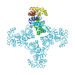
1V59
 | | Crystal structure of yeast lipoamide dehydrogenase complexed with NAD+ | | 分子名称: | Dihydrolipoamide dehydrogenase, FLAVIN-ADENINE DINUCLEOTIDE, NICOTINAMIDE-ADENINE-DINUCLEOTIDE, ... | | 著者 | Adachi, W, Suzuki, K, Tsunoda, M, Sekiguchi, T, Reed, L.J, Takenaka, A. | | 登録日 | 2003-11-21 | | 公開日 | 2005-02-15 | | 最終更新日 | 2023-12-27 | | 実験手法 | X-RAY DIFFRACTION (2.2 Å) | | 主引用文献 | Crystal structure of yeast lipoamide dehydrogenase complexed with NAD+
To be Published
|
|
1ZS6
 | | structure of human nucleoside-diphosphate kinase 3 | | 分子名称: | ADENOSINE-5'-DIPHOSPHATE, Nucleoside diphosphate kinase 3 | | 著者 | Choe, J, Dimov, S, Arrowsmith, C, Edwards, A, Sundstrom, M, Bochkarev, A, Park, H, Structural Genomics Consortium (SGC) | | 登録日 | 2005-05-23 | | 公開日 | 2005-06-07 | | 最終更新日 | 2023-08-23 | | 実験手法 | X-RAY DIFFRACTION (2.3 Å) | | 主引用文献 | Structure of human nucleoside-diphosphate kinase 3
To be Published
|
|
1ZSY
 | | The structure of human mitochondrial 2-enoyl thioester reductase (CGI-63) | | 分子名称: | GLYCEROL, MITOCHONDRIAL 2-ENOYL THIOESTER REDUCTASE, SULFATE ION | | 著者 | Lukacik, P, Shafqat, N, Kavanagh, K.L, Johansson, C, Smee, C, Edwards, A, Arrowsmith, C, Sundstrom, M, von Delft, F, Oppermann, U, Structural Genomics Consortium (SGC) | | 登録日 | 2005-05-25 | | 公開日 | 2005-06-07 | | 最終更新日 | 2023-08-23 | | 実験手法 | X-RAY DIFFRACTION (1.75 Å) | | 主引用文献 | The structure of human mitochondrial 2-enoyl thioester reductase (CGI-63)
To be Published
|
|
1ZD8
 | | Structure of human adenylate kinase 3 like 1 | | 分子名称: | GTP:AMP phosphotransferase mitochondrial | | 著者 | Choe, J, Atanassova, A, Arrowsmith, C, Edwards, A, Sundstrom, M, Bochkarev, A, Park, H, Structural Genomics Consortium (SGC) | | 登録日 | 2005-04-14 | | 公開日 | 2005-05-17 | | 最終更新日 | 2023-08-23 | | 実験手法 | X-RAY DIFFRACTION (1.48 Å) | | 主引用文献 | Structure of human adenylate kinase 3 like 1
to be published
|
|
1ZBZ
 | | High-Resolution Crystal Structure of Compound I intermediate of Cytochrome c Peroxidase (CcP) | | 分子名称: | Cytochrome c peroxidase, PROTOPORPHYRIN IX CONTAINING FE | | 著者 | Bonagura, C.A, Bhaskar, B, Shimizu, H, Li, H, Sundaramoorthy, M, McRee, D.E, Goodin, D.B, Poulos, T.L. | | 登録日 | 2005-04-09 | | 公開日 | 2005-05-03 | | 最終更新日 | 2023-10-25 | | 実験手法 | X-RAY DIFFRACTION (1.29 Å) | | 主引用文献 | High-resolution crystal structures and spectroscopy of native and compound I cytochrome c peroxidase
Biochemistry, 42, 2003
|
|
217D
 | |
1ZSX
 | | Crystal Structure Of Human Potassium Channel Kv Beta-subunit (KCNAB2) | | 分子名称: | CHLORIDE ION, NADP NICOTINAMIDE-ADENINE-DINUCLEOTIDE PHOSPHATE, Voltage-gated potassium channel beta-2 subunit | | 著者 | Wennerstrand, P, Johannsson, I, Ugochukwu, E, Kavanagh, K, Edwards, A, Arrowsmith, C, Williams, L, Sundstrom, M, Guo, K, von Delft, F, Oppermann, U, Structural Genomics Consortium (SGC) | | 登録日 | 2005-05-25 | | 公開日 | 2005-06-07 | | 最終更新日 | 2023-08-23 | | 実験手法 | X-RAY DIFFRACTION (1.9 Å) | | 主引用文献 | Crystal Structure Of Human Potassium Channel Kv Beta-subunit (KCNAB2)
To be Published
|
|
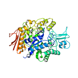
3AXH
 | | Crystal structure of isomaltase in complex with isomaltose | | 分子名称: | CALCIUM ION, Oligo-1,6-glucosidase IMA1, alpha-D-glucopyranose-(1-6)-alpha-D-glucopyranose | | 著者 | Yamamoto, K, Miyake, H, Kusunoki, M, Osaki, S. | | 登録日 | 2011-04-06 | | 公開日 | 2011-10-05 | | 最終更新日 | 2023-11-01 | | 実験手法 | X-RAY DIFFRACTION (1.8 Å) | | 主引用文献 | Steric hindrance by 2 amino acid residues determines the substrate specificity of isomaltase from Saccharomyces cerevisiae
J.Biosci.Bioeng., 112, 2011
|
|
3AXI
 | | Crystal structure of isomaltase in complex with maltose | | 分子名称: | CALCIUM ION, Oligo-1,6-glucosidase IMA1, alpha-D-glucopyranose | | 著者 | Yamamoto, K, Miyake, H, Kusunoki, M, Osaki, S. | | 登録日 | 2011-04-06 | | 公開日 | 2011-10-05 | | 最終更新日 | 2023-11-01 | | 実験手法 | X-RAY DIFFRACTION (1.4 Å) | | 主引用文献 | Steric hindrance by 2 amino acid residues determines the substrate specificity of isomaltase from Saccharomyces cerevisiae
J.Biosci.Bioeng., 112, 2011
|
|
2PXX
 | | Human putative methyltransferase MGC2408 | | 分子名称: | S-ADENOSYL-L-HOMOCYSTEINE, UNKNOWN ATOM OR ION, Uncharacterized protein MGC2408 | | 著者 | Tempel, W, Wu, H, Dombrovsky, L, Zeng, H, Zhu, H, Loppnau, P, Weigelt, J, Sundstrom, M, Arrowsmith, C.H, Edwards, A.M, Bochkarev, A, Plotnikov, A.N, Structural Genomics Consortium (SGC) | | 登録日 | 2007-05-14 | | 公開日 | 2007-05-22 | | 最終更新日 | 2024-02-21 | | 実験手法 | X-RAY DIFFRACTION (1.3 Å) | | 主引用文献 | An intact SAM-dependent methyltransferase fold is encoded by the human endothelin-converting enzyme-2 gene.
Proteins, 74, 2009
|
|
216D
 | |


