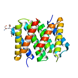
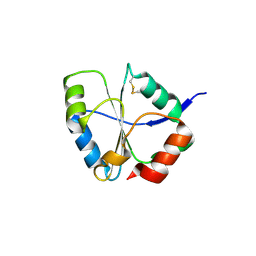
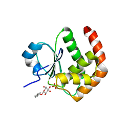
3MP9
 
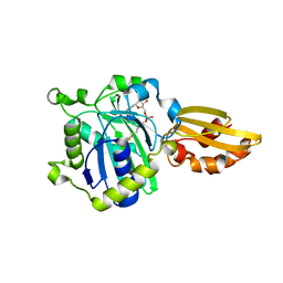
 | | Structure of Streptococcal protein G B1 domain at pH 3.0 | | 分子名称: | FORMIC ACID, Immunoglobulin G-binding protein G | | 著者 | Tomlinson, J.H, Green, V.L, Baker, P.J, Williamson, M.P. | | 登録日 | 2010-04-26 | | 公開日 | 2011-02-23 | | 最終更新日 | 2023-09-06 | | 実験手法 | X-RAY DIFFRACTION (1.2 Å) | | 主引用文献 | Structural origins of pH-dependent chemical shifts in the B1 domain of protein G.
Proteins, 78, 2010
|
|
5IZ2
 | |
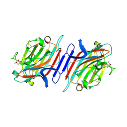
2MZW
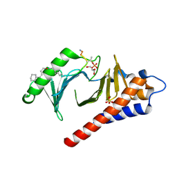
 | | Staphylococcus aureus FusB:EF-GC3 complex | | 分子名称: | Elongation factor G, Far1, ZINC ION | | 著者 | Tomlinson, J.H, Thompson, G.S, Kalverda, A.P, Zhuravleva, A, O'Neill, A. | | 登録日 | 2015-02-25 | | 公開日 | 2016-01-27 | | 最終更新日 | 2024-05-15 | | 実験手法 | SOLUTION NMR | | 主引用文献 | A target-protection mechanism of antibiotic resistance at atomic resolution: insights into FusB-type fusidic acid resistance.
Sci Rep, 6, 2016
|
|
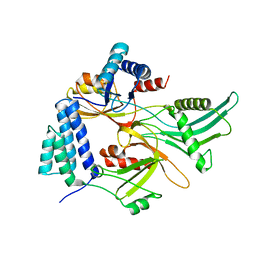
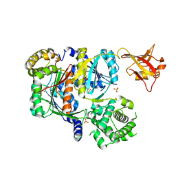
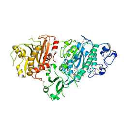
4K87
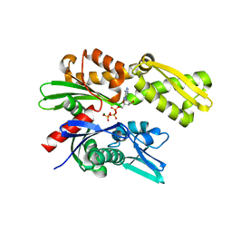
 | | Crystal structure of human prolyl-tRNA synthetase (substrate bound form) | | 分子名称: | ADENOSINE, PROLINE, Proline--tRNA ligase, ... | | 著者 | Hwang, K.Y, Son, J.H, Lee, E.H. | | 登録日 | 2013-04-18 | | 公開日 | 2013-10-09 | | 最終更新日 | 2023-11-08 | | 実験手法 | X-RAY DIFFRACTION (2.301 Å) | | 主引用文献 | Conformational changes in human prolyl-tRNA synthetase upon binding of the substrates proline and ATP and the inhibitor halofuginone.
Acta Crystallogr.,Sect.D, 69, 2013
|
|
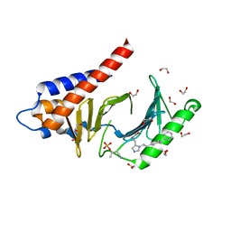
4K88
 | | Crystal structure of human prolyl-tRNA synthetase (halofuginone bound form) | | 分子名称: | 7-bromo-6-chloro-3-{3-[(2R,3S)-3-hydroxypiperidin-2-yl]-2-oxopropyl}quinazolin-4(3H)-one, Proline--tRNA ligase, ZINC ION | | 著者 | Hwang, K.Y, Son, J.H, Lee, E.H. | | 登録日 | 2013-04-18 | | 公開日 | 2013-10-09 | | 最終更新日 | 2024-03-20 | | 実験手法 | X-RAY DIFFRACTION (2.619 Å) | | 主引用文献 | Conformational changes in human prolyl-tRNA synthetase upon binding of the substrates proline and ATP and the inhibitor halofuginone.
Acta Crystallogr.,Sect.D, 69, 2013
|
|
4K86
 | |
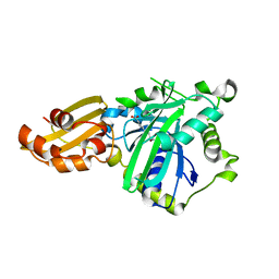
1UC7
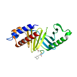
 | | Crystal structure of DsbDgamma | | 分子名称: | Thiol:disulfide interchange protein dsbD | | 著者 | Kim, J.H, Kim, S.J, Jeong, D.G, Son, J.H, Ryu, S.E. | | 登録日 | 2003-04-09 | | 公開日 | 2004-04-27 | | 最終更新日 | 2023-12-27 | | 実験手法 | X-RAY DIFFRACTION (1.9 Å) | | 主引用文献 | Crystal structure of DsbDgamma reveals the mechanism of redox potential shift and substrate specificity(1)
FEBS LETT., 543, 2003
|
|
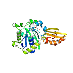
1XM2
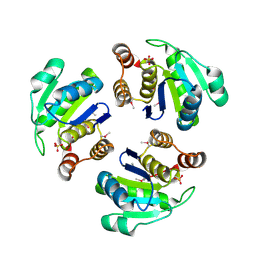 | | Crystal structure of Human PRL-1 | | 分子名称: | SULFATE ION, Tyrosine Phosphatase | | 著者 | Jeong, D.G, Kim, S.J, Kim, J.H, Son, J.H, Ryu, S.E. | | 登録日 | 2004-10-01 | | 公開日 | 2005-01-25 | | 最終更新日 | 2021-11-10 | | 実験手法 | X-RAY DIFFRACTION (2.7 Å) | | 主引用文献 | Trimeric structure of PRL-1 phosphatase reveals an active enzyme conformation and regulation mechanisms
J.Mol.Biol., 345, 2005
|
|
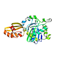
1ZZW
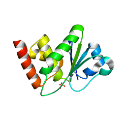 | | Crystal Structure of catalytic domain of Human MAP Kinase Phosphatase 5 | | 分子名称: | 1,2-ETHANEDIOL, Dual specificity protein phosphatase 10, SULFATE ION | | 著者 | Jeong, D.G, Yoon, T.S, Kim, J.H, Shim, M.Y, Jeong, S.K, Son, J.H, Ryu, S.E, Kim, S.J. | | 登録日 | 2005-06-14 | | 公開日 | 2006-07-04 | | 最終更新日 | 2024-03-13 | | 実験手法 | X-RAY DIFFRACTION (1.6 Å) | | 主引用文献 | Crystal Structure of the Catalytic Domain of Human MAP Kinase Phosphatase 5: Structural Insight into Constitutively Active Phosphatase.
J.Mol.Biol., 360, 2006
|
|
1YZ4
 | | Crystal structure of DUSP15 | | 分子名称: | SULFATE ION, dual specificity phosphatase-like 15 isoform a, octyl beta-D-glucopyranoside | | 著者 | Kim, S.J, Ryu, S.E, Jeong, D.G, Yoon, T.S, Kim, J.H, Cho, Y.H, Jeong, S.K, Lee, J.W, Son, J.H. | | 登録日 | 2005-02-28 | | 公開日 | 2005-11-01 | | 最終更新日 | 2024-05-29 | | 実験手法 | X-RAY DIFFRACTION (2.4 Å) | | 主引用文献 | Crystal structure of the catalytic domain of human VHY, a dual-specificity protein phosphatase
Proteins, 61, 2005
|
|
6CWY
 | | Crystal structure of SUMO E1 in complex with an allosteric inhibitor | | 分子名称: | GLYCEROL, MAGNESIUM ION, SULFATE ION, ... | | 著者 | Lv, Z, Yuan, L, Atkison, J.H, Williams, K.M, Olsen, S.K. | | 登録日 | 2018-04-01 | | 公開日 | 2019-01-16 | | 最終更新日 | 2023-10-04 | | 実験手法 | X-RAY DIFFRACTION (2.462 Å) | | 主引用文献 | Molecular mechanism of a covalent allosteric inhibitor of SUMO E1 activating enzyme.
Nat Commun, 9, 2018
|
|
1JXN
 | | Crystal Structure of the Lectin I from Ulex europaeus in complex with the methyl glycoside of alpha-L-fucose | | 分子名称: | (4R)-2-METHYLPENTANE-2,4-DIOL, CALCIUM ION, MANGANESE (II) ION, ... | | 著者 | Audette, G.F, Olson, D.J.H, Ross, A.R.S, Quail, J.W, Delbaere, L.T.J. | | 登録日 | 2001-09-07 | | 公開日 | 2002-12-06 | | 最終更新日 | 2023-08-16 | | 実験手法 | X-RAY DIFFRACTION (2.3 Å) | | 主引用文献 | Examination of the Structural Basis for O(H) Blood Group Specificity by Ulex europaeus Lectin I
Can.J.Chem., 80, 2002
|
|
4RCP
 | | Crystal structure of Plk1 Polo-box domain in complex with PL-2 | | 分子名称: | 1,2-ETHANEDIOL, PL-2, Serine/threonine-protein kinase PLK1 | | 著者 | Lee, W.C, Song, J.H, Kim, H.Y. | | 登録日 | 2014-09-16 | | 公開日 | 2014-11-12 | | 最終更新日 | 2015-01-21 | | 実験手法 | X-RAY DIFFRACTION (1.6 Å) | | 主引用文献 | A new class of peptidomimetics targeting the polo-box domain of polo-like kinase 1.
J.Med.Chem., 58, 2015
|
|
3U66
 | | Crystal structure of T6SS SciP/TssL from Escherichia Coli Enteroaggregative 042 | | 分子名称: | GLYCEROL, Putative type VI secretion protein | | 著者 | Durand, E, Aschtgen, M.S, Zoued, A, Spinelli, S, Watson, P.J.H, Cambillau, C, Cascales, E. | | 登録日 | 2011-10-12 | | 公開日 | 2012-03-07 | | 最終更新日 | 2017-11-08 | | 実験手法 | X-RAY DIFFRACTION (2.63 Å) | | 主引用文献 | Structural characterization and oligomerization of the TssL protein, a component shared by bacterial type VI and type IVb secretion systems.
J.Biol.Chem., 287, 2012
|
|
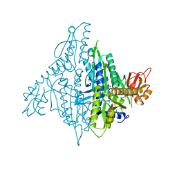
5KXA
 | | Selective Inhibition of Autotaxin is Effective in Mouse Models of Liver Fibrosis | | 分子名称: | 2-acetamido-2-deoxy-beta-D-glucopyranose, 3-[6-chloranyl-2-cyclopropyl-1-(1-ethylpyrazol-4-yl)-7-fluoranyl-indol-3-yl]sulfanyl-2-fluoranyl-benzoic acid, CALCIUM ION, ... | | 著者 | Stein, A.J, Bain, G, Hutchinson, J.H, Evans, J.F. | | 登録日 | 2016-07-20 | | 公開日 | 2016-11-09 | | 最終更新日 | 2023-10-04 | | 実験手法 | X-RAY DIFFRACTION (2.59 Å) | | 主引用文献 | Selective Inhibition of Autotaxin Is Efficacious in Mouse Models of Liver Fibrosis.
J. Pharmacol. Exp. Ther., 360, 2017
|
|
5IDH
 | |
5ICH
 | |
5IBY
 | |
5ICL
 | |
4LKM
 | | Crystal structure of Plk1 Polo-box domain in complex with PL-74 | | 分子名称: | GLYCEROL, PL-74, SULFATE ION, ... | | 著者 | Lee, W.C, Song, J.H, Kim, H.Y. | | 登録日 | 2013-07-08 | | 公開日 | 2013-12-11 | | 最終更新日 | 2023-11-15 | | 実験手法 | X-RAY DIFFRACTION (2 Å) | | 主引用文献 | Exploring the binding nature of pyrrolidine pocket-dependent interactions in the polo-box domain of polo-like kinase 1
Plos One, 8, 2013
|
|
4LKL
 | |
1Y6A
 | | Crystal structure of VEGFR2 in complex with a 2-anilino-5-aryl-oxazole inhibitor | | 分子名称: | N-[5-(ETHYLSULFONYL)-2-METHOXYPHENYL]-5-[3-(2-PYRIDINYL)PHENYL]-1,3-OXAZOL-2-AMINE, Vascular endothelial growth factor receptor 2 | | 著者 | Harris, P.A, Cheung, M, Hunter, R.N, Brown, M.L, Veal, J.M, Nolte, R.T, Wang, L, Liu, W, Crosby, R.M, Johnson, J.H, Epperly, A.H, Kumar, R, Luttrell, D.K, Stafford, J.A. | | 登録日 | 2004-12-05 | | 公開日 | 2005-06-07 | | 最終更新日 | 2024-02-14 | | 実験手法 | X-RAY DIFFRACTION (2.1 Å) | | 主引用文献 | Discovery and evaluation of 2-anilino-5-aryloxazoles as a novel class of VEGFR2 kinase inhibitors.
J.Med.Chem., 48, 2005
|
|
1Y6B
 | | Crystal structure of VEGFR2 in complex with a 2-anilino-5-aryl-oxazole inhibitor | | 分子名称: | N-(CYCLOPROPYLMETHYL)-4-(METHYLOXY)-3-({5-[3-(3-PYRIDINYL)PHENYL]-1,3-OXAZOL-2-YL}AMINO)BENZENESULFONAMIDE, Vascular endothelial growth factor receptor 2 | | 著者 | Harris, P.A, Cheung, M, Hunter, R.N, Brown, M.L, Veal, J.M, Nolte, R.T, Wang, L, Liu, W, Crosby, R.M, Johnson, J.H, Epperly, A.H, Kumar, R, Luttrell, D.K, Stafford, J.A. | | 登録日 | 2004-12-05 | | 公開日 | 2005-06-07 | | 最終更新日 | 2024-02-14 | | 実験手法 | X-RAY DIFFRACTION (2.1 Å) | | 主引用文献 | Discovery and evaluation of 2-anilino-5-aryloxazoles as a novel class of VEGFR2 kinase inhibitors.
J.Med.Chem., 48, 2005
|
|
5UMB
 | | Crystal structure of ATPase domain of Malaria GRP78 with ADP bound | | 分子名称: | ADENOSINE-5'-DIPHOSPHATE, Chaperone DnaK, MAGNESIUM ION, ... | | 著者 | Chen, Y, Antoshchenko, T, Pizarro, J.C, Song, J.H, Park, H. | | 登録日 | 2017-01-26 | | 公開日 | 2022-07-27 | | 最終更新日 | 2023-10-04 | | 実験手法 | X-RAY DIFFRACTION (2.3 Å) | | 主引用文献 | Repurposing drugs to target the malaria parasite unfolding protein response.
Sci Rep, 8, 2018
|
|
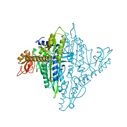
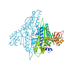
6NYA
 | | Crystal Structure of ubiquitin E1 (Uba1) in complex with Ubc3 (Cdc34) and ubiquitin | | 分子名称: | 1,2-ETHANEDIOL, ADENOSINE-5'-TRIPHOSPHATE, MAGNESIUM ION, ... | | 著者 | Olsen, S.K, Williams, K.M, Atkison, J.H. | | 登録日 | 2019-02-11 | | 公開日 | 2019-08-07 | | 最終更新日 | 2023-10-11 | | 実験手法 | X-RAY DIFFRACTION (2.065 Å) | | 主引用文献 | Structural insights into E1 recognition and the ubiquitin-conjugating activity of the E2 enzyme Cdc34.
Nat Commun, 10, 2019
|
|


