6B2T
 
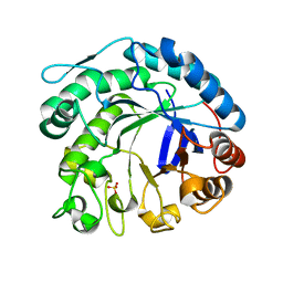
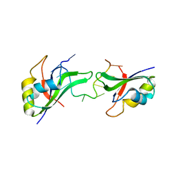
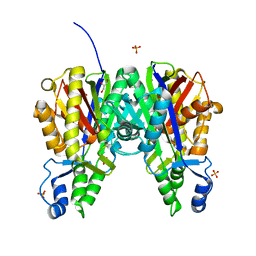 | | Crystal structure of Xanthomonas campestris OleA H285D | | 分子名称: | 3-oxoacyl-[ACP] synthase III, GLYCEROL, PHOSPHATE ION | | 著者 | Jensen, M.R, Goblirsch, B.R, Esler, M.A, Christenson, J.K, Mohamed, F.A, Wackett, L.P, Wilmot, C.M. | | 登録日 | 2017-09-20 | | 公開日 | 2018-02-28 | | 最終更新日 | 2020-01-01 | | 実験手法 | X-RAY DIFFRACTION (2.8 Å) | | 主引用文献 | The role of OleA His285 in orchestration of long-chain acyl-coenzyme A substrates.
FEBS Lett., 592, 2018
|
|
6CWY
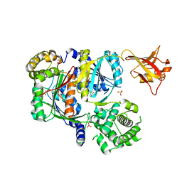 | | Crystal structure of SUMO E1 in complex with an allosteric inhibitor | | 分子名称: | GLYCEROL, MAGNESIUM ION, SULFATE ION, ... | | 著者 | Lv, Z, Yuan, L, Atkison, J.H, Williams, K.M, Olsen, S.K. | | 登録日 | 2018-04-01 | | 公開日 | 2019-01-16 | | 最終更新日 | 2023-10-04 | | 実験手法 | X-RAY DIFFRACTION (2.462 Å) | | 主引用文献 | Molecular mechanism of a covalent allosteric inhibitor of SUMO E1 activating enzyme.
Nat Commun, 9, 2018
|
|
5C6K
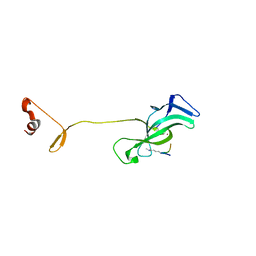
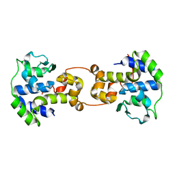 | | Bacteriophage P2 integrase catalytic domain | | 分子名称: | Integrase | | 著者 | Skaar, K, Claesson, M, Odegrip, R, Eriksson, J, Hogbom, M, Haggard-Ljungquist, E, Stenmark, P. | | 登録日 | 2015-06-23 | | 公開日 | 2015-10-21 | | 最終更新日 | 2024-01-10 | | 実験手法 | X-RAY DIFFRACTION (1.9 Å) | | 主引用文献 | Crystal structure of the bacteriophage P2 integrase catalytic domain.
Febs Lett., 589, 2015
|
|
6CO2
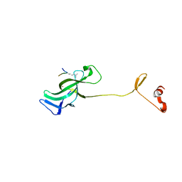
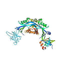 | | Structure of an engineered protein (NUDT16TI) in complex with 53BP1 Tudor domains | | 分子名称: | NUDT16-Tudor-interacting (NUDT16TI), TP53-binding protein 1 | | 著者 | Botuyan, M.V, Thompson, J.R, Cui, G, Mer, G. | | 登録日 | 2018-03-10 | | 公開日 | 2018-06-06 | | 最終更新日 | 2023-10-04 | | 実験手法 | X-RAY DIFFRACTION (2.49 Å) | | 主引用文献 | Mechanism of 53BP1 activity regulation by RNA-binding TIRR and a designer protein.
Nat. Struct. Mol. Biol., 25, 2018
|
|
2C0H
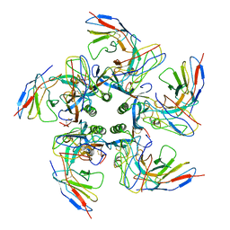
 | | X-ray structure of beta-mannanase from blue mussel Mytilus edulis | | 分子名称: | MANNAN ENDO-1,4-BETA-MANNOSIDASE, SULFATE ION | | 著者 | Larsson, A.M, Anderson, L, Xu, B, Munoz, I.G, Uson, I, Janson, J.-C, Stalbrand, H, Stahlberg, J. | | 登録日 | 2005-09-02 | | 公開日 | 2006-02-22 | | 最終更新日 | 2024-05-08 | | 実験手法 | X-RAY DIFFRACTION (1.6 Å) | | 主引用文献 | Three-Dimensional Crystal Structure and Enzymic Characterization of Beta-Mannanase Man5A from Blue Mussel Mytilus Edulis.
J.Mol.Biol., 357, 2006
|
|
7LMM
 | |
7LMK
 | |
7K7I
 | | Density-fitted Model Structure of Antibody Variable Domains of TyTx4 in Complex with PltB pentamer of Typhoid Toxin | | 分子名称: | Fab Heavy Chain Variable Domain, Fab Light Chain Variable Domain, Putative pertussis-like toxin subunit | | 著者 | Nguyen, T, Feathers, J.R, Fromme, J.C, Song, J. | | 登録日 | 2020-09-22 | | 公開日 | 2021-09-01 | | 最終更新日 | 2021-09-22 | | 実験手法 | ELECTRON MICROSCOPY (3.13 Å) | | 主引用文献 | The structural basis of Salmonella A 2 B 5 toxin neutralization by antibodies targeting the glycan-receptor binding subunits.
Cell Rep, 36, 2021
|
|
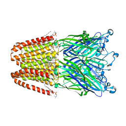
3P50
 | | Structure of propofol bound to a pentameric ligand-gated ion channel, GLIC | | 分子名称: | 2,6-BIS(1-METHYLETHYL)PHENOL, DIUNDECYL PHOSPHATIDYL CHOLINE, DODECYL-BETA-D-MALTOSIDE, ... | | 著者 | Nury, H, Van Renterghem, C, Weng, Y, Tran, A, Baaden, M, Dufresne, V, Changeux, J.P, Sonner, J.M, Delarue, M, Corringer, P.J. | | 登録日 | 2010-10-07 | | 公開日 | 2011-01-19 | | 最終更新日 | 2023-11-01 | | 実験手法 | X-RAY DIFFRACTION (3.3 Å) | | 主引用文献 | X-ray structures of general anaesthetics bound to a pentameric ligand-gated ion channel
Nature, 469, 2011
|
|
3P4W
 | | Structure of desflurane bound to a pentameric ligand-gated ion channel, GLIC | | 分子名称: | (2S)-2-(difluoromethoxy)-1,1,1,2-tetrafluoroethane, DIUNDECYL PHOSPHATIDYL CHOLINE, DODECYL-BETA-D-MALTOSIDE, ... | | 著者 | Nury, H, Van Renterghem, C, Weng, Y, Tran, A, Baaden, M, Dufresne, V, Changeux, J.P, Sonner, J.M, Delarue, M, Corringer, P.J. | | 登録日 | 2010-10-07 | | 公開日 | 2011-01-19 | | 最終更新日 | 2023-11-01 | | 実験手法 | X-RAY DIFFRACTION (3.2 Å) | | 主引用文献 | X-ray structures of general anaesthetics bound to a pentameric ligand-gated ion channel
Nature, 469, 2011
|
|
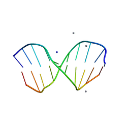
1ZF3
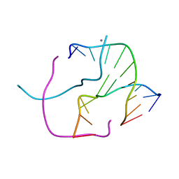 | | ATC Four-stranded DNA Holliday Junction | | 分子名称: | 5'-D(*CP*CP*GP*AP*TP*AP*TP*CP*GP*G)-3', CALCIUM ION | | 著者 | Hays, F.A, Teegarden, A.T, Jones, Z.J.R, Harms, M, Raup, D, Watson, J, Cavaliere, E, Ho, P.S. | | 登録日 | 2005-04-19 | | 公開日 | 2005-05-10 | | 最終更新日 | 2024-04-03 | | 実験手法 | X-RAY DIFFRACTION (1.84 Å) | | 主引用文献 | How sequence defines structure: a crystallographic map of DNA structure and conformation.
Proc.Natl.Acad.Sci.Usa, 102, 2005
|
|
1ZFH
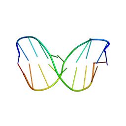 | | TTA Duplex B-DNA | | 分子名称: | 5'-D(*CP*CP*TP*AP*AP*TP*TP*AP*GP*G)-3' | | 著者 | Hays, F.A, Teegarden, A.T, Jones, Z.J.R, Harms, M, Raup, D, Watson, J, Cavaliere, E, Ho, P.S. | | 登録日 | 2005-04-20 | | 公開日 | 2005-05-10 | | 最終更新日 | 2024-04-03 | | 実験手法 | X-RAY DIFFRACTION (2.51 Å) | | 主引用文献 | How sequence defines structure: a crystallographic map of DNA structure and conformation.
Proc.Natl.Acad.Sci.Usa, 102, 2005
|
|
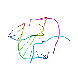
1ZEZ
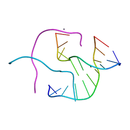 | | ACC Holliday Junction | | 分子名称: | 5'-D(*CP*CP*GP*GP*TP*AP*CP*CP*GP*G)-3', CALCIUM ION | | 著者 | Hays, F.A, Teegarden, A.T, Jones, Z.J.R, Harms, M, Raup, D, Watson, J, Cavaliere, E, Ho, P.S. | | 登録日 | 2005-04-19 | | 公開日 | 2005-05-10 | | 最終更新日 | 2024-04-03 | | 実験手法 | X-RAY DIFFRACTION (2 Å) | | 主引用文献 | How sequence defines structure: a crystallographic map of DNA structure and conformation.
Proc.Natl.Acad.Sci.Usa, 102, 2005
|
|
1ZF7
 | | GAC Duplex B-DNA | | 分子名称: | 5'-D(*CP*CP*GP*TP*CP*GP*AP*CP*GP*G)-3', CALCIUM ION, SODIUM ION | | 著者 | Hays, F.A, Teegarden, A.T, Jones, Z.J.R, Harms, M, Raup, D, Watson, J, Cavaliere, E, Ho, P.S. | | 登録日 | 2005-04-20 | | 公開日 | 2005-05-10 | | 最終更新日 | 2024-04-03 | | 実験手法 | X-RAY DIFFRACTION (1.05 Å) | | 主引用文献 | How sequence defines structure: a crystallographic map of DNA structure and conformation.
Proc.Natl.Acad.Sci.Usa, 102, 2005
|
|
3P8D
 | | Crystal structure of the second Tudor domain of human PHF20 (homodimer form) | | 分子名称: | Medulloblastoma antigen MU-MB-50.72 | | 著者 | Cui, G, Lee, J, Thompson, J.R, Botuyan, M.V, Mer, G. | | 登録日 | 2010-10-13 | | 公開日 | 2011-06-22 | | 最終更新日 | 2012-09-26 | | 実験手法 | X-RAY DIFFRACTION (2 Å) | | 主引用文献 | PHF20 is an effector protein of p53 double lysine methylation that stabilizes and activates p53.
Nat.Struct.Mol.Biol., 19, 2012
|
|
4AXP
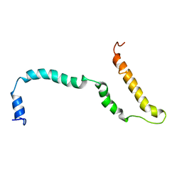 | | NMR structure of Hsp12, a protein induced by and required for dietary restriction-induced lifespan extension in yeast. | | 分子名称: | 12 KDA HEAT SHOCK PROTEIN | | 著者 | Herbert, A.P, Riesen, M, Bloxam, L, Kosmidou, E, Wareing, B.M, Johnson, J.R, Phelan, M.M, Pennington, S.R, Lian, L.Y, Morgan, A. | | 登録日 | 2012-06-13 | | 公開日 | 2012-08-08 | | 最終更新日 | 2024-05-15 | | 実験手法 | SOLUTION NMR | | 主引用文献 | NMR Structure of Hsp12, a Protein Induced by and Required for Dietary Restriction-Induced Lifespan Extension in Yeast.
Plos One, 7, 2012
|
|
1ZF4
 | | ATC Four-stranded DNA Holliday Junction | | 分子名称: | 5'-D(*CP*CP*GP*AP*TP*AP*TP*CP*GP*G)-3', SODIUM ION | | 著者 | Hays, F.A, Teegarden, A.T, Jones, Z.J.R, Harms, M, Raup, D, Watson, J, Cavaliere, E, Ho, P.S. | | 登録日 | 2005-04-19 | | 公開日 | 2005-05-10 | | 最終更新日 | 2024-04-03 | | 実験手法 | X-RAY DIFFRACTION (1.65 Å) | | 主引用文献 | How sequence defines structure: a crystallographic map of DNA structure and conformation.
Proc.Natl.Acad.Sci.Usa, 102, 2005
|
|
1ZFF
 | | TTC Duplex B-DNA | | 分子名称: | 5'-D(*CP*CP*GP*AP*AP*TP*TP*CP*GP*G)-3' | | 著者 | Hays, F.A, Teegarden, A.T, Jones, Z.J.R, Harms, M, Raup, D, Watson, J, Cavaliere, E, Ho, P.S. | | 登録日 | 2005-04-20 | | 公開日 | 2005-05-10 | | 最終更新日 | 2024-04-03 | | 実験手法 | X-RAY DIFFRACTION (0.94 Å) | | 主引用文献 | How sequence defines structure: a crystallographic map of DNA structure and conformation.
Proc.Natl.Acad.Sci.Usa, 102, 2005
|
|
2O8O
 | |
3NMM
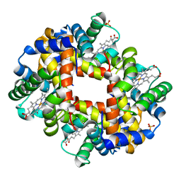 | | Human Hemoglobin A mutant alpha H58W deoxy-form | | 分子名称: | Hemoglobin subunit alpha, Hemoglobin subunit beta, PROTOPORPHYRIN IX CONTAINING FE, ... | | 著者 | Birukou, I, Soman, J, Olson, J.S. | | 登録日 | 2010-06-22 | | 公開日 | 2010-07-14 | | 最終更新日 | 2023-09-06 | | 実験手法 | X-RAY DIFFRACTION (1.6 Å) | | 主引用文献 | Blocking the gate to ligand entry in human hemoglobin.
J.Biol.Chem., 286, 2011
|
|
1ZEY
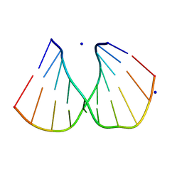 | | CGG A-DNA | | 分子名称: | 5'-D(*CP*CP*CP*CP*GP*CP*GP*GP*GP*G)-3', SODIUM ION | | 著者 | Hays, F.A, Teegarden, A.T, Jones, Z.J.R, Harms, M, Raup, D, Watson, J, Cavaliere, E, Ho, P.S. | | 登録日 | 2005-04-19 | | 公開日 | 2005-05-10 | | 最終更新日 | 2024-04-03 | | 実験手法 | X-RAY DIFFRACTION (1.7 Å) | | 主引用文献 | How sequence defines structure: a crystallographic map of DNA structure and conformation.
Proc.Natl.Acad.Sci.Usa, 102, 2005
|
|
1ZF6
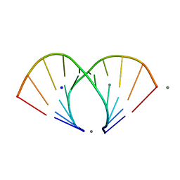 | | TGG DUPLEX A-DNA | | 分子名称: | 5'-D(*CP*CP*CP*CP*AP*TP*GP*GP*GP*G)-3', CALCIUM ION, SODIUM ION | | 著者 | Hays, F.A, Teegarden, A.T, Jones, Z.J.R, Harms, M, Raup, D, Watson, J, Cavaliere, E, Ho, P.S. | | 登録日 | 2005-04-19 | | 公開日 | 2005-05-10 | | 最終更新日 | 2024-04-03 | | 実験手法 | X-RAY DIFFRACTION (1.5 Å) | | 主引用文献 | How sequence defines structure: a crystallographic map of DNA structure and conformation.
Proc.Natl.Acad.Sci.Usa, 102, 2005
|
|
3OPE
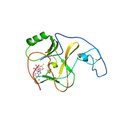 | |
3PKP
 | | Q83S Variant of S. Enterica RmlA with dATP | | 分子名称: | 2'-DEOXYADENOSINE 5'-TRIPHOSPHATE, Glucose-1-phosphate thymidylyltransferase, MAGNESIUM ION | | 著者 | Chang, A, Moretti, R, Bingman, C.A, Thorson, J.S, Phillips Jr, G.N, Center for Eukaryotic Structural Genomics (CESG) | | 登録日 | 2010-11-11 | | 公開日 | 2011-01-12 | | 最終更新日 | 2024-02-21 | | 実験手法 | X-RAY DIFFRACTION (2.6 Å) | | 主引用文献 | Expanding the Nucleotide and Sugar 1-Phosphate Promiscuity of Nucleotidyltransferase RmlA via Directed Evolution.
J.Biol.Chem., 286, 2011
|
|
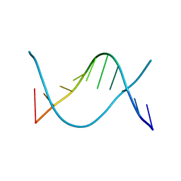
3NNH
 | | Crystal Structure of the CUGBP1 RRM1 with GUUGUUUUGUUU RNA | | 分子名称: | CUGBP Elav-like family member 1, RNA (5'-R(*GP*UP*UP*GP*UP*UP*UP*UP*GP*UP*UP*U)-3') | | 著者 | Teplova, M, Song, J, Gaw, H, Teplov, A, Patel, D.J. | | 登録日 | 2010-06-23 | | 公開日 | 2010-10-27 | | 最終更新日 | 2023-09-06 | | 実験手法 | X-RAY DIFFRACTION (2.7501 Å) | | 主引用文献 | Structural Insights into RNA Recognition by the Alternate-Splicing Regulator CUG-Binding Protein 1.
Structure, 18, 2010
|
|


