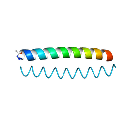
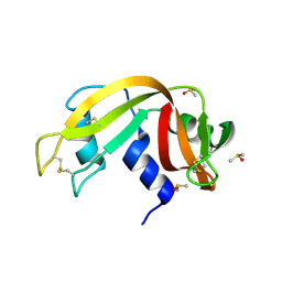
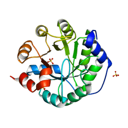
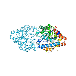
6UNT
 
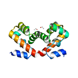 | | Barrier-to-autointegration factor soaked in DMSO: 1 of 14 in MSCS set | | 分子名称: | Barrier-to-autointegration factor, ETHANOL | | 著者 | Agarwal, S, Smith, M, De La Rosa, I, Kliment, A.V, Swartz, P, Segura-Totten, M, Mattos, C. | | 登録日 | 2019-10-13 | | 公開日 | 2020-10-07 | | 最終更新日 | 2024-03-13 | | 実験手法 | X-RAY DIFFRACTION (1.75 Å) | | 主引用文献 | Development of a structure-analysis pipeline using multiple-solvent crystal structures of barrier-to-autointegration factor.
Acta Crystallogr D Struct Biol, 76, 2020
|
|
1PIH
 | | THE THREE DIMENSIONAL STRUCTURE OF THE PARAMAGNETIC PROTEIN HIPIP I FROM E.HALOPHILA THROUGH NUCLEAR MAGNETIC RESONANCE | | 分子名称: | HIGH POTENTIAL IRON SULFUR PROTEIN, IRON/SULFUR CLUSTER | | 著者 | Banci, L, Bertini, I, Eltis, L.D, Felli, I, Kastrau, D.H.W, Luchinat, C, Piccioli, M, Pierattelli, R, Smith, M. | | 登録日 | 1994-08-03 | | 公開日 | 1994-12-20 | | 最終更新日 | 2024-05-22 | | 実験手法 | SOLUTION NMR | | 主引用文献 | The three-dimensional structure in solution of the paramagnetic high-potential iron-sulfur protein I from Ectothiorhodospira halophila through nuclear magnetic resonance.
Eur.J.Biochem., 225, 1994
|
|
1PIJ
 | | THE THREE DIMENSIONAL STRUCTURE OF THE PARAMAGNETIC PROTEIN HIPIP I FROM E.HALOPHILA THROUGH NUCLEAR MAGNETIC RESONANCE | | 分子名称: | HIGH POTENTIAL IRON SULFUR PROTEIN, IRON/SULFUR CLUSTER | | 著者 | Banci, L, Bertini, I, Eltis, L.D, Felli, I.C, Kastrau, D.H.W, Luchinat, C, Piccioli, M, Pierattelli, R, Smith, M. | | 登録日 | 1994-11-11 | | 公開日 | 1995-02-07 | | 最終更新日 | 2024-05-22 | | 実験手法 | SOLUTION NMR | | 主引用文献 | The three-dimensional structure in solution of the paramagnetic high-potential iron-sulfur protein I from Ectothiorhodospira halophila through nuclear magnetic resonance.
Eur.J.Biochem., 225, 1994
|
|
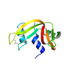
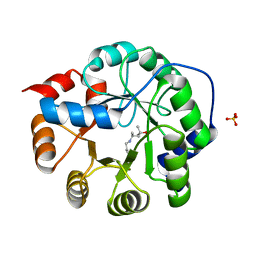
6O2E
 | |
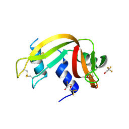
6O2F
 | |
1NZ2
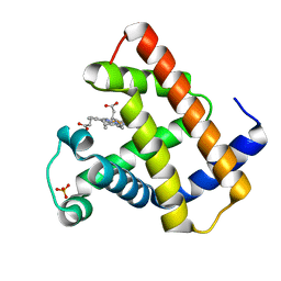 | | K45E Variant of Horse Heart Myoglobin | | 分子名称: | Myoglobin, PROTOPORPHYRIN IX CONTAINING FE, SULFATE ION | | 著者 | Hunter, C.L, Maurus, R, Mauk, M.R, Lee, H, Raven, E.L, Tong, H, Nguyen, N, Smith, M, Brayer, G.D, Mauk, A.G. | | 登録日 | 2003-02-15 | | 公開日 | 2003-04-08 | | 最終更新日 | 2023-08-16 | | 実験手法 | X-RAY DIFFRACTION (1.9 Å) | | 主引用文献 | Introduction and characterization of a functionally linked metal ion binding site at
the exposed heme edge of myoglobin
Proc.Natl.Acad.Sci.USA, 100, 2003
|
|
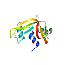
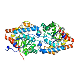
3EV3
 | | Crystal Structure of Ribonuclease A in 70% t-Butanol | | 分子名称: | Ribonuclease pancreatic, TERTIARY-BUTYL ALCOHOL | | 著者 | Dechene, M, Wink, G, Smith, M, Swartz, P, Mattos, C. | | 登録日 | 2008-10-12 | | 公開日 | 2009-06-23 | | 最終更新日 | 2023-12-27 | | 実験手法 | X-RAY DIFFRACTION (1.68 Å) | | 主引用文献 | Multiple solvent crystal structures of ribonuclease A: An assessment of the method
Proteins, 76, 2009
|
|
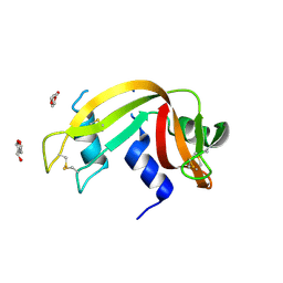
3EUY
 | | Crystal Structure of Ribonuclease A in 50% Dioxane | | 分子名称: | 1,4-DIETHYLENE DIOXIDE, Ribonuclease pancreatic | | 著者 | Dechene, M, Wink, G, Smith, M, Swartz, P, Mattos, C. | | 登録日 | 2008-10-12 | | 公開日 | 2009-06-23 | | 最終更新日 | 2023-12-27 | | 実験手法 | X-RAY DIFFRACTION (1.95 Å) | | 主引用文献 | Multiple solvent crystal structures of ribonuclease A: An assessment of the method
Proteins, 76, 2009
|
|
3EV6
 | | Crystal Structure of Ribonuclease A in 50% R,S,R-Bisfuranol | | 分子名称: | (3R,3aS,6aR)-hexahydrofuro[2,3-b]furan-3-ol, Ribonuclease pancreatic | | 著者 | Dechene, M, Wink, G, Smith, M, Swartz, P, Mattos, C. | | 登録日 | 2008-10-12 | | 公開日 | 2009-06-23 | | 最終更新日 | 2023-12-27 | | 実験手法 | X-RAY DIFFRACTION (1.76 Å) | | 主引用文献 | Multiple solvent crystal structures of ribonuclease A: An assessment of the method
Proteins, 76, 2009
|
|
3EV0
 | | Crystal Structure of Ribonuclease A in 70% Dimethyl Sulfoxide | | 分子名称: | DIMETHYL SULFOXIDE, Ribonuclease pancreatic | | 著者 | Dechene, M, Wink, G, Smith, M, Swartz, P, Mattos, C. | | 登録日 | 2008-10-12 | | 公開日 | 2009-06-23 | | 最終更新日 | 2023-12-27 | | 実験手法 | X-RAY DIFFRACTION (1.76 Å) | | 主引用文献 | Multiple solvent crystal structures of ribonuclease A: An assessment of the method
Proteins, 76, 2009
|
|
3EUX
 | | Crystal Structure of Crosslinked Ribonuclease A | | 分子名称: | Ribonuclease pancreatic | | 著者 | Dechene, M, Wink, G, Smith, M, Swartz, P, Mattos, C. | | 登録日 | 2008-10-12 | | 公開日 | 2009-06-23 | | 最終更新日 | 2023-12-27 | | 実験手法 | X-RAY DIFFRACTION (1.65 Å) | | 主引用文献 | Multiple solvent crystal structures of ribonuclease A: An assessment of the method
Proteins, 76, 2009
|
|
3EV4
 | | Crystal Structure of Ribonuclease A in 50% Trifluoroethanol | | 分子名称: | Ribonuclease pancreatic, TRIFLUOROETHANOL | | 著者 | Dechene, M, Wink, G, Smith, M, Swartz, P, Mattos, C. | | 登録日 | 2008-10-12 | | 公開日 | 2009-06-23 | | 最終更新日 | 2023-12-27 | | 実験手法 | X-RAY DIFFRACTION (1.93 Å) | | 主引用文献 | Multiple solvent crystal structures of ribonuclease A: An assessment of the method
Proteins, 76, 2009
|
|
3EUZ
 | | Crystal Structure of Ribonuclease A in 50% Dimethylformamide | | 分子名称: | DIMETHYLFORMAMIDE, Ribonuclease pancreatic | | 著者 | Dechene, M, Wink, G, Smith, M, Swartz, P, Mattos, C. | | 登録日 | 2008-10-12 | | 公開日 | 2009-06-23 | | 最終更新日 | 2023-12-27 | | 実験手法 | X-RAY DIFFRACTION (1.84 Å) | | 主引用文献 | Multiple solvent crystal structures of ribonuclease A: An assessment of the method
Proteins, 76, 2009
|
|
3EV1
 | | Crystal Structure of Ribonuclease A in 70% Hexanediol | | 分子名称: | HEXANE-1,6-DIOL, Ribonuclease pancreatic | | 著者 | Dechene, M, Wink, G, Smith, M, Swartz, P, Mattos, C. | | 登録日 | 2008-10-12 | | 公開日 | 2009-06-23 | | 最終更新日 | 2023-12-27 | | 実験手法 | X-RAY DIFFRACTION (2 Å) | | 主引用文献 | Multiple solvent crystal structures of ribonuclease A: An assessment of the method
Proteins, 76, 2009
|
|
3EV5
 | | Crystal Structure of Ribonuclease A in 1M Trimethylamine N-Oxide | | 分子名称: | Ribonuclease pancreatic, trimethylamine oxide | | 著者 | Dechene, M, Wink, G, Smith, M, Swartz, P, Mattos, C. | | 登録日 | 2008-10-12 | | 公開日 | 2009-06-23 | | 最終更新日 | 2023-12-27 | | 実験手法 | X-RAY DIFFRACTION (1.68 Å) | | 主引用文献 | Multiple solvent crystal structures of ribonuclease A: An assessment of the method
Proteins, 76, 2009
|
|
3EV2
 | | Crystal Structure of Ribonuclease A in 70% Isopropanol | | 分子名称: | ISOPROPYL ALCOHOL, Ribonuclease pancreatic | | 著者 | Dechene, M, Wink, G, Smith, M, Swartz, P, Mattos, C. | | 登録日 | 2008-10-12 | | 公開日 | 2009-06-23 | | 最終更新日 | 2023-12-27 | | 実験手法 | X-RAY DIFFRACTION (2.02 Å) | | 主引用文献 | Multiple solvent crystal structures of ribonuclease A: An assessment of the method
Proteins, 76, 2009
|
|
6MYN
 | | Crystal structure of murine NF-kappaB inducing kinase (NIK) bound to inhibitor R7 | | 分子名称: | (5s,7s)-9-fluoro-10-[(3R)-3-hydroxy-3-(5-methyl-1,2-oxazol-3-yl)but-1-yn-1-yl]-N~3~-methyl-6,7-dihydro-5H-5,7-methanoimidazo[2,1-a][2]benzazepine-2,3-dicarboxamide, Mitogen-activated protein kinase kinase kinase 14, SULFATE ION | | 著者 | Harris, S.F, Smith, M, Barker, J. | | 登録日 | 2018-11-01 | | 公開日 | 2019-08-07 | | 最終更新日 | 2024-03-13 | | 実験手法 | X-RAY DIFFRACTION (2.744 Å) | | 主引用文献 | Structure Based Design of Potent Selective Inhibitors of Protein Kinase D1 (PKD1).
Acs Med.Chem.Lett., 10, 2019
|
|
3O6Y
 | | Robust computational design, optimization, and structural characterization of retroaldol enzymes | | 分子名称: | Retro-Aldolase, SULFATE ION | | 著者 | Althoff, E.A, Wang, L, Jiang, L, Moody, J, Bolduc, J, Lassila, J.K, Wang, Z.Z, Smith, M, Hari, S, Herschlag, D, Stoddard, B.L, Baker, D. | | 登録日 | 2010-07-29 | | 公開日 | 2011-06-29 | | 最終更新日 | 2023-09-06 | | 実験手法 | X-RAY DIFFRACTION (2.091 Å) | | 主引用文献 | Structural analyses of covalent enzyme-substrate analog complexes reveal strengths and limitations of de novo enzyme design.
J.Mol.Biol., 415, 2012
|
|
3NXF
 | | Robust computational design, optimization, and structural characterization of retroaldol enzymes | | 分子名称: | Retro-Aldolase, SULFATE ION | | 著者 | Althoff, E.A, Jiang, L, Wang, L, Lassila, J.K, Moody, J, Bolduc, J, Wang, Z.Z, Smith, M, Hari, S, Herschlag, D, Stoddard, B.L, Baker, D. | | 登録日 | 2010-07-13 | | 公開日 | 2011-06-29 | | 最終更新日 | 2023-09-06 | | 実験手法 | X-RAY DIFFRACTION (2.4 Å) | | 主引用文献 | Structural analyses of covalent enzyme-substrate analog complexes reveal strengths and limitations of de novo enzyme design.
J.Mol.Biol., 415, 2012
|
|
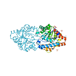
5HQ3
 | | Stable, high-expression variant of human acetylcholinesterase | | 分子名称: | 2-(N-MORPHOLINO)-ETHANESULFONIC ACID, Acetylcholinesterase, O-ETHYLMETHYLPHOSPHONIC ACID ESTER GROUP | | 著者 | Goldenzweig, A, Goldsmith, M, Hill, S.E, Gertman, O, Laurino, P, Ashani, Y, Dym, O, Albeck, S, Unger, T, Prilusky, J, Lieberman, R.L, Aharoni, A, Silman, I, Sussman, J.L, Tawfik, D.S, Fleishman, S.J. | | 登録日 | 2016-01-21 | | 公開日 | 2016-07-27 | | 最終更新日 | 2024-01-10 | | 実験手法 | X-RAY DIFFRACTION (2.6 Å) | | 主引用文献 | Automated Structure- and Sequence-Based Design of Proteins for High Bacterial Expression and Stability.
Mol.Cell, 63, 2016
|
|
6FQE
 | | Phosphotriesterase PTE_A53_4 | | 分子名称: | (4~{S},6~{R})-2,2,6-trimethyl-1,3-dioxan-4-ol, DI(HYDROXYETHYL)ETHER, FORMIC ACID, ... | | 著者 | Dym, O, Aggarwal, N, Albeck, S, Unger, T, Hamer Rogotner, S, Silman, I, Leader, H, Ashani, Y, Goldsmith, M, Greisen, P, Tawfik, D, Sussman, L.J. | | 登録日 | 2018-02-14 | | 公開日 | 2019-03-13 | | 最終更新日 | 2024-01-17 | | 実験手法 | X-RAY DIFFRACTION (1.75 Å) | | 主引用文献 | Phosphotriesterase
PTE_A53_4
To Be Published
|
|
6G1J
 | | Phosphotriesterase PTE_C23M_1 | | 分子名称: | 1-ethyl-1-methyl-cyclohexane, FORMIC ACID, GLYCEROL, ... | | 著者 | Dym, O, Aggarwal, N, Albeck, S, Unger, T, Hamer Rogotner, S, Silman, I, Leader, H, Ashani, Y, Goldsmith, M, Greisen, P, Tawfik, D, Sussman, L.J. | | 登録日 | 2018-03-21 | | 公開日 | 2019-04-10 | | 最終更新日 | 2024-01-17 | | 実験手法 | X-RAY DIFFRACTION (2.1 Å) | | 主引用文献 | Phosphotriesterase
PTE_C23M_1
To Be Published
|
|
6FWE
 | | Phosphotriesterase PTE_C23_6 | | 分子名称: | 1-[methoxy(methyl)phosphoryl]oxyethane, 1-ethyl-1-methyl-cyclohexane, FORMIC ACID, ... | | 著者 | Dym, O, Aggarwal, N, Albeck, S, Unger, T, Hamer Rogotner, S, Silman, I, Leader, H, Ashani, Y, Goldsmith, M, Greisen, P, Tawfik, D, Sussman, L.J. | | 登録日 | 2018-03-06 | | 公開日 | 2019-03-27 | | 最終更新日 | 2024-01-17 | | 実験手法 | X-RAY DIFFRACTION (1.774 Å) | | 主引用文献 | Phosphotriesterase
PTE_C23_6
To Be Published
|
|
6GBL
 | | Repertoires of functionally diverse enzymes through computational design at epistatic active-site positions | | 分子名称: | 1,2-ETHANEDIOL, CACODYLATE ION, FORMIC ACID, ... | | 著者 | Khersonsky, O, Lipsh, R, Avizemer, Z, Goldsmith, M, Ashani, Y, Leader, H, Dym, O, Rogotner, S, Trudeau, D, Tawfik, D.S, Fleishman, S.J. | | 登録日 | 2018-04-15 | | 公開日 | 2018-10-24 | | 最終更新日 | 2024-01-17 | | 実験手法 | X-RAY DIFFRACTION (1.95 Å) | | 主引用文献 | Automated Design of Efficient and Functionally Diverse Enzyme Repertoires.
Mol. Cell, 72, 2018
|
|
6FU6
 | | Phosphotriesterase PTE_C23_2 | | 分子名称: | FORMIC ACID, POLYACRYLIC ACID, Parathion hydrolase, ... | | 著者 | Dym, O, Aggarwal, N, Albeck, S, Unger, T, Hamer Rogotner, S, Silman, I, Leader, H, Ashani, Y, Goldsmith, M, Greisen, P, Tawfik, D, Sussman, L.J. | | 登録日 | 2018-02-26 | | 公開日 | 2019-03-20 | | 最終更新日 | 2024-01-17 | | 実験手法 | X-RAY DIFFRACTION (1.95 Å) | | 主引用文献 | Phosphotriesterase
PTE_A53_4
To Be Published
|
|


