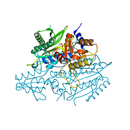
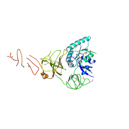
3EDU
 
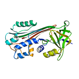
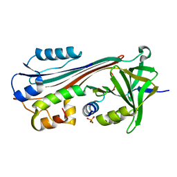
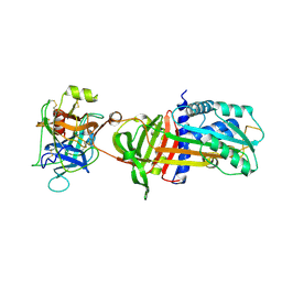
 | | Crystal structure of the ankyrin-binding domain of human erythroid spectrin | | 分子名称: | Spectrin beta chain, erythrocyte | | 著者 | Simonovic, M, Stabach, P, Simonovic, I, Steitz, T.A, Morrow, J.S. | | 登録日 | 2008-09-03 | | 公開日 | 2009-02-10 | | 最終更新日 | 2024-02-21 | | 実験手法 | X-RAY DIFFRACTION (2.1 Å) | | 主引用文献 | The structure of the ankyrin-binding site of {beta}-spectrin reveals how tandem spectrin-repeats generate unique ligand-binding properties
Blood, 113, 2009
|
|
1C8O
 | |
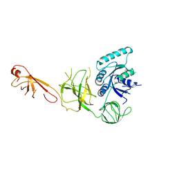
2FOT
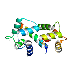 | | Crystal structure of the complex between calmodulin and alphaII-spectrin | | 分子名称: | CALCIUM ION, Calmodulin, alpha-II spectrin Spectrin | | 著者 | Simonovic, M, Zhang, Z, Cianci, C.D, Steitz, T.A, Morrow, J.S. | | 登録日 | 2006-01-13 | | 公開日 | 2006-09-05 | | 最終更新日 | 2023-08-30 | | 実験手法 | X-RAY DIFFRACTION (2.45 Å) | | 主引用文献 | Structure of the calmodulin alphaII-spectrin complex provides insight into the regulation of cell plasticity.
J.Biol.Chem., 281, 2006
|
|
1JBE
 | |
3CME
 | |
3CMA
 | |
1M93
 | |
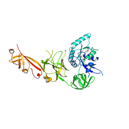
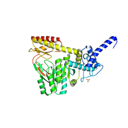
1IMV
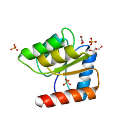
 | | 2.85 A crystal structure of PEDF | | 分子名称: | 2-acetamido-2-deoxy-beta-D-glucopyranose, PIGMENT EPITHELIUM-DERIVED FACTOR | | 著者 | Simonovic, M, Gettins, P.G.W, Volz, K. | | 登録日 | 2001-05-11 | | 公開日 | 2001-09-26 | | 最終更新日 | 2023-08-16 | | 実験手法 | X-RAY DIFFRACTION (2.85 Å) | | 主引用文献 | Crystal structure of human PEDF, a potent anti-angiogenic and neurite growth-promoting factor.
Proc.Natl.Acad.Sci.USA, 98, 2001
|
|
1J8E
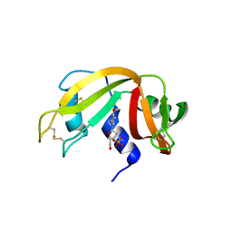
 | | Crystal structure of ligand-binding repeat CR7 from LRP | | 分子名称: | CALCIUM ION, LOW-DENSITY LIPOPROTEIN RECEPTOR-RELATED PROTEIN 1 | | 著者 | Simonovic, M, Dolmer, K, Huang, W, Strickland, D.K, Volz, K, Gettins, P.G.W. | | 登録日 | 2001-05-21 | | 公開日 | 2001-12-19 | | 最終更新日 | 2021-10-27 | | 実験手法 | X-RAY DIFFRACTION (1.85 Å) | | 主引用文献 | Calcium coordination and pH dependence of the calcium affinity of ligand-binding repeat CR7 from the LRP. Comparison with related domains from the LRP and the LDL receptor.
Biochemistry, 40, 2001
|
|
4EO4
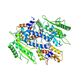
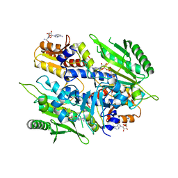 | | Crystal structure of the yeast mitochondrial threonyl-tRNA synthetase (MST1) in complex with seryl sulfamoyl adenylate | | 分子名称: | 5'-O-(N-(L-SERYL)-SULFAMOYL)ADENOSINE, Threonine--tRNA ligase, mitochondrial, ... | | 著者 | Peterson, K.M, Ling, J, Simonovic, I, Soll, D, Simonovic, M. | | 登録日 | 2012-04-13 | | 公開日 | 2012-07-11 | | 最終更新日 | 2024-02-28 | | 実験手法 | X-RAY DIFFRACTION (2.87 Å) | | 主引用文献 | The mechanism of pre-transfer editing in yeast mitochondrial threonyl-tRNA synthetase.
J.Biol.Chem., 287, 2012
|
|
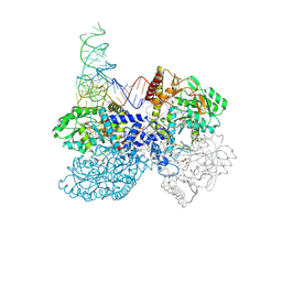
7ZJX
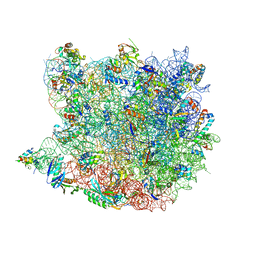
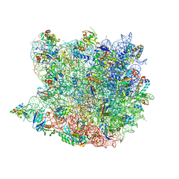
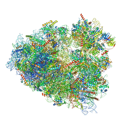 | | Rabbit 80S ribosome programmed with SECIS and SBP2 | | 分子名称: | 18S rRNA, 28S rRNA, 40S Ribosomal protein eS19, ... | | 著者 | Hilal, T, Simonovic, M, Spahn, C.M.T. | | 登録日 | 2022-04-12 | | 公開日 | 2022-09-07 | | 最終更新日 | 2024-04-24 | | 実験手法 | ELECTRON MICROSCOPY (3.1 Å) | | 主引用文献 | Structure of the mammalian ribosome as it decodes the selenocysteine UGA codon.
Science, 376, 2022
|
|
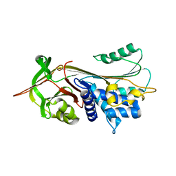
3LXO
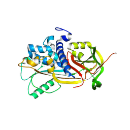
 | | The crystal structure of ribonuclease A in complex with thymidine-3'-monophosphate | | 分子名称: | Ribonuclease pancreatic, THYMIDINE-3'-PHOSPHATE | | 著者 | Doucet, N, Jayasundera, T.B, Simonovic, M, Loria, J.P. | | 登録日 | 2010-02-25 | | 公開日 | 2010-04-28 | | 最終更新日 | 2011-07-13 | | 実験手法 | X-RAY DIFFRACTION (1.549 Å) | | 主引用文献 | The crystal structure of ribonuclease A in complex with thymidine-3'-monophosphate provides further insight into ligand binding.
Proteins, 78, 2010
|
|
8G9Z
 | | High-resolution crystal structure of the human selenomethionine-derived SepSecS-tRNASec complex | | 分子名称: | (4S)-2-METHYL-2,4-PENTANEDIOL, (5-HYDROXY-4,6-DIMETHYLPYRIDIN-3-YL)METHYL DIHYDROGEN PHOSPHATE, O-phosphoseryl-tRNA(Sec) selenium transferase, ... | | 著者 | Puppala, A, Simonovic, M, Castillo Suchkou, J. | | 登録日 | 2023-02-22 | | 公開日 | 2023-04-05 | | 最終更新日 | 2023-05-17 | | 実験手法 | X-RAY DIFFRACTION (2.07 Å) | | 主引用文献 | Structural basis for the tRNA-dependent activation of the terminal complex of selenocysteine synthesis in humans.
Nucleic Acids Res., 51, 2023
|
|
3UGQ
 | | Crystal structure of the apo form of the yeast mitochondrial threonyl-tRNA synthetase determined at 2.1 Angstrom resolution | | 分子名称: | POTASSIUM ION, SULFATE ION, Threonyl-tRNA synthetase, ... | | 著者 | Peterson, K.M, Ling, J, Simonovic, I, Cho, C, Soll, D, Simonovic, M. | | 登録日 | 2011-11-02 | | 公開日 | 2012-02-22 | | 最終更新日 | 2024-02-28 | | 実験手法 | X-RAY DIFFRACTION (2.1 Å) | | 主引用文献 | Yeast mitochondrial threonyl-tRNA synthetase recognizes tRNA isoacceptors by distinct mechanisms and promotes CUN codon reassignment.
Proc.Natl.Acad.Sci.USA, 109, 2012
|
|
3UGT
 | | Crystal structure of the yeast mitochondrial threonyl-tRNA synthetase - orthorhombic crystal form | | 分子名称: | Threonyl-tRNA synthetase, mitochondrial, ZINC ION | | 著者 | Peterson, K.M, Ling, J, Simonovic, I, Cho, C, Soll, D, Simonovic, M. | | 登録日 | 2011-11-02 | | 公開日 | 2012-02-22 | | 最終更新日 | 2024-02-28 | | 実験手法 | X-RAY DIFFRACTION (3.6 Å) | | 主引用文献 | Yeast mitochondrial threonyl-tRNA synthetase recognizes tRNA isoacceptors by distinct mechanisms and promotes CUN codon reassignment.
Proc.Natl.Acad.Sci.USA, 109, 2012
|
|
3UH0
 | | Crystal structure of the yeast mitochondrial threonyl-tRNA synthetase (MST1) in complex with threonyl sulfamoyl adenylate | | 分子名称: | 5'-O-(N-(L-THREONYL)-SULFAMOYL)ADENOSINE, SULFATE ION, Threonyl-tRNA synthetase, ... | | 著者 | Peterson, K.M, Ling, J, Simonovic, I, Cho, C, Soll, D, Simonovic, M. | | 登録日 | 2011-11-03 | | 公開日 | 2012-02-22 | | 最終更新日 | 2024-02-28 | | 実験手法 | X-RAY DIFFRACTION (2 Å) | | 主引用文献 | Yeast mitochondrial threonyl-tRNA synthetase recognizes tRNA isoacceptors by distinct mechanisms and promotes CUN codon reassignment.
Proc.Natl.Acad.Sci.USA, 109, 2012
|
|
7ZJW
 | | Rabbit 80S ribosome as it decodes the Sec-UGA codon | | 分子名称: | 18S rRNA, 28S rRNA, 40S Ribosomal protein eS19, ... | | 著者 | Hilal, T, Simonovic, M, Spahn, C.M.T. | | 登録日 | 2022-04-12 | | 公開日 | 2022-10-19 | | 最終更新日 | 2024-04-24 | | 実験手法 | ELECTRON MICROSCOPY (2.8 Å) | | 主引用文献 | Structure of the mammalian ribosome as it decodes the selenocysteine UGA codon.
Science, 376, 2022
|
|
3HL2
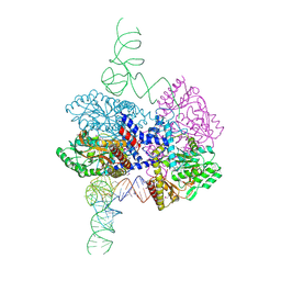
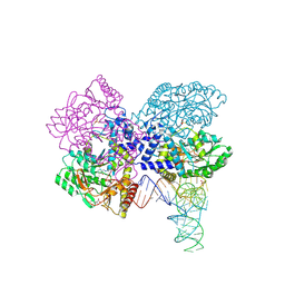 | | The crystal structure of the human SepSecS-tRNASec complex | | 分子名称: | (5-HYDROXY-4,6-DIMETHYLPYRIDIN-3-YL)METHYL DIHYDROGEN PHOSPHATE, Monothiophosphate, O-phosphoseryl-tRNA(Sec) selenium transferase, ... | | 著者 | Palioura, S, Steitz, T.A, Soll, D, Simonovic, M. | | 登録日 | 2009-05-26 | | 公開日 | 2009-10-06 | | 最終更新日 | 2023-09-06 | | 実験手法 | X-RAY DIFFRACTION (2.81 Å) | | 主引用文献 | The human SepSecS-tRNASec complex reveals the mechanism of selenocysteine formation.
Science, 325, 2009
|
|
5IZL
 | |
5IZK
 | |
5IZM
 | |
7L1T
 | | Crystal structure of human holo SepSecS | | 分子名称: | (5-HYDROXY-4,6-DIMETHYLPYRIDIN-3-YL)METHYL DIHYDROGEN PHOSPHATE, O-phosphoseryl-tRNA(Sec) selenium transferase, PHOSPHATE ION | | 著者 | Puppala, A, Castillo Suchkou, J, Simonovic, M. | | 登録日 | 2020-12-15 | | 公開日 | 2022-02-09 | | 最終更新日 | 2023-10-25 | | 実験手法 | X-RAY DIFFRACTION (2.25 Å) | | 主引用文献 | Structural basis for the tRNA-dependent activation of the terminal complex of selenocysteine synthesis in humans.
Nucleic Acids Res., 2023
|
|
7MDL
 | | High-resolution crystal structure of human SepSecS-tRNASec complex | | 分子名称: | (5-HYDROXY-4,6-DIMETHYLPYRIDIN-3-YL)METHYL DIHYDROGEN PHOSPHATE, CITRATE ANION, O-phosphoseryl-tRNA(Sec) selenium transferase, ... | | 著者 | Puppala, A, French, R.L, Simonovic, M. | | 登録日 | 2021-04-05 | | 公開日 | 2022-11-09 | | 最終更新日 | 2023-10-25 | | 実験手法 | X-RAY DIFFRACTION (2.32 Å) | | 主引用文献 | Structural basis for the tRNA-dependent activation of the terminal complex of selenocysteine synthesis in humans.
Nucleic Acids Res., 2023
|
|
1OO8
 | |
1OPH
 | | NON-COVALENT COMPLEX BETWEEN ALPHA-1-PI-PITTSBURGH AND S195A TRYPSIN | | 分子名称: | Alpha-1-antitrypsin precursor, Trypsinogen, cationic precursor | | 著者 | Dementiev, A, Simonovic, M, Volz, K, Gettins, P.G. | | 登録日 | 2003-03-05 | | 公開日 | 2003-08-05 | | 最終更新日 | 2023-08-16 | | 実験手法 | X-RAY DIFFRACTION (2.3 Å) | | 主引用文献 | Canonical inhibitor-like interactions explain reactivity of alpha1-proteinase inhibitor Pittsburgh and antithrombin with proteinases
J.Biol.Chem., 278, 2003
|
|


