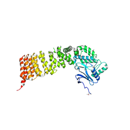
3WAV
 
 | | Crystal Structure of Autotaxin in Complex with Compound 10 | | 分子名称: | (5Z)-5-(3,4-dichlorobenzylidene)-2-(4-methylpiperazin-1-yl)-1,3-thiazol-4(5H)-one, 1,2-ETHANEDIOL, 2-acetamido-2-deoxy-beta-D-glucopyranose-(1-4)-2-acetamido-2-deoxy-beta-D-glucopyranose, ... | | 著者 | Nishimasu, H, Ishitani, R, Nureki, O. | | 登録日 | 2013-05-09 | | 公開日 | 2013-07-31 | | 最終更新日 | 2023-11-08 | | 実験手法 | X-RAY DIFFRACTION (1.797 Å) | | 主引用文献 | Screening and X-ray Crystal Structure-based Optimization of Autotaxin (ENPP2) Inhibitors, Using a Newly Developed Fluorescence Probe
Acs Chem.Biol., 8, 2013
|
|
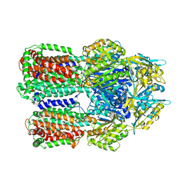
7D5V
 | | Structure of the C646A mutant of peptidylarginine deiminase type III (PAD3) | | 分子名称: | 1,2-ETHANEDIOL, GLYCEROL, Protein-arginine deiminase type-3 | | 著者 | Akimoto, M, Mashimo, R, Unno, M. | | 登録日 | 2020-09-28 | | 公開日 | 2021-06-02 | | 最終更新日 | 2023-11-29 | | 実験手法 | X-RAY DIFFRACTION (2.102 Å) | | 主引用文献 | Structures of human peptidylarginine deiminase type III provide insights into substrate recognition and inhibitor design.
Arch.Biochem.Biophys., 708, 2021
|
|
5XSZ
 | | Crystal structure of zebrafish lysophosphatidic acid receptor LPA6 | | 分子名称: | (2R)-2,3-dihydroxypropyl (9Z)-octadec-9-enoate, Lysophosphatidic acid receptor 6a,Endolysin,Lysophosphatidic acid receptor 6a | | 著者 | Taniguchi, R, Nishizawa, T, Ishitani, R, Nureki, O. | | 登録日 | 2017-06-16 | | 公開日 | 2017-08-16 | | 最終更新日 | 2023-11-22 | | 実験手法 | X-RAY DIFFRACTION (3.2 Å) | | 主引用文献 | Structural insights into ligand recognition by the lysophosphatidic acid receptor LPA6
Nature, 548, 2017
|
|
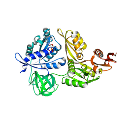
3VQX
 | | Crystal structure of the catalytic domain of pyrrolysyl-tRNA synthetase in triclinic crystal form | | 分子名称: | ADENOSINE MONOPHOSPHATE, PHOSPHATE ION, Pyrrolysine--tRNA ligase, ... | | 著者 | Yanagisawa, T, Sumida, T, Ishii, R, Yokoyama, S, RIKEN Structural Genomics/Proteomics Initiative (RSGI) | | 登録日 | 2012-04-02 | | 公開日 | 2013-01-02 | | 最終更新日 | 2023-11-08 | | 実験手法 | X-RAY DIFFRACTION (2.3 Å) | | 主引用文献 | A novel crystal form of pyrrolysyl-tRNA synthetase reveals the pre- and post-aminoacyl-tRNA synthesis conformational states of the adenylate and aminoacyl moieties and an asparagine residue in the catalytic site
Acta Crystallogr.,Sect.D, 69, 2013
|
|
3VVP
 | |
3VQW
 | | Crystal structure of the SeMet substituted catalytic domain of pyrrolysyl-tRNA synthetase | | 分子名称: | MAGNESIUM ION, PHOSPHOAMINOPHOSPHONIC ACID-ADENYLATE ESTER, Pyrrolysine--tRNA ligase | | 著者 | Yanagisawa, T, Sumida, T, Ishii, R, Yokoyama, S, RIKEN Structural Genomics/Proteomics Initiative (RSGI) | | 登録日 | 2012-04-01 | | 公開日 | 2013-01-02 | | 最終更新日 | 2013-09-04 | | 実験手法 | X-RAY DIFFRACTION (2.4 Å) | | 主引用文献 | A novel crystal form of pyrrolysyl-tRNA synthetase reveals the pre- and post-aminoacyl-tRNA synthesis conformational states of the adenylate and aminoacyl moieties and an asparagine residue in the catalytic site
Acta Crystallogr.,Sect.D, 69, 2013
|
|
3VQY
 | | Crystal structure of the catalytic domain of pyrrolysyl-tRNA synthetase in complex with BocLys and AMPPNP (form 2) | | 分子名称: | MAGNESIUM ION, N~6~-(tert-butoxycarbonyl)-L-lysine, PHOSPHOAMINOPHOSPHONIC ACID-ADENYLATE ESTER, ... | | 著者 | Yanagisawa, T, Sumida, T, Ishii, R, Yokoyama, S, RIKEN Structural Genomics/Proteomics Initiative (RSGI) | | 登録日 | 2012-04-02 | | 公開日 | 2013-01-02 | | 最終更新日 | 2023-11-08 | | 実験手法 | X-RAY DIFFRACTION (2.4 Å) | | 主引用文献 | A novel crystal form of pyrrolysyl-tRNA synthetase reveals the pre- and post-aminoacyl-tRNA synthesis conformational states of the adenylate and aminoacyl moieties and an asparagine residue in the catalytic site
Acta Crystallogr.,Sect.D, 69, 2013
|
|
3VVR
 | | Crystal structure of MATE in complex with MaD5 | | 分子名称: | (2R)-2,3-dihydroxypropyl (9Z)-octadec-9-enoate, Putative uncharacterized protein, macrocyclic peptide | | 著者 | Tanaka, Y, Ishitani, R, Nureki, O. | | 登録日 | 2012-07-27 | | 公開日 | 2013-04-03 | | 最終更新日 | 2023-11-08 | | 実験手法 | X-RAY DIFFRACTION (3 Å) | | 主引用文献 | Structural basis for the drug extrusion mechanism by a MATE multidrug transporter.
Nature, 496, 2013
|
|
3VWA
 | | Crystal structure of Cex1p | | 分子名称: | Cytoplasmic export protein 1 | | 著者 | Nozawa, K, Ishitani, R, Nureki, O. | | 登録日 | 2012-08-13 | | 公開日 | 2013-06-05 | | 実験手法 | X-RAY DIFFRACTION (2.2 Å) | | 主引用文献 | Crystal structure of Cex1p reveals the mechanism of tRNA trafficking between nucleus and cytoplasm
Nucleic Acids Res., 41, 2013
|
|
3VVS
 | | Crystal structure of MATE in complex with MaD3S | | 分子名称: | (2R)-2,3-dihydroxypropyl (9Z)-octadec-9-enoate, Putative uncharacterized protein, macrocyclic peptide | | 著者 | Tanaka, Y, Ishitani, R, Nureki, O. | | 登録日 | 2012-07-27 | | 公開日 | 2013-04-03 | | 最終更新日 | 2023-11-08 | | 実験手法 | X-RAY DIFFRACTION (2.6 Å) | | 主引用文献 | Structural basis for the drug extrusion mechanism by a MATE multidrug transporter.
Nature, 496, 2013
|
|
3VVO
 | |
3VVN
 | |
3WO7
 | | Crystal structure of YidC from Bacillus halodurans (form II) | | 分子名称: | COPPER (II) ION, Membrane protein insertase YidC 2 | | 著者 | Kumazaki, K, Tsukazaki, T, Ishitani, R, Nureki, O. | | 登録日 | 2013-12-20 | | 公開日 | 2014-04-23 | | 最終更新日 | 2024-04-03 | | 実験手法 | X-RAY DIFFRACTION (3.201 Å) | | 主引用文献 | Structural basis of Sec-independent membrane protein insertion by YidC.
Nature, 509, 2014
|
|
3WBN
 | | Crystal structure of MATE in complex with MaL6 | | 分子名称: | (2R)-2,3-dihydroxypropyl (9Z)-octadec-9-enoate, MaL6, Putative uncharacterized protein | | 著者 | Tanaka, Y, Ishitani, R, Nureki, O. | | 登録日 | 2013-05-20 | | 公開日 | 2013-06-12 | | 最終更新日 | 2023-11-08 | | 実験手法 | X-RAY DIFFRACTION (2.45 Å) | | 主引用文献 | Structural basis for the drug extrusion mechanism by a MATE multidrug transporter.
Nature, 496, 2013
|
|
3WO6
 | | Crystal structure of YidC from Bacillus halodurans (form I) | | 分子名称: | (2R)-2,3-dihydroxypropyl (9Z)-octadec-9-enoate, CADMIUM ION, Membrane protein insertase YidC 2 | | 著者 | Kumazaki, K, Tsukazaki, T, Ishitani, R, Nureki, O. | | 登録日 | 2013-12-20 | | 公開日 | 2014-04-23 | | 最終更新日 | 2024-04-03 | | 実験手法 | X-RAY DIFFRACTION (2.403 Å) | | 主引用文献 | Structural basis of Sec-independent membrane protein insertion by YidC.
Nature, 509, 2014
|
|
3W4T
 | | Crystal structure of MATE P26A mutant | | 分子名称: | (2R)-2,3-dihydroxypropyl (9Z)-octadec-9-enoate, Putative uncharacterized protein | | 著者 | Tanaka, Y, Ishitani, R, Nureki, O. | | 登録日 | 2013-01-16 | | 公開日 | 2013-04-03 | | 最終更新日 | 2024-03-20 | | 実験手法 | X-RAY DIFFRACTION (2.096 Å) | | 主引用文献 | Structural basis for the drug extrusion mechanism by a MATE multidrug transporter.
Nature, 496, 2013
|
|
3UG9
 | | Crystal Structure of the Closed State of Channelrhodopsin | | 分子名称: | Archaeal-type opsin 1, Archaeal-type opsin 2, OLEIC ACID, ... | | 著者 | Kato, H.E, Ishitani, R, Nureki, O. | | 登録日 | 2011-11-02 | | 公開日 | 2012-01-25 | | 最終更新日 | 2017-08-09 | | 実験手法 | X-RAY DIFFRACTION (2.3 Å) | | 主引用文献 | Crystal structure of the channelrhodopsin light-gated cation channel
Nature, 482, 2012
|
|
3W9H
 | | Structural basis for the inhibition of bacterial multidrug exporters | | 分子名称: | Acriflavine resistance protein B, [{2-[({[(3R)-1-{8-[(4-tert-butyl-1,3-thiazol-2-yl)carbamoyl]-4-oxo-3-[(E)-2-(1H-tetrazol-5-yl)ethenyl]-4H-pyrido[1,2-a]pyrimidin-2-yl}piperidin-3-yl]oxy}carbonyl)amino]ethyl}(dimethyl)ammonio]acetate | | 著者 | Sakurai, K, Nagata, C, Nakashima, R, Yamaguchi, A. | | 登録日 | 2013-04-04 | | 公開日 | 2013-07-03 | | 最終更新日 | 2023-11-08 | | 実験手法 | X-RAY DIFFRACTION (3.05 Å) | | 主引用文献 | Structural basis for the inhibition of bacterial multidrug exporters
Nature, 500, 2013
|
|
3WQH
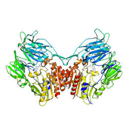 | | Crystal Structure of human DPP-IV in complex with Anagliptin | | 分子名称: | 2-acetamido-2-deoxy-beta-D-glucopyranose, Dipeptidyl peptidase 4, N-[2-({2-[(2S)-2-cyanopyrrolidin-1-yl]-2-oxoethyl}amino)-2-methylpropyl]-2-methylpyrazolo[1,5-a]pyrimidine-6-carboxamide | | 著者 | Watanabe, Y.S, Okada, S, Motoyama, T, Takahashi, R, Adachi, H, Oka, M. | | 登録日 | 2014-01-27 | | 公開日 | 2015-07-15 | | 最終更新日 | 2023-11-08 | | 実験手法 | X-RAY DIFFRACTION (2.85 Å) | | 主引用文献 | Anagliptin, a potent dipeptidyl peptidase IV inhibitor: its single-crystal structure and enzyme interactions.
J Enzyme Inhib Med Chem, 30, 2015
|
|
2ZBZ
 | | Crystal structure of vitamin D hydroxylase cytochrome P450 105A1 (R84A mutant) in complex with 1,25-dihydroxyvitamin D3 | | 分子名称: | 5-{2-[1-(5-HYDROXY-1,5-DIMETHYL-HEXYL)-7A-METHYL-OCTAHYDRO-INDEN-4-YLIDENE]-ETHYLIDENE}-4-METHYLENE-CYCLOHEXANE-1,3-DIOL, Cytochrome P450-SU1, PROTOPORPHYRIN IX CONTAINING FE | | 著者 | Sugimoto, H, Shinkyo, R, Hayashi, K, Yoneda, S, Yamada, M, Kamakura, M, Ikushiro, S, Shiro, Y, Sakaki, T. | | 登録日 | 2007-10-30 | | 公開日 | 2008-04-08 | | 最終更新日 | 2023-11-01 | | 実験手法 | X-RAY DIFFRACTION (1.9 Å) | | 主引用文献 | Crystal Structure of CYP105A1 (P450SU-1) in Complex with 1alpha,25-Dihydroxyvitamin D3
Biochemistry, 47, 2008
|
|
2ZBY
 | | Crystal structure of vitamin D hydroxylase cytochrome P450 105A1 (R84A mutant) | | 分子名称: | Cytochrome P450-SU1, PROTOPORPHYRIN IX CONTAINING FE | | 著者 | Sugimoto, H, Shinkyo, R, Hayashi, K, Yoneda, S, Yamada, M, Kamakura, M, Ikushiro, S, Shiro, Y, Sakaki, T. | | 登録日 | 2007-10-30 | | 公開日 | 2008-04-08 | | 最終更新日 | 2023-11-01 | | 実験手法 | X-RAY DIFFRACTION (1.6 Å) | | 主引用文献 | Crystal Structure of CYP105A1 (P450SU-1) in Complex with 1alpha,25-Dihydroxyvitamin D3
Biochemistry, 47, 2008
|
|
5ZSU
 | | Structure of the human homo-hexameric LRRC8A channel at 4.25 Angstroms | | 分子名称: | Volume-regulated anion channel subunit LRRC8A | | 著者 | Kasuya, G, Nakane, T, Yokoyama, T, Shirouzu, M, Ishitani, R, Nureki, O. | | 登録日 | 2018-04-29 | | 公開日 | 2018-08-15 | | 最終更新日 | 2018-09-26 | | 実験手法 | ELECTRON MICROSCOPY (4.25 Å) | | 主引用文献 | Cryo-EM structures of the human volume-regulated anion channel LRRC8.
Nat. Struct. Mol. Biol., 25, 2018
|
|
2ZBX
 | | Crystal structure of vitamin D hydroxylase cytochrome P450 105A1 (wild type) with imidazole bound | | 分子名称: | Cytochrome P450-SU1, IMIDAZOLE, PROTOPORPHYRIN IX CONTAINING FE | | 著者 | Sugimoto, H, Shinkyo, R, Hayashi, K, Yoneda, S, Yamada, M, Kamakura, M, Ikushiro, S, Shiro, Y, Sakaki, T. | | 登録日 | 2007-10-30 | | 公開日 | 2008-04-08 | | 最終更新日 | 2024-04-03 | | 実験手法 | X-RAY DIFFRACTION (1.5 Å) | | 主引用文献 | Crystal Structure of CYP105A1 (P450SU-1) in Complex with 1alpha,25-Dihydroxyvitamin D3
Biochemistry, 47, 2008
|
|
2YWH
 | | Crystal structure of GDP-bound LepA from Aquifex aeolicus | | 分子名称: | GTP-binding protein LepA, GUANOSINE-5'-DIPHOSPHATE, MAGNESIUM ION | | 著者 | Kawazoe, M, Takemoto, C, Kaminishi, T, Nishino, A, Nakayama-Ushikoshi, R, Hanawa-Suetsugu, K, Terada, T, Shirouzu, M, Yokoyama, S, RIKEN Structural Genomics/Proteomics Initiative (RSGI) | | 登録日 | 2007-04-20 | | 公開日 | 2008-04-29 | | 最終更新日 | 2023-10-25 | | 実験手法 | X-RAY DIFFRACTION (2.24 Å) | | 主引用文献 | Crystal structures of GTP-binding protein LepA from Aquifex aeolicus.
To be Published
|
|
2YWF
 | | Crystal structure of GMPPNP-bound LepA from Aquifex aeolicus | | 分子名称: | GTP-binding protein lepA, MAGNESIUM ION, PHOSPHOAMINOPHOSPHONIC ACID-GUANYLATE ESTER | | 著者 | Kawazoe, M, Takemoto, C, Kaminishi, T, Nishino, A, Nakayama-Ushikoshi, R, Hanawa-Suetsugu, K, Terada, T, Shirouzu, M, Yokoyama, S, RIKEN Structural Genomics/Proteomics Initiative (RSGI) | | 登録日 | 2007-04-20 | | 公開日 | 2008-04-29 | | 最終更新日 | 2023-10-25 | | 実験手法 | X-RAY DIFFRACTION (2.24 Å) | | 主引用文献 | Crystal structures of GTP-binding protein LepA from Aquifex aeolicus
To be Published
|
|


