Loading
PDBj
MenuPDBj@FacebookPDBj@TwitterPDBj@YouTubewwPDB FoundationwwPDB
RCSB PDBPDBeBMRBAdv. SearchSearch help
PDB: 369 results

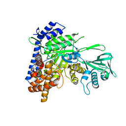
5CC3
DownloadVisualize
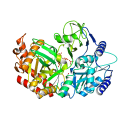
BU of 5cc3 by Molmil
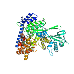
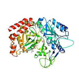
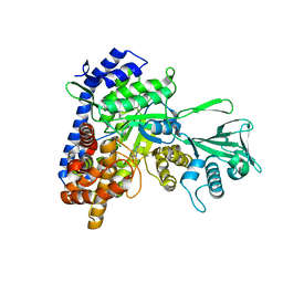
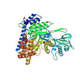
Crystal structure of Mycobacterium tuberculosis malate synthase in complex with 6-bromo-1H-indole-2-carboxylic acid
Descriptor: 6-bromanyl-1H-indole-2-carboxylic acid, MAGNESIUM ION, Malate synthase G
Authors:Huang, H.-L, Sacchettini, J.C.
Deposit date:2015-07-01
Release date:2016-08-10
Last modified:2023-09-27
Method:X-RAY DIFFRACTION (2.204 Å)
Cite:Crystal structures of fragment-bound malate synthase from Mycobacterium tuberculosis provide insights into mechanism and potential inhibitor designs
To Be Published
5C9W
DownloadVisualize
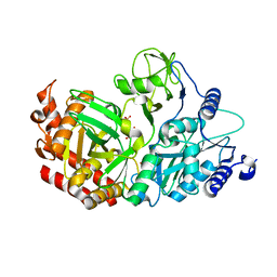
BU of 5c9w by Molmil
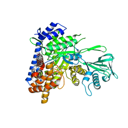
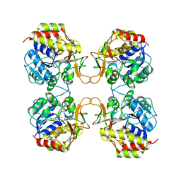
Crystal structure of Mycobacterium tuberculosis malate synthase in complex with (Z)-N-(2-bromophenyl)-2-(hydroxyimino)acetamide
Descriptor: (2E)-N-(2-bromophenyl)-2-(hydroxyimino)acetamide, MAGNESIUM ION, Malate synthase G
Authors:Huang, H.-L, Sacchettini, J.C.
Deposit date:2015-06-29
Release date:2016-08-10
Last modified:2023-09-27
Method:X-RAY DIFFRACTION (2.0928 Å)
Cite:Mycobacterium tuberculosis Malate Synthase Structures with Fragments Reveal a Portal for Substrate/Product Exchange.
J. Biol. Chem., 291, 2016
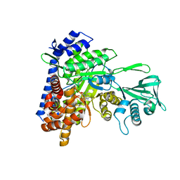
4WPV
DownloadVisualize
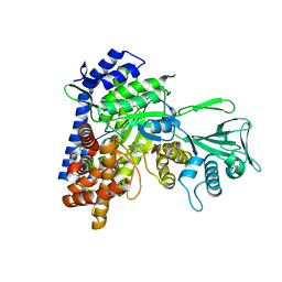
BU of 4wpv by Molmil
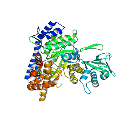
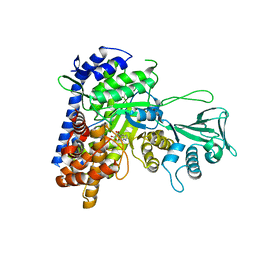
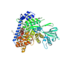
Crystal Structure of E83A mutant of Mtb PEPCK in complex with Zn2+ and phosphate ion
Descriptor: GLYCEROL, PHOSPHATE ION, Phosphoenolpyruvate carboxykinase [GTP], ...
Authors:Kim, H.L, Sacchettini, J.C.
Deposit date:2014-10-21
Release date:2015-11-04
Last modified:2023-09-27
Method:X-RAY DIFFRACTION (1.672 Å)
Cite:Crystal Structure of E83A from Mtb PEPCK in complex with Zn2+ and phosphate ion
To Be Published
4WPT
DownloadVisualize
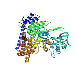
BU of 4wpt by Molmil
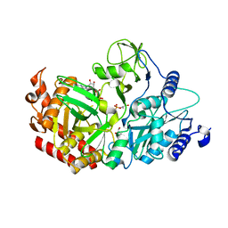
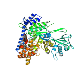
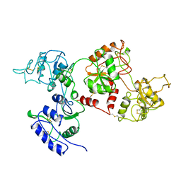
Crystal Structure of Mtb PEPCK in complex with PEP
Descriptor: GLYCEROL, PHOSPHATE ION, PHOSPHOENOLPYRUVATE, ...
Authors:Kim, H.L, Krieger, I.V, Sacchettini, J.C.
Deposit date:2014-10-21
Release date:2015-11-04
Last modified:2023-09-27
Method:X-RAY DIFFRACTION (1.6 Å)
Cite:Crystal Structure of Mtb PEPCK in complex with PEP
To Be Published
5CC7
DownloadVisualize
BU of 5cc7 by Molmil
Crystal structure of Mycobacterium tuberculosis malate synthase in complex with 1H-indole-6-carboxylic acid
Descriptor: 1H-indole-6-carboxylic acid, MAGNESIUM ION, Malate synthase G
Authors:Huang, H.-L, Sacchettini, J.C.
Deposit date:2015-07-01
Release date:2016-08-10
Last modified:2023-09-27
Method:X-RAY DIFFRACTION (2.12 Å)
Cite:Crystal structures of fragment-bound malate synthase from Mycobacterium tuberculosis provide insights into mechanism and potential inhibitor designs
To Be Published
5CEW
DownloadVisualize
BU of 5cew by Molmil
Crystal structure of Mycobacterium tuberculosis malate synthase in complex with 2-(pyridin-4-yl)thiazolidine-4-carboxylic acid
Descriptor: (2S,4S)-2-(pyridin-4-yl)-1,3-thiazolidine-4-carboxylic acid, MAGNESIUM ION, Malate synthase G
Authors:Huang, H.-L, Sacchettini, J.C.
Deposit date:2015-07-07
Release date:2016-08-10
Last modified:2023-09-27
Method:X-RAY DIFFRACTION (2.03 Å)
Cite:Mycobacterium tuberculosis Malate Synthase Structures with Fragments Reveal a Portal for Substrate/Product Exchange.
J. Biol. Chem., 291, 2016
5CBJ
DownloadVisualize
BU of 5cbj by Molmil
Crystal structure of Mycobacterium tuberculosis malate synthase in complex with 3-(phenylthio)acrylic acid
Descriptor: (2E)-3-(phenylsulfanyl)prop-2-enoic acid, MAGNESIUM ION, Malate synthase G
Authors:Huang, H.-L, Sacchettini, J.C.
Deposit date:2015-06-30
Release date:2016-08-10
Last modified:2023-09-27
Method:X-RAY DIFFRACTION (1.957 Å)
Cite:Mycobacterium tuberculosis Malate Synthase Structures with Fragments Reveal a Portal for Substrate/Product Exchange.
J. Biol. Chem., 291, 2016
5C9R
DownloadVisualize
BU of 5c9r by Molmil
Crystal structure of Mycobacterium tuberculosis malate synthase in complex with 3-((4-chlorophenyl)thio)propanoic acid
Descriptor: 3-[(4-chlorophenyl)sulfanyl]propanoic acid, MAGNESIUM ION, Malate synthase G
Authors:Huang, H.-L, Sacchettini, J.C.
Deposit date:2015-06-28
Release date:2016-07-27
Last modified:2023-09-27
Method:X-RAY DIFFRACTION (2 Å)
Cite:Mycobacterium tuberculosis Malate Synthase Structures with Fragments Reveal a Portal for Substrate/Product Exchange.
J. Biol. Chem., 291, 2016
5CCZ
DownloadVisualize
BU of 5ccz by Molmil
Crystal structure of Mycobacterium tuberculosis malate synthase in complex with 3-(4-fluorophenyl)-4-methyl-1H-pyrazol-5-amine
Descriptor: 3-(4-fluorophenyl)-4-methyl-1H-pyrazol-5-amine, MAGNESIUM ION, Malate synthase G
Authors:Huang, H.-L, Sacchettini, J.C.
Deposit date:2015-07-02
Release date:2016-08-10
Last modified:2023-09-27
Method:X-RAY DIFFRACTION (2.136 Å)
Cite:Mycobacterium tuberculosis Malate Synthase Structures with Fragments Reveal a Portal for Substrate/Product Exchange.
J. Biol. Chem., 291, 2016
4WPU
DownloadVisualize
BU of 4wpu by Molmil
Crystal Structure of E83A mutant of Mtb PEPCK in complex with PEP and GDP
Descriptor: GLYCEROL, GUANOSINE-5'-DIPHOSPHATE, PHOSPHOENOLPYRUVATE, ...
Authors:Kim, H.L, Sacchettini, J.C.
Deposit date:2014-10-21
Release date:2015-11-04
Last modified:2023-12-27
Method:X-RAY DIFFRACTION (2.26 Å)
Cite:Crystal Structure of E83A mutant of Mtb PEPCK in complex with PEP and GDP
To Be Published
4WOU
DownloadVisualize
BU of 4wou by Molmil
Crystal Structure of Mtb PEPCK in complex with GDP and metals
Descriptor: GLYCEROL, GUANOSINE-5'-DIPHOSPHATE, MANGANESE (II) ION, ...
Authors:Kim, H.L, Sacchettini, J.C.
Deposit date:2014-10-16
Release date:2015-11-04
Last modified:2023-12-27
Method:X-RAY DIFFRACTION (2.122 Å)
Cite:Crystal Structure of Mtb PEPCK in complex with GDP and metals
To Be Published
2MAS
DownloadVisualize
BU of 2mas by Molmil
PURINE NUCLEOSIDE HYDROLASE WITH A TRANSITION STATE INHIBITOR
Descriptor: 2-(4-AMINO-PHENYL)-5-HYDROXYMETHYL-PYRROLIDINE-3,4-DIOL, CALCIUM ION, INOSINE-URIDINE NUCLEOSIDE N-RIBOHYDROLASE
Authors:Degano, M, Schramm, V.L, Sacchettini, J.C.
Deposit date:1996-10-17
Release date:1997-08-12
Last modified:2024-05-22
Method:X-RAY DIFFRACTION (2.3 Å)
Cite:Trypanosomal nucleoside hydrolase. A novel mechanism from the structure with a transition-state inhibitor.
Biochemistry, 37, 1998
5DRI
DownloadVisualize
BU of 5dri by Molmil
Crystal structure of Mycobacterium tuberculosis malate synthase in complex with 2-hydroxy-4-(1H-indol-5-yl)-4-oxobut-2-enoic acid inhibitor
Descriptor: (2Z)-2-hydroxy-4-(1H-indol-5-yl)-4-oxobut-2-enoic acid, MAGNESIUM ION, Malate synthase G
Authors:Krieger, I.V, Huang, H.-L, Sacchettini, J.C.
Deposit date:2015-09-15
Release date:2016-09-28
Last modified:2023-09-27
Method:X-RAY DIFFRACTION (2.8 Å)
Cite:Mycobacterium tuberculosis Malate Synthase Structures with Fragments Reveal a Portal for Substrate/Product Exchange.
J. Biol. Chem., 291, 2016
5DX7
DownloadVisualize
BU of 5dx7 by Molmil
Crystal structure of Mycobacterium tuberculosis malate synthase in complex with 5-(5-chlorothiophen-2-yl)isoxazole-3-carboxylic acid
Descriptor: 5-(5-chlorothiophen-2-yl)-1,2-oxazole-3-carboxylic acid, MAGNESIUM ION, Malate synthase G
Authors:Huang, H.-L, Sacchettini, J.C.
Deposit date:2015-09-23
Release date:2016-10-05
Last modified:2023-09-27
Method:X-RAY DIFFRACTION (2.1 Å)
Cite:Mycobacterium tuberculosis Malate Synthase Structures with Fragments Reveal a Portal for Substrate/Product Exchange.
J. Biol. Chem., 291, 2016
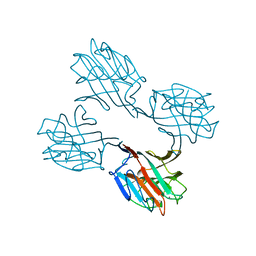
1SBD
DownloadVisualize
BU of 1sbd by Molmil
SOYBEAN AGGLUTININ COMPLEXED WITH 2,4-PENTASACCHARIDE
Descriptor: CALCIUM ION, MANGANESE (II) ION, SOYBEAN AGGLUTININ, ...
Authors:Dessen, A, Olsen, L.R, Gupta, D, Sabesan, S, Brewer, C.F, Sacchettini, J.C.
Deposit date:1995-07-13
Release date:1998-04-22
Last modified:2024-02-14
Method:X-RAY DIFFRACTION (2.52 Å)
Cite:X-ray crystallographic studies of unique cross-linked lattices between four isomeric biantennary oligosaccharides and soybean agglutinin.
Biochemistry, 36, 1997
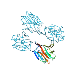
1SBE
DownloadVisualize
BU of 1sbe by Molmil
SOYBEAN AGGLUTININ FROM GLYCINE MAX
Descriptor: 2-acetamido-2-deoxy-beta-D-glucopyranose, CALCIUM ION, MANGANESE (II) ION, ...
Authors:Dessen, A, Olsen, L.R, Gupta, D, Sabesan, S, Sacchettini, J.C, Brewer, C.F.
Deposit date:1995-07-13
Release date:1998-04-22
Last modified:2024-10-09
Method:X-RAY DIFFRACTION (2.8 Å)
Cite:X-ray crystallographic studies of unique cross-linked lattices between four isomeric biantennary oligosaccharides and soybean agglutinin.
Biochemistry, 36, 1997
5ECV
DownloadVisualize
BU of 5ecv by Molmil
Crystal structure of Mycobacterium tuberculosis malate synthase in complex with 2-hydroxy-4-(4-methyl-1H-indol-5-yl)-4-oxobut-2-enoic acid
Descriptor: (~{Z})-4-(4-methyl-1~{H}-indol-5-yl)-2-oxidanyl-4-oxidanylidene-but-2-enoic acid, MAGNESIUM ION, Malate synthase G
Authors:Krieger, I.V, Huang, H.-L, Sacchettini, J.C.
Deposit date:2015-10-20
Release date:2016-10-26
Last modified:2023-09-27
Method:X-RAY DIFFRACTION (2.085 Å)
Cite:Mycobacterium tuberculosis Malate Synthase Structures with Fragments Reveal a Portal for Substrate/Product Exchange.
J. Biol. Chem., 291, 2016
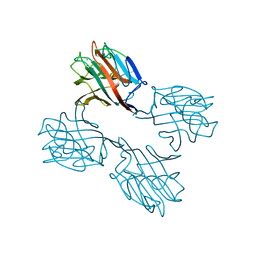
1SBF
DownloadVisualize
BU of 1sbf by Molmil
SOYBEAN AGGLUTININ
Descriptor: CALCIUM ION, MANGANESE (II) ION, SOYBEAN AGGLUTININ, ...
Authors:Olsen, L.R, Dessen, A, Gupta, D, Sabesan, S, Brewer, C.F, Sacchettini, J.C.
Deposit date:1997-10-21
Release date:1998-04-22
Last modified:2024-05-22
Method:X-RAY DIFFRACTION (2.43 Å)
Cite:X-ray crystallographic studies of unique cross-linked lattices between four isomeric biantennary oligosaccharides and soybean agglutinin.
Biochemistry, 36, 1997
4XRB
DownloadVisualize
BU of 4xrb by Molmil
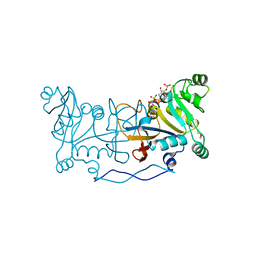
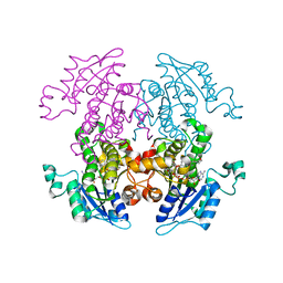
Crystal structure of Rv2671 from Mycobacterium tuberculosis
Descriptor: DI(HYDROXYETHYL)ETHER, NADPH DIHYDRO-NICOTINAMIDE-ADENINE-DINUCLEOTIDE PHOSPHATE, RV2671
Authors:Cheng, Y.S, Sacchettini, J.C, TB Structural Genomics Consortium (TBSGC)
Deposit date:2015-01-20
Release date:2016-02-24
Last modified:2023-09-27
Method:X-RAY DIFFRACTION (1.9 Å)
Cite:Structural Insights into Mycobacterium tuberculosis Rv2671 Protein as a Dihydrofolate Reductase Functional Analogue Contributing to para-Aminosalicylic Acid Resistance.
Biochemistry, 55, 2016
5E9X
DownloadVisualize
BU of 5e9x by Molmil
Crystal structure of Mycobacterium tuberculosis malate synthase in complex with 2-chloro-6H-thieno[2,3-b]pyrrole-5-carboxylic acid
Descriptor: 2-chloranyl-6~{H}-thieno[2,3-b]pyrrole-5-carboxylic acid, MAGNESIUM ION, Malate synthase G
Authors:Huang, H.-L, Sacchettini, J.C.
Deposit date:2015-10-15
Release date:2016-10-26
Last modified:2023-09-27
Method:X-RAY DIFFRACTION (1.943 Å)
Cite:Mycobacterium tuberculosis Malate Synthase Structures with Fragments Reveal a Portal for Substrate/Product Exchange.
J. Biol. Chem., 291, 2016
5DRC
DownloadVisualize
BU of 5drc by Molmil
Crystal structure of Mycobacterium tuberculosis malate synthase in complex with 2-hydroxy-4-(1H-indol-3-yl)-4-oxobut-2-enoic acid
Descriptor: (2Z)-2-hydroxy-4-(1H-indol-3-yl)-4-oxobut-2-enoic acid, MAGNESIUM ION, Malate synthase G
Authors:Huang, H.-L, Sacchettini, J.C.
Deposit date:2015-09-15
Release date:2016-09-28
Last modified:2024-10-23
Method:X-RAY DIFFRACTION (2.18 Å)
Cite:Mycobacterium tuberculosis Malate Synthase Structures with Fragments Reveal a Portal for Substrate/Product Exchange.
J. Biol. Chem., 291, 2016
1AIV
DownloadVisualize
BU of 1aiv by Molmil
APO OVOTRANSFERRIN
Descriptor: 2-acetamido-2-deoxy-alpha-D-glucopyranose-(1-4)-2-acetamido-2-deoxy-beta-D-glucopyranose, OVOTRANSFERRIN
Authors:Kurokawa, H, Dewan, J.C, Mikami, B, Sacchettini, J.C, Hirose, M.
Deposit date:1997-04-28
Release date:1998-04-29
Last modified:2023-08-02
Method:X-RAY DIFFRACTION (3 Å)
Cite:Crystal structure of hen apo-ovotransferrin. Both lobes adopt an open conformation upon loss of iron
J.Biol.Chem., 274, 1999
1ZXL
DownloadVisualize
BU of 1zxl by Molmil
Synthesis, Biological Activity, and X-Ray Crystal Structural Analysis of Diaryl Ether Inhibitors of Malarial Enoyl ACP Reductase. Part 1:4'-Substituted Triclosan Derivatives
Descriptor: N-[3-CHLORO-4-(4-CHLORO-2-HYDROXYPHENOXY)PHENYL]MORPHOLINE-4-CARBOXAMIDE, NICOTINAMIDE-ADENINE-DINUCLEOTIDE, enoyl-acyl carrier reductase
Authors:Freundlich, J.S, Anderson, J.W, Sarantakis, D, Shieh, H.M, Yu, M, Lucumi, E, Kuo, M, Schiehser, G.A, Jacobus, D.P, Jacobs Jr, W.R, Fidock, D.A, Sacchettini, J.C.
Deposit date:2005-06-08
Release date:2006-06-13
Last modified:2023-08-23
Method:X-RAY DIFFRACTION (3 Å)
Cite:Synthesis, biological activity, and X-ray crystal structural analysis of diaryl ether inhibitors of malarial enoyl acyl carrier protein reductase. Part 1: 4'-Substituted triclosan derivatives.
Bioorg.Med.Chem.Lett., 15, 2005
1UBV
DownloadVisualize
BU of 1ubv by Molmil
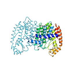
STRUCTURE OF FARNESYL PYROPHOSPHATE SYNTHETASE
Descriptor: FARNESYL DIPHOSPHATE SYNTHASE
Authors:Tarshis, L.C, Proteau, P, Poulter, C.D, Sacchettini, J.C.
Deposit date:1996-10-14
Release date:1997-03-12
Last modified:2024-02-14
Method:X-RAY DIFFRACTION (2.5 Å)
Cite:Regulation of product chain length by isoprenyl diphosphate synthases.
Proc.Natl.Acad.Sci.USA, 93, 1996
1UBW
DownloadVisualize
BU of 1ubw by Molmil
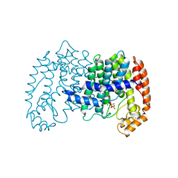
STRUCTURE OF FARNESYL PYROPHOSPHATE SYNTHETASE
Descriptor: FARNESYL DIPHOSPHATE SYNTHASE, GERANYL DIPHOSPHATE, MAGNESIUM ION
Authors:Tarshis, L.C, Proteau, P, Poulter, C.D, Sacchettini, J.C.
Deposit date:1996-10-14
Release date:1997-03-12
Last modified:2024-02-14
Method:X-RAY DIFFRACTION (2.5 Å)
Cite:Regulation of product chain length by isoprenyl diphosphate synthases.
Proc.Natl.Acad.Sci.USA, 93, 1996

226707

건을2024-10-30부터공개중

PDB statisticsPDBj update infoContact PDBjnumon