4CYA
 
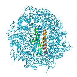 | |
4CYB
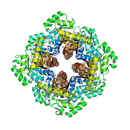
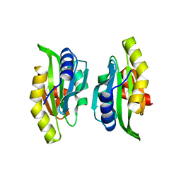
 | | DpsC from Streptomyces coelicolor | | 分子名称: | FE (III) ION, PUTATIVE DNA PROTECTION PROTEIN, SODIUM ION | | 著者 | Townsend, P.D, Hitchings, M.D, Del Sol, R, Pohl, E. | | 登録日 | 2014-04-10 | | 公開日 | 2014-06-25 | | 最終更新日 | 2024-05-08 | | 実験手法 | X-RAY DIFFRACTION (1.78 Å) | | 主引用文献 | A Tale of Tails: Deciphering the Contribution of Terminal Tails to the Biochemical Properties of Two Dps Proteins from Streptomyces Coelicolor
Cell.Mol.Life Sci., 71, 2014
|
|
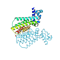
7OXX
 | | CrabP2 mutant R30AK31A | | 分子名称: | Cellular retinoic acid-binding protein 2, SODIUM ION | | 著者 | Tomlinson, C.W.E, Basle, A, Pohl, E. | | 登録日 | 2021-06-23 | | 公開日 | 2022-07-13 | | 最終更新日 | 2024-01-31 | | 実験手法 | X-RAY DIFFRACTION (1.33 Å) | | 主引用文献 | Structural requirements for the specific binding of CRABP2 to cyclin D3
To Be Published
|
|
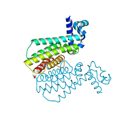
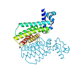
1FX0
 | |
1GU2
 | | Crystal structure of oxidized cytochrome c'' from Methylophilus methylotrophus | | 分子名称: | CYTOCHROME C'', HEME C | | 著者 | Enguita, F.J, Pohl, E, Rodrigues, A, Santos, H, Carrondo, M.A. | | 登録日 | 2002-01-22 | | 公開日 | 2003-01-16 | | 最終更新日 | 2019-07-24 | | 実験手法 | X-RAY DIFFRACTION (1.19 Å) | | 主引用文献 | Structural Evidence for a Proton Transfer Pathway Coupled with Haem Reduction of Cytochrome C" from Methylophilus Methylotrophus.
J.Biol.Inorg.Chem., 11, 2006
|
|
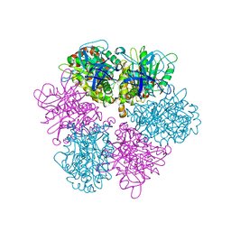
1HF0
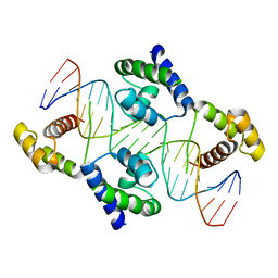 | | Crystal structure of the DNA-binding domain of Oct-1 bound to DNA as a dimer | | 分子名称: | DNA 5'-D(*CP*AP*CP*AP*TP*TP*TP*GP*AP*AP*AP*GP*GP* CP*AP*AP*AP*TP*GP*GP*AP*G)-3', DNA 5'-D(*CP*TP*CP*CP*AP*TP*TP*TP*GP*CP*CP*TP*TP* TP*CP*AP*AP*AP*TP*GP*TP*G)-3', OCTAMER-BINDING TRANSCRIPTION FACTOR 1 | | 著者 | Remenyi, A, Tomilin, A, Pohl, E, Scholer, H.R, Wilmanns, M. | | 登録日 | 2000-11-27 | | 公開日 | 2001-11-10 | | 最終更新日 | 2024-05-08 | | 実験手法 | X-RAY DIFFRACTION (2.7 Å) | | 主引用文献 | Differential Dimer Activities of the Transcription Factor Oct-1 by DNA-Induced Interface Swapping
Mol.Cell, 8, 2001
|
|
7PJO
 | |
7AA1
 | | Structural comparison of cellular retinoic acid binding proteins I and II in the presence and absence of natural and synthetic ligands | | 分子名称: | 4-[2-(5,5,8,8-tetramethyl-6,7-dihydroquinoxalin-2-yl)ethynyl]benzoic acid, Cellular retinoic acid-binding protein 2 | | 著者 | Tomlinson, C.W.E, Cornish, K.A.S, Pohl, E. | | 登録日 | 2020-09-02 | | 公開日 | 2021-02-17 | | 最終更新日 | 2024-01-31 | | 実験手法 | X-RAY DIFFRACTION (1.71 Å) | | 主引用文献 | Structure-functional relationship of cellular retinoic acid-binding proteins I and II interacting with natural and synthetic ligands.
Acta Crystallogr D Struct Biol, 77, 2021
|
|
7A9Y
 | | Structural comparison of cellular retinoic acid binding protein I and II in the presence and absence of natural and synthetic ligands | | 分子名称: | Cellular retinoic acid-binding protein 1, GLYCEROL, MYRISTIC ACID, ... | | 著者 | Tomlinson, C.W.E, Cornish, K.A.S, Pohl, E. | | 登録日 | 2020-09-02 | | 公開日 | 2021-02-17 | | 最終更新日 | 2024-01-31 | | 実験手法 | X-RAY DIFFRACTION (1.64 Å) | | 主引用文献 | Structure-functional relationship of cellular retinoic acid-binding proteins I and II interacting with natural and synthetic ligands.
Acta Crystallogr D Struct Biol, 77, 2021
|
|
7A9Z
 | | Structural comparison of cellular retinoic acid binding protein I and II in the presence and absence of natural and synthetic ligands | | 分子名称: | 4-[2-(5,5,8,8-tetramethyl-6,7-dihydroquinoxalin-2-yl)ethynyl]benzoic acid, Cellular retinoic acid-binding protein 1 | | 著者 | Tomlinson, C.W.E, Cornish, K.A.S, Pohl, E. | | 登録日 | 2020-09-02 | | 公開日 | 2021-02-17 | | 最終更新日 | 2024-01-31 | | 実験手法 | X-RAY DIFFRACTION (2.41 Å) | | 主引用文献 | Structure-functional relationship of cellular retinoic acid-binding proteins I and II interacting with natural and synthetic ligands.
Acta Crystallogr D Struct Biol, 77, 2021
|
|
7AA0
 | | Structural comparison of cellular retinoic acid binding protein I and II in the presence and absence of natural and synthetic ligands | | 分子名称: | (~{E})-3-[4-(4,4-dimethyl-1-propan-2-yl-2,3-dihydroquinolin-6-yl)phenyl]prop-2-enoic acid, Cellular retinoic acid-binding protein 2 | | 著者 | Tomlinson, C.W.E, Cornish, K.A.S, Pohl, E. | | 登録日 | 2020-09-02 | | 公開日 | 2021-02-17 | | 最終更新日 | 2024-01-31 | | 実験手法 | X-RAY DIFFRACTION (1.82 Å) | | 主引用文献 | Structure-functional relationship of cellular retinoic acid-binding proteins I and II interacting with natural and synthetic ligands.
Acta Crystallogr D Struct Biol, 77, 2021
|
|
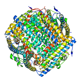
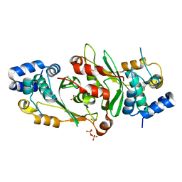
1M5W
 | | 1.96 A Crystal Structure of Pyridoxine 5'-Phosphate Synthase in Complex with 1-deoxy-D-xylulose phosphate | | 分子名称: | 1-DEOXY-D-XYLULOSE-5-PHOSPHATE, PHOSPHATE ION, Pyridoxal phosphate biosynthetic protein pdxJ | | 著者 | Yeh, J.I, Du, S, Pohl, E, Cane, D.E. | | 登録日 | 2002-07-10 | | 公開日 | 2003-07-15 | | 最終更新日 | 2024-02-14 | | 実験手法 | X-RAY DIFFRACTION (1.96 Å) | | 主引用文献 | Multistate Binding in Pyridoxine 5'-Phosphate Synthase: 1.96 A Crystal Structure in
Complex with 1-deoxy-D-xylulose phosphate
Biochemistry, 41, 2002
|
|
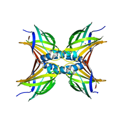
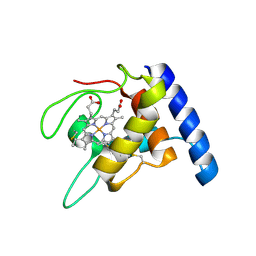
1M4J
 | | CRYSTAL STRUCTURE OF THE N-TERMINAL ADF-H DOMAIN OF MOUSE TWINFILIN ISOFORM-1 | | 分子名称: | A6 gene product | | 著者 | Paavilainen, V.O, Merckel, M.C, Falck, S, Ojala, P.J, Pohl, E, Wilmanns, M, Lappalainen, P. | | 登録日 | 2002-07-03 | | 公開日 | 2002-11-13 | | 最終更新日 | 2024-02-14 | | 実験手法 | X-RAY DIFFRACTION (1.6 Å) | | 主引用文献 | Structural Conservation Between the Actin Monomer-binding Sites of Twinfilin and Actin-depolymerizing Factor (ADF)/Cofilin
J.Biol.Chem., 277, 2002
|
|
7NGO
 | |
7NGJ
 | |
7NGR
 | |
7NGY
 | |
7NGT
 | |
7NGN
 | |
7NGD
 | |
7NGW
 | |
7NGI
 | |
7NGK
 | |
7NGU
 | |
7NGS
 | |


