Loading
PDBj
メニューPDBj@FacebookPDBj@TwitterPDBj@YouTubewwPDB FoundationwwPDB
RCSB PDBPDBeBMRBAdv. SearchSearch help
PDB: 120 件

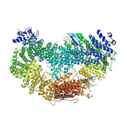
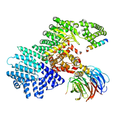
5WBI
DownloadVisualize
BU of 5wbi by Molmil
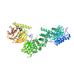
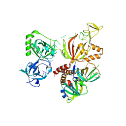
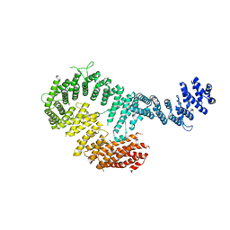
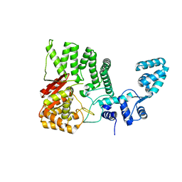
Crystal structure of the Arabidopsis thaliana Raptor
分子名称: Regulatory-associated protein of TOR 1
著者Pavletich, N.P, Jiang, X.
登録日2017-06-29
公開日2017-12-20
最終更新日2024-03-13
実験手法X-RAY DIFFRACTION (3 Å)
主引用文献Mechanisms of mTORC1 activation by RHEB and inhibition by PRAS40.
Nature, 552, 2017
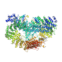
5WBU
DownloadVisualize
BU of 5wbu by Molmil
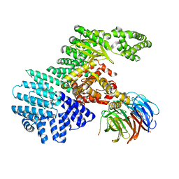
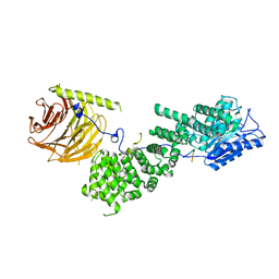
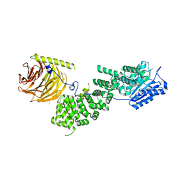
Crystal structure of mTOR(deltaN)-mLST8-PRAS40(alpha-helix & beta-strand) complex
分子名称: Proline-rich AKT1 substrate 1, Serine/threonine-protein kinase mTOR, Target of rapamycin complex subunit LST8
著者Pavletich, N.P, Yang, H.
登録日2017-06-29
公開日2017-12-20
最終更新日2023-10-04
実験手法X-RAY DIFFRACTION (3.42 Å)
主引用文献Mechanisms of mTORC1 activation by RHEB and inhibition by PRAS40.
Nature, 552, 2017
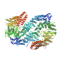
5WBJ
DownloadVisualize
BU of 5wbj by Molmil
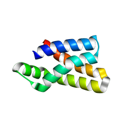
Crystal structure of the arabidopsis thaliana Raptor in complex with the TOS peptide of human 4EBP1
分子名称: Eukaryotic translation initiation factor 4E-binding protein 1, Regulatory-associated protein of TOR 1
著者Pavletich, N.P, Jiang, X.
登録日2017-06-29
公開日2017-12-20
最終更新日2023-10-04
実験手法X-RAY DIFFRACTION (3 Å)
主引用文献Mechanisms of mTORC1 activation by RHEB and inhibition by PRAS40.
Nature, 552, 2017
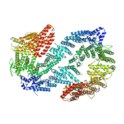
5WBY
DownloadVisualize
BU of 5wby by Molmil
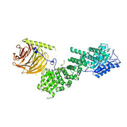
Crystal structure of mTOR(deltaN)-mLST8-PRAS40(beta-strand) complex
分子名称: Proline-rich AKT1 substrate 1, Serine/threonine-protein kinase mTOR, Target of rapamycin complex subunit LST8
著者Pavletich, N.P, Yang, H.
登録日2017-06-29
公開日2017-12-20
最終更新日2023-10-04
実験手法X-RAY DIFFRACTION (3.1 Å)
主引用文献Mechanisms of mTORC1 activation by RHEB and inhibition by PRAS40.
Nature, 552, 2017
2GLI
DownloadVisualize
BU of 2gli by Molmil
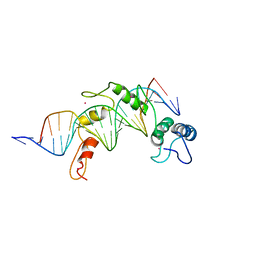
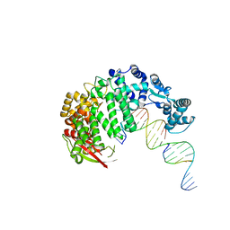
FIVE-FINGER GLI/DNA COMPLEX
分子名称: COBALT (II) ION, DNA (5'-D(*AP*CP*GP*TP*GP*GP*AP*CP*CP*AP*CP*CP*CP*AP*AP*GP*AP*CP*GP*AP*A)-3'), DNA (5'-D(*TP*TP*TP*CP*GP*TP*CP*TP*TP*GP*GP*GP*TP*GP*GP*TP*CP*CP*AP*CP*G)-3'), ...
著者Pavletich, N.P, Pabo, C.O.
登録日1993-11-09
公開日1993-11-09
最終更新日2024-02-14
実験手法X-RAY DIFFRACTION (2.6 Å)
主引用文献Crystal structure of a five-finger GLI-DNA complex: new perspectives on zinc fingers.
Science, 261, 1993
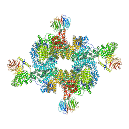
6BCX
DownloadVisualize
BU of 6bcx by Molmil
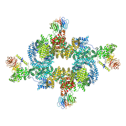
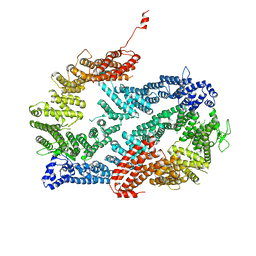
mTORC1 structure refined to 3.0 angstroms
分子名称: ADENOSINE-5'-TRIPHOSPHATE, Eukaryotic translation initiation factor 4E-binding protein 1, MAGNESIUM ION, ...
著者Pavletich, N.P, Yang, H.
登録日2017-10-20
公開日2017-12-20
最終更新日2024-03-13
実験手法ELECTRON MICROSCOPY (3.23 Å)
主引用文献Mechanisms of mTORC1 activation by RHEB and inhibition by PRAS40.
Nature, 552, 2017
6BCU
DownloadVisualize
BU of 6bcu by Molmil
Cryo-EM structure of the activated RHEB-mTORC1 refined to 3.4 angstrom
分子名称: ADENOSINE-5'-TRIPHOSPHATE, Eukaryotic translation initiation factor 4E-binding protein 1, GTP-binding protein Rheb, ...
著者Pavletich, N.P, Yang, H.
登録日2017-10-20
公開日2017-12-20
最終更新日2024-03-13
実験手法ELECTRON MICROSCOPY (3.8 Å)
主引用文献Mechanisms of mTORC1 activation by RHEB and inhibition by PRAS40.
Nature, 552, 2017
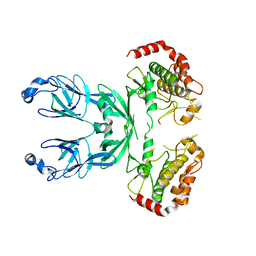
3I6W
DownloadVisualize
BU of 3i6w by Molmil
Structure and Activation Mechanism of the CHK2 DNA-Damage Checkpoint Kinase
分子名称: Serine/threonine-protein kinase Chk2
著者Pavletich, N.P.
登録日2009-07-07
公開日2009-11-10
最終更新日2023-09-06
実験手法X-RAY DIFFRACTION (3.25 Å)
主引用文献Structure and activation mechanism of the CHK2 DNA damage checkpoint kinase.
Mol.Cell, 35, 2009
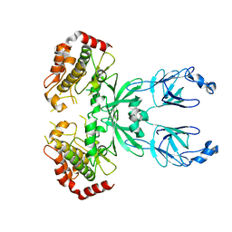
3I6U
DownloadVisualize
BU of 3i6u by Molmil
Structure and Activation Mechanism of the CHK2 DNA-Damage Checkpoint Kinase
分子名称: Serine/threonine-protein kinase Chk2
著者Pavletich, N.P.
登録日2009-07-07
公開日2009-11-10
最終更新日2023-09-06
実験手法X-RAY DIFFRACTION (3 Å)
主引用文献Structure and activation mechanism of the CHK2 DNA damage checkpoint kinase.
Mol.Cell, 35, 2009
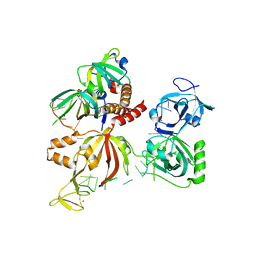
4GOP
DownloadVisualize
BU of 4gop by Molmil
Structure and Conformational Change of a Replication Protein A Heterotrimer Bound to ssDNA
分子名称: DNA (25-MER), Putative uncharacterized protein, ZINC ION
著者Pavletich, N.P, Jie, F.
登録日2012-08-20
公開日2012-11-28
実験手法X-RAY DIFFRACTION (3.1 Å)
主引用文献Structure and conformational change of a replication protein A heterotrimer bound to ssDNA.
Genes Dev., 26, 2012
4GNX
DownloadVisualize
BU of 4gnx by Molmil
Structure of U. maydis Replication protein A bound to ssDNA
分子名称: DNA (25-MER), Putative uncharacterized protein, ZINC ION
著者Pavletich, N.P, Fan, J.
登録日2012-08-17
公開日2013-02-20
最終更新日2024-10-30
実験手法X-RAY DIFFRACTION (2.8 Å)
主引用文献Structure of
To be Published
5WBL
DownloadVisualize
BU of 5wbl by Molmil
Crystal structure of the Arabidopsis thaliana Raptor in complex with the TOS peptide of human PRAS40
分子名称: Proline-rich AKT1 substrate 1, Regulatory-associated protein of TOR 1
著者Pavletich, N.P, Jiang, X.
登録日2017-06-29
公開日2017-12-20
最終更新日2023-10-04
実験手法X-RAY DIFFRACTION (3.35 Å)
主引用文献Mechanisms of mTORC1 activation by RHEB and inhibition by PRAS40.
Nature, 552, 2017
5WBH
DownloadVisualize
BU of 5wbh by Molmil
Structure of the FRB domain of mTOR bound to a substrate recruitment peptide of S6K1
分子名称: Ribosomal protein S6 kinase beta-1, Serine/threonine-protein kinase mTOR
著者Pavletich, N.P, Yang, H.
登録日2017-06-29
公開日2017-12-20
最終更新日2023-10-04
実験手法X-RAY DIFFRACTION (1.75 Å)
主引用文献Mechanisms of mTORC1 activation by RHEB and inhibition by PRAS40.
Nature, 552, 2017
5WBK
DownloadVisualize
BU of 5wbk by Molmil
Crystal structure of the arabidopsis thaliana Raptor in complex with the TOS peptide of human S6K1
分子名称: Regulatory-associated protein of TOR 1, Ribosomal protein S6 kinase beta-1
著者Pavletich, N.P, Jiang, X.
登録日2017-06-29
公開日2017-12-20
最終更新日2023-10-04
実験手法X-RAY DIFFRACTION (3.11 Å)
主引用文献Mechanisms of mTORC1 activation by RHEB and inhibition by PRAS40.
Nature, 552, 2017
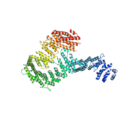
6VAF
DownloadVisualize
BU of 6vaf by Molmil
Structure of mono-ubiquitinated FANCD2 bound to non-ubiquitinated FANCI and to DNA
分子名称: DNA (29-MER), Fanconi anemia group D2 protein, Fanconi anemia, ...
著者Pavletich, N.P.
登録日2019-12-17
公開日2020-03-18
最終更新日2024-03-06
実験手法ELECTRON MICROSCOPY (3.9 Å)
主引用文献DNA clamp function of the monoubiquitinated Fanconi anaemia ID complex.
Nature, 580, 2020
6VAE
DownloadVisualize
BU of 6vae by Molmil
Mono-ubiquitinated Fanconi Anemia ID complex bound to ICL DNA
分子名称: DNA (29-MER), Fanconi anemia group D2 protein, Fanconi anemia, ...
著者Pavletich, N.P.
登録日2019-12-17
公開日2020-03-18
最終更新日2020-04-22
実験手法ELECTRON MICROSCOPY (3.5 Å)
主引用文献DNA clamp function of the monoubiquitinated Fanconi anaemia ID complex.
Nature, 580, 2020
6VAA
DownloadVisualize
BU of 6vaa by Molmil
Structure of the Fanconi Anemia ID complex bound to ICL DNA
分子名称: DNA (26-MER), DNA (5'-D(*TP*TP*TP*TP*TP*TP*TP*TP*TP*TP*TP*TP*TP*TP*TP*T)-3'), DNA (5'-D(P*AP*AP*AP*AP*AP*AP*AP*AP*AP*AP*AP*AP*AP*AP*A)-3'), ...
著者Pavletich, N.P.
登録日2019-12-17
公開日2020-03-18
最終更新日2024-03-06
実験手法ELECTRON MICROSCOPY (3.4 Å)
主引用文献DNA clamp function of the monoubiquitinated Fanconi anaemia ID complex.
Nature, 580, 2020
6VAD
DownloadVisualize
BU of 6vad by Molmil
Fanconi Anemia ID complex
分子名称: Fanconi anemia group D2 protein, Fanconi anemia, complementation group I
著者Pavletich, N.P.
登録日2019-12-17
公開日2020-03-18
最終更新日2024-03-06
実験手法ELECTRON MICROSCOPY (3.3 Å)
主引用文献DNA clamp function of the monoubiquitinated Fanconi anaemia ID complex.
Nature, 580, 2020
3S51
DownloadVisualize
BU of 3s51 by Molmil
Structure of FANCI
分子名称: Fanconi anemia group I protein homolog
著者Pavletich, N.P.
登録日2011-05-20
公開日2011-07-27
最終更新日2024-02-28
実験手法X-RAY DIFFRACTION (3.3 Å)
主引用文献Structure of the FANCI-FANCD2 complex: insights into the Fanconi anemia DNA repair pathway.
Science, 333, 2011
3S4W
DownloadVisualize
BU of 3s4w by Molmil
Structure of the FANCI-FANCD2 complex
分子名称: Fanconi anemia group D2 protein homolog, Fanconi anemia group I protein homolog
著者Pavletich, N.P.
登録日2011-05-20
公開日2011-07-27
最終更新日2023-09-13
実験手法X-RAY DIFFRACTION (3.408 Å)
主引用文献Structure of the FANCI-FANCD2 complex: insights into the Fanconi anemia DNA repair pathway.
Science, 333, 2011
3S4Z
DownloadVisualize
BU of 3s4z by Molmil
Structure of a Y DNA-FANCI complex
分子名称: dna repair 1
著者Pavletich, N.P.
登録日2011-05-20
公開日2011-07-27
最終更新日2024-02-28
実験手法X-RAY DIFFRACTION (7.8 Å)
主引用文献Structure of the FANCI-FANCD2 complex: insights into the Fanconi anemia DNA repair pathway.
Science, 333, 2011
4RIA
DownloadVisualize
BU of 4ria by Molmil
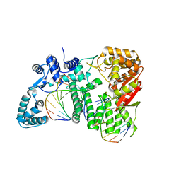
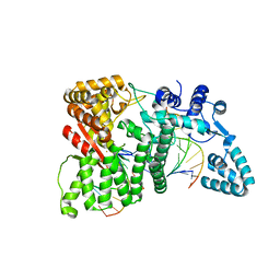
FAN1 Nuclease bound to 5' phosphorylated nicked DNA
分子名称: BARIUM ION, DNA (5'-D(*TP*TP*TP*TP*TP*TP*G*AP*GP*GP*CP*GP*TP*G)-3'), DNA (5'-D(P*AP*GP*AP*CP*TP*CP*CP*TP*C)-3'), ...
著者Pavletich, N.P, Wang, R.
登録日2014-10-05
公開日2014-12-10
最終更新日2024-02-28
実験手法X-RAY DIFFRACTION (3 Å)
主引用文献DNA repair. Mechanism of DNA interstrand cross-link processing by repair nuclease FAN1.
Science, 346, 2014
4RID
DownloadVisualize
BU of 4rid by Molmil
Human FAN1 nuclease
分子名称: Fanconi-associated nuclease 1
著者Pavletich, N.P, Wang, R.
登録日2014-10-05
公開日2014-12-03
最終更新日2024-02-28
実験手法X-RAY DIFFRACTION (3.3 Å)
主引用文献DNA repair. Mechanism of DNA interstrand cross-link processing by repair nuclease FAN1.
Science, 346, 2014
4RIC
DownloadVisualize
BU of 4ric by Molmil
FAN1 Nuclease bound to 5' hydroxyl (dT-dT) single flap DNA
分子名称: CALCIUM ION, DNA (5'-D(*GP*CP*TP*GP*AP*GP*GP*AP*GP*TP*CP*T)-3'), DNA (5'-D(*TP*TP*AP*GP*CP*CP*AP*CP*GP*CP*CP*TP*AP*GP*AP*CP*TP*CP*CP*TP*C)-3'), ...
著者Pavletich, N.P, Wang, R.
登録日2014-10-05
公開日2014-12-03
最終更新日2024-02-28
実験手法X-RAY DIFFRACTION (2.8 Å)
主引用文献DNA repair. Mechanism of DNA interstrand cross-link processing by repair nuclease FAN1.
Science, 346, 2014
4RI9
DownloadVisualize
BU of 4ri9 by Molmil
FAN1 Nuclease bound to 5' phosphorylated p(dT)/3'(dT-dT-dT-dT-dT-dT-dT-dT) double flap DNA
分子名称: BARIUM ION, DNA (5'-D(*TP*TP*TP*TP*TP*TP*GP*AP*GP*GP*CP*GP*TP*G)-3'), DNA (5'-D(P*AP*GP*AP*CP*TP*CP*CP*TP*CP*TP*TP*TP*TP*TP*TP*TP*T)-3'), ...
著者Pavletich, N.P, Wang, R.
登録日2014-10-05
公開日2014-12-10
最終更新日2024-02-28
実験手法X-RAY DIFFRACTION (2.9 Å)
主引用文献DNA repair. Mechanism of DNA interstrand cross-link processing by repair nuclease FAN1.
Science, 346, 2014

226707

件を2024-10-30に公開中

PDB statisticsPDBj update infoContact PDBjnumon