7BYK
 
 | |
6JLA
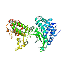
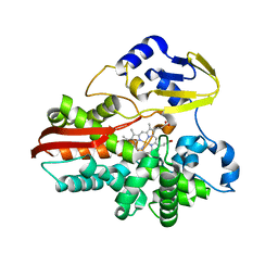
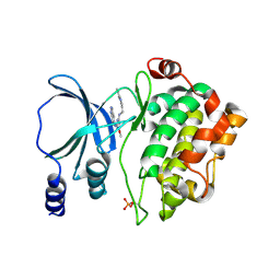
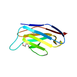
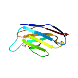
 | | Crystal structure of a mouse ependymin related protein | | 分子名称: | 2-acetamido-2-deoxy-beta-D-glucopyranose, 2-acetamido-2-deoxy-beta-D-glucopyranose-(1-4)-[alpha-L-fucopyranose-(1-6)]2-acetamido-2-deoxy-beta-D-glucopyranose, Mammalian ependymin-related protein 1 | | 著者 | Park, S. | | 登録日 | 2019-03-04 | | 公開日 | 2020-03-04 | | 最終更新日 | 2020-09-16 | | 実験手法 | X-RAY DIFFRACTION (2.4 Å) | | 主引用文献 | Structures of three ependymin-related proteins suggest their function as a hydrophobic molecule binder.
Iucrj, 6, 2019
|
|
8SQF
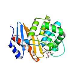 | | OXA-48 bound to inhibitor CDD-2725 | | 分子名称: | (1M)-3'-(benzyloxy)-5-hydroxy[1,1'-biphenyl]-3,4'-dicarboxylic acid, BICARBONATE ION, Beta-lactamase | | 著者 | Park, S, Judge, A, Fan, J, Sankaran, B, Prasad, B.V.V, Palzkill, T. | | 登録日 | 2023-05-04 | | 公開日 | 2024-01-03 | | 最終更新日 | 2024-01-17 | | 実験手法 | X-RAY DIFFRACTION (2.3 Å) | | 主引用文献 | Exploiting the Carboxylate-Binding Pocket of beta-Lactamase Enzymes Using a Focused DNA-Encoded Chemical Library.
J.Med.Chem., 67, 2024
|
|
1IO8
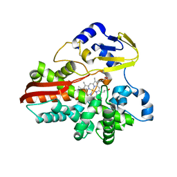
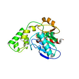
 | | Thermophilic cytochrome P450 (CYP119) from sulfolobus solfataricus: High resolution structural origin of its thermostability and functional properties | | 分子名称: | CYTOCHROME P450 CYP119, PROTOPORPHYRIN IX CONTAINING FE | | 著者 | Park, S.-Y, Yamane, K, Adachi, S, Shiro, Y, Sligar, S.G, RIKEN Structural Genomics/Proteomics Initiative (RSGI) | | 登録日 | 2001-02-08 | | 公開日 | 2001-03-07 | | 最終更新日 | 2024-04-03 | | 実験手法 | X-RAY DIFFRACTION (2 Å) | | 主引用文献 | Thermophilic cytochrome P450 (CYP119) from Sulfolobus solfataricus: high resolution structure and functional properties
J.Inorg.Biochem., 91, 2002
|
|
1IO9
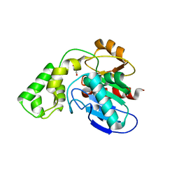
 | | THERMOPHILIC CYTOCHROME P450 (CYP119) FROM SULFOLOBUS SOLFATARICUS: HIGH RESOLUTION STRUCTURAL ORIGIN OF ITS THERMOSTABILITY AND FUNCTIONAL PROPERTIES | | 分子名称: | CYTOCHROME P450 CYP119, PROTOPORPHYRIN IX CONTAINING FE | | 著者 | Park, S.-Y, Yamane, K, Adachi, S, Shiro, Y, Sligar, S.G, RIKEN Structural Genomics/Proteomics Initiative (RSGI) | | 登録日 | 2001-02-08 | | 公開日 | 2001-03-07 | | 最終更新日 | 2024-04-03 | | 実験手法 | X-RAY DIFFRACTION (2.05 Å) | | 主引用文献 | Thermophilic cytochrome P450 (CYP119) from Sulfolobus solfataricus: high resolution structure and functional properties.
J.Inorg.Biochem., 91, 2002
|
|
1IO7
 | | THERMOPHILIC CYTOCHROME P450 (CYP119) FROM SULFOLOBUS SOLFATARICUS: HIGH RESOLUTION STRUCTURAL ORIGIN OF ITS THERMOSTABILITY AND FUNCTIONAL PROPERTIES | | 分子名称: | CYTOCHROME P450 CYP119, PROTOPORPHYRIN IX CONTAINING FE | | 著者 | Park, S.-Y, Yamane, K, Adachi, S, Shiro, Y, Sligar, S.G, RIKEN Structural Genomics/Proteomics Initiative (RSGI) | | 登録日 | 2001-02-08 | | 公開日 | 2001-02-28 | | 最終更新日 | 2023-12-27 | | 実験手法 | X-RAY DIFFRACTION (1.5 Å) | | 主引用文献 | Thermophilic cytochrome P450 (CYP119) from Sulfolobus solfataricus: high resolution structure and functional properties.
J.Inorg.Biochem., 91, 2002
|
|
7CBV
 | |
5I0B
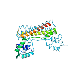
 | | Structure of PAK4 | | 分子名称: | 6-bromo-2-[1-methyl-3-(propan-2-yl)-1H-pyrazol-4-yl]-1H-imidazo[4,5-b]pyridine, Serine/threonine-protein kinase PAK 4 | | 著者 | Park, S.Y. | | 登録日 | 2016-02-03 | | 公開日 | 2016-12-14 | | 最終更新日 | 2023-11-08 | | 実験手法 | X-RAY DIFFRACTION (3.09 Å) | | 主引用文献 | The discovery and the structural basis of an imidazo[4,5-b]pyridine-based p21-activated kinase 4 inhibitor
Bioorg. Med. Chem. Lett., 26, 2016
|
|
2ROM
 | |
7XYZ
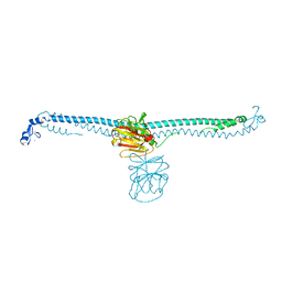
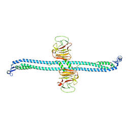
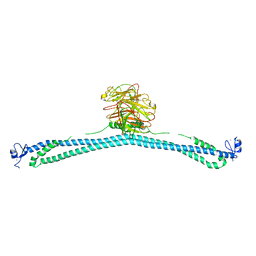
 | | TRIM E3 ubiquitin ligase | | 分子名称: | Tripartite motif-containing protein 72, ZINC ION | | 著者 | Park, S.H, Song, H.K. | | 登録日 | 2022-06-02 | | 公開日 | 2023-07-12 | | 最終更新日 | 2023-11-29 | | 実験手法 | X-RAY DIFFRACTION (4.62 Å) | | 主引用文献 | Structure and activation of the RING E3 ubiquitin ligase TRIM72 on the membrane.
Nat.Struct.Mol.Biol., 30, 2023
|
|
4NJD
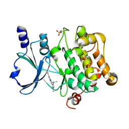
 | | Structure of p21-activated kinase 4 with a novel inhibitor KY-04031 | | 分子名称: | N-(1H-indazol-5-yl)-N'-[2-(1H-indol-3-yl)ethyl]-6-methoxy-1,3,5-triazine-2,4-diamine, Serine/threonine-protein kinase PAK 4 | | 著者 | Park, S. | | 登録日 | 2013-11-09 | | 公開日 | 2014-05-21 | | 最終更新日 | 2022-08-24 | | 実験手法 | X-RAY DIFFRACTION (2.5 Å) | | 主引用文献 | Discovery and the structural basis of a novel p21-activated kinase 4 inhibitor.
Cancer Lett., 349, 2014
|
|
7XV2
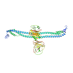
 | | TRIM E3 ubiquitin ligase | | 分子名称: | Tripartite motif-containing protein 72, ZINC ION | | 著者 | Park, S.H, Song, H.K. | | 登録日 | 2022-05-20 | | 公開日 | 2023-05-24 | | 最終更新日 | 2023-11-29 | | 実験手法 | X-RAY DIFFRACTION (2.75 Å) | | 主引用文献 | Structure and activation of the RING E3 ubiquitin ligase TRIM72 on the membrane.
Nat.Struct.Mol.Biol., 30, 2023
|
|
7XZ1
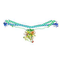
 | | TRIM E3 ubiquitin ligase | | 分子名称: | Tripartite motif-containing protein 72, ZINC ION | | 著者 | Park, S.H, Song, H.K. | | 登録日 | 2022-06-02 | | 公開日 | 2023-07-12 | | 最終更新日 | 2023-11-29 | | 実験手法 | X-RAY DIFFRACTION (5.2 Å) | | 主引用文献 | Structure and activation of the RING E3 ubiquitin ligase TRIM72 on the membrane.
Nat.Struct.Mol.Biol., 30, 2023
|
|
7XZ0
 | | TRIM E3 ubiquitin ligase | | 分子名称: | Tripartite motif-containing protein 72, ZINC ION | | 著者 | Park, S.H, Song, H.K. | | 登録日 | 2022-06-02 | | 公開日 | 2023-07-12 | | 最終更新日 | 2023-11-29 | | 実験手法 | X-RAY DIFFRACTION (3.28 Å) | | 主引用文献 | Structure and activation of the RING E3 ubiquitin ligase TRIM72 on the membrane.
Nat.Struct.Mol.Biol., 30, 2023
|
|
7XZ2
 | | TRIM E3 ubiquitin ligase | | 分子名称: | Tripartite motif-containing protein 72, ZINC ION | | 著者 | Park, S.H, Song, H.K. | | 登録日 | 2022-06-02 | | 公開日 | 2023-07-12 | | 最終更新日 | 2023-11-29 | | 実験手法 | X-RAY DIFFRACTION (3.5 Å) | | 主引用文献 | Structure and activation of the RING E3 ubiquitin ligase TRIM72 on the membrane.
Nat.Struct.Mol.Biol., 30, 2023
|
|
7XYY
 | | TRIM E3 ubiquitin ligase WT | | 分子名称: | Tripartite motif-containing protein 72, ZINC ION | | 著者 | Park, S.H, Song, H.K. | | 登録日 | 2022-06-02 | | 公開日 | 2023-07-12 | | 最終更新日 | 2023-11-29 | | 実験手法 | X-RAY DIFFRACTION (7.1 Å) | | 主引用文献 | Structure and activation of the RING E3 ubiquitin ligase TRIM72 on the membrane.
Nat.Struct.Mol.Biol., 30, 2023
|
|
6LXX
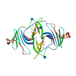
 | | Frog EPDR1 with an Ir atom | | 分子名称: | CALCIUM ION, CHLORIDE ION, Ependymin-related 1, ... | | 著者 | Park, S, Park, J. | | 登録日 | 2020-02-12 | | 公開日 | 2021-02-17 | | 最終更新日 | 2023-11-29 | | 実験手法 | X-RAY DIFFRACTION (2.4 Å) | | 主引用文献 | A Single Soaked Iridium (IV) Ion Observed in the Frog Ependymin-Related Protein.
Bull.Korean Chem.Soc., 41, 2020
|
|
3BD5
 | |
3BD4
 | |
3BD3
 | |
1ROM
 | |
6IY6
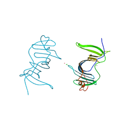
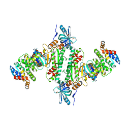 | | Crystal structure of human cytosolic aspartyl-tRNA synthetase (DRS) in complex with glutathion-S transferase (GST) domains from Aminoacyl tRNA synthase complex-interacting multifunctional protein 2 (AIMP2) and glutamyl-prolyl-tRNA synthetase (EPRS) | | 分子名称: | Aminoacyl tRNA synthase complex-interacting multifunctional protein 2, Aspartate--tRNA ligase, cytoplasmic, ... | | 著者 | Park, S.H, Hahn, H, Han, B.W. | | 登録日 | 2018-12-13 | | 公開日 | 2019-09-11 | | 最終更新日 | 2023-11-22 | | 実験手法 | X-RAY DIFFRACTION (3.6 Å) | | 主引用文献 | The DRS-AIMP2-EPRS subcomplex acts as a pivot in the multi-tRNA synthetase complex.
Iucrj, 6, 2019
|
|
3BF7
 | |
3BF8
 | |
7MBM
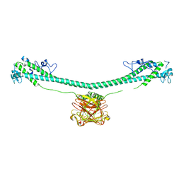
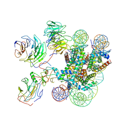 | | Cryo-EM structure of MLL1-NCP (H3K4M) complex, mode01 | | 分子名称: | DNA (145-MER), Histone H2A, Histone H2B 1.1, ... | | 著者 | Park, S.H, Ayoub, A, Lee, Y.T, Dou, Y, Cho, U. | | 登録日 | 2021-04-01 | | 公開日 | 2021-12-29 | | 最終更新日 | 2022-01-19 | | 実験手法 | ELECTRON MICROSCOPY | | 主引用文献 | Regulation of MLL1 Methyltransferase Activity in Two Distinct Nucleosome Binding Modes.
Biochemistry, 61, 2022
|
|


