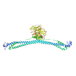
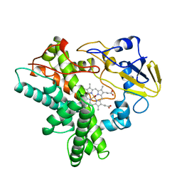
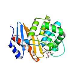
5ZOP
 
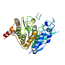 | | Crystal structure of histone deacetylase 4 (HDAC4) in complex with a SMRT corepressor SP2 fragment | | 分子名称: | Histone deacetylase 4, POTASSIUM ION, SMRT corepressor SP2 fragment, ... | | 著者 | Park, S.Y, Hwang, H.J, Kim, J.S. | | 登録日 | 2018-04-13 | | 公開日 | 2018-10-10 | | 最終更新日 | 2023-11-22 | | 実験手法 | X-RAY DIFFRACTION (2.698 Å) | | 主引用文献 | Structural basis of the specific interaction of SMRT corepressor with histone deacetylase 4.
Nucleic Acids Res., 46, 2018
|
|
5Y7D
 | | Crystal structure of human Endothelial-overexpressed LPS associated factor 1 | | 分子名称: | CHLORIDE ION, GLYCEROL, Protein CXorf40A, ... | | 著者 | Park, S.H, Kim, M.J, Park, J.S, Kim, H.J, Han, B.W. | | 登録日 | 2017-08-17 | | 公開日 | 2018-08-22 | | 最終更新日 | 2024-03-27 | | 実験手法 | X-RAY DIFFRACTION (1.71 Å) | | 主引用文献 | Crystal Structure of Human EOLA1 Implies Its Possibility of RNA Binding.
Molecules, 24, 2019
|
|
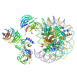
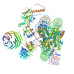
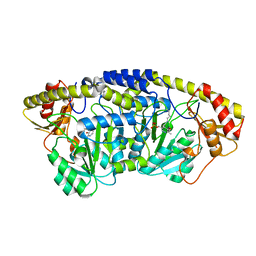
6PWX
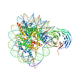 | | Cryo-EM structure of RbBP5 bound to the nucleosome | | 分子名称: | DNA (146-MER), Histone H2A type 1, Histone H2B 1.1, ... | | 著者 | Park, S.H, Ayoub, A, Lee, Y.T, Xu, J, Zhang, W, Zhang, B, Zhang, Y, Cianfrocco, M.A, Su, M, Dou, Y, Cho, U. | | 登録日 | 2019-07-23 | | 公開日 | 2019-12-18 | | 最終更新日 | 2024-03-20 | | 実験手法 | ELECTRON MICROSCOPY (4.2 Å) | | 主引用文献 | Cryo-EM structure of the human MLL1 core complex bound to the nucleosome.
Nat Commun, 10, 2019
|
|
7XYZ
 | | TRIM E3 ubiquitin ligase | | 分子名称: | Tripartite motif-containing protein 72, ZINC ION | | 著者 | Park, S.H, Song, H.K. | | 登録日 | 2022-06-02 | | 公開日 | 2023-07-12 | | 最終更新日 | 2023-11-29 | | 実験手法 | X-RAY DIFFRACTION (4.62 Å) | | 主引用文献 | Structure and activation of the RING E3 ubiquitin ligase TRIM72 on the membrane.
Nat.Struct.Mol.Biol., 30, 2023
|
|
7XV2
 | | TRIM E3 ubiquitin ligase | | 分子名称: | Tripartite motif-containing protein 72, ZINC ION | | 著者 | Park, S.H, Song, H.K. | | 登録日 | 2022-05-20 | | 公開日 | 2023-05-24 | | 最終更新日 | 2023-11-29 | | 実験手法 | X-RAY DIFFRACTION (2.75 Å) | | 主引用文献 | Structure and activation of the RING E3 ubiquitin ligase TRIM72 on the membrane.
Nat.Struct.Mol.Biol., 30, 2023
|
|
7XZ1
 | | TRIM E3 ubiquitin ligase | | 分子名称: | Tripartite motif-containing protein 72, ZINC ION | | 著者 | Park, S.H, Song, H.K. | | 登録日 | 2022-06-02 | | 公開日 | 2023-07-12 | | 最終更新日 | 2023-11-29 | | 実験手法 | X-RAY DIFFRACTION (5.2 Å) | | 主引用文献 | Structure and activation of the RING E3 ubiquitin ligase TRIM72 on the membrane.
Nat.Struct.Mol.Biol., 30, 2023
|
|
7XZ0
 | | TRIM E3 ubiquitin ligase | | 分子名称: | Tripartite motif-containing protein 72, ZINC ION | | 著者 | Park, S.H, Song, H.K. | | 登録日 | 2022-06-02 | | 公開日 | 2023-07-12 | | 最終更新日 | 2023-11-29 | | 実験手法 | X-RAY DIFFRACTION (3.28 Å) | | 主引用文献 | Structure and activation of the RING E3 ubiquitin ligase TRIM72 on the membrane.
Nat.Struct.Mol.Biol., 30, 2023
|
|
7XZ2
 | | TRIM E3 ubiquitin ligase | | 分子名称: | Tripartite motif-containing protein 72, ZINC ION | | 著者 | Park, S.H, Song, H.K. | | 登録日 | 2022-06-02 | | 公開日 | 2023-07-12 | | 最終更新日 | 2023-11-29 | | 実験手法 | X-RAY DIFFRACTION (3.5 Å) | | 主引用文献 | Structure and activation of the RING E3 ubiquitin ligase TRIM72 on the membrane.
Nat.Struct.Mol.Biol., 30, 2023
|
|
7XYY
 | | TRIM E3 ubiquitin ligase WT | | 分子名称: | Tripartite motif-containing protein 72, ZINC ION | | 著者 | Park, S.H, Song, H.K. | | 登録日 | 2022-06-02 | | 公開日 | 2023-07-12 | | 最終更新日 | 2023-11-29 | | 実験手法 | X-RAY DIFFRACTION (7.1 Å) | | 主引用文献 | Structure and activation of the RING E3 ubiquitin ligase TRIM72 on the membrane.
Nat.Struct.Mol.Biol., 30, 2023
|
|
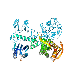
4NJD
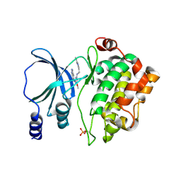 | | Structure of p21-activated kinase 4 with a novel inhibitor KY-04031 | | 分子名称: | N-(1H-indazol-5-yl)-N'-[2-(1H-indol-3-yl)ethyl]-6-methoxy-1,3,5-triazine-2,4-diamine, Serine/threonine-protein kinase PAK 4 | | 著者 | Park, S. | | 登録日 | 2013-11-09 | | 公開日 | 2014-05-21 | | 最終更新日 | 2022-08-24 | | 実験手法 | X-RAY DIFFRACTION (2.5 Å) | | 主引用文献 | Discovery and the structural basis of a novel p21-activated kinase 4 inhibitor.
Cancer Lett., 349, 2014
|
|
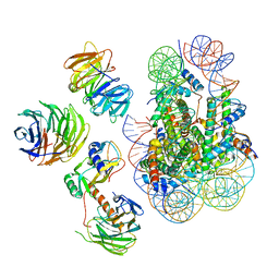
6W5N
 | | Cryo-EM structure of MLL1 in complex with RbBP5, WDR5, SET1, and ASH2L bound to the nucleosome (Class05) | | 分子名称: | DNA (147-MER), Histone H2A type 1, Histone H2B 1.1, ... | | 著者 | Park, S.H, Lee, Y.T, Ayoub, A, Dou, Y, Cho, U. | | 登録日 | 2020-03-13 | | 公開日 | 2021-03-31 | | 最終更新日 | 2024-05-29 | | 実験手法 | ELECTRON MICROSCOPY (6 Å) | | 主引用文献 | Mechanism for DPY30 and ASH2L intrinsically disordered regions to modulate the MLL/SET1 activity on chromatin.
Nat Commun, 12, 2021
|
|
4YUT
 | | Crystal structure of photoactivated adenylyl cyclase of a cyanobacteriaOscillatoria acuminata in orthorhombic form | | 分子名称: | FLAVIN MONONUCLEOTIDE, Family 3 adenylate cyclase | | 著者 | Park, S.-Y, Ohki, M, Sugiyama, K, Kawai, F, Iseki, M. | | 登録日 | 2015-03-19 | | 公開日 | 2016-06-01 | | 最終更新日 | 2024-03-20 | | 実験手法 | X-RAY DIFFRACTION (2.9 Å) | | 主引用文献 | Structural insight into photoactivation of an adenylate cyclase from a photosynthetic cyanobacterium
Proc.Natl.Acad.Sci.USA, 113, 2016
|
|
6W5I
 | | Cryo-EM structure of MLL1 in complex with RbBP5, WDR5, SET1, and ASH2L bound to the nucleosome (Class01) | | 分子名称: | DNA (147-MER), Histone H2A, Histone H2B 1.1, ... | | 著者 | Park, S.H, Lee, Y.T, Ayoub, A, Dou, Y, Cho, U. | | 登録日 | 2020-03-13 | | 公開日 | 2021-03-31 | | 最終更新日 | 2024-05-29 | | 実験手法 | ELECTRON MICROSCOPY (6.9 Å) | | 主引用文献 | Mechanism for DPY30 and ASH2L intrinsically disordered regions to modulate the MLL/SET1 activity on chromatin.
Nat Commun, 12, 2021
|
|
6W5M
 | | Cryo-EM structure of MLL1 in complex with RbBP5, WDR5, SET1, and ASH2L bound to the nucleosome (Class02) | | 分子名称: | DNA (147-MER), Histone H2A type 1, Histone H2B 1.1, ... | | 著者 | Park, S.H, Lee, Y.T, Ayoub, A, Dou, Y, Cho, U. | | 登録日 | 2020-03-13 | | 公開日 | 2021-03-31 | | 最終更新日 | 2024-05-29 | | 実験手法 | ELECTRON MICROSCOPY (4.6 Å) | | 主引用文献 | Mechanism for DPY30 and ASH2L intrinsically disordered regions to modulate the MLL/SET1 activity on chromatin.
Nat Commun, 12, 2021
|
|
4YUS
 | | Crystal structure of photoactivated adenylyl cyclase of a cyanobacteriaOscillatoria acuminata in hexagonal form | | 分子名称: | FLAVIN MONONUCLEOTIDE, Family 3 adenylate cyclase | | 著者 | Park, S.-Y, Ohki, M, Sugiyama, K, Kawai, F, Iseki, M. | | 登録日 | 2015-03-19 | | 公開日 | 2016-06-01 | | 最終更新日 | 2024-03-20 | | 実験手法 | X-RAY DIFFRACTION (1.8 Å) | | 主引用文献 | Structural insight into photoactivation of an adenylate cyclase from a photosynthetic cyanobacterium
Proc.Natl.Acad.Sci.USA, 113, 2016
|
|
8SQG
 | | OXA-48 bound to inhibitor CDD-2801 | | 分子名称: | (1M)-3'-(benzyloxy)-5-[2-(methylamino)-2-oxoethoxy][1,1'-biphenyl]-3,4'-dicarboxylic acid, BICARBONATE ION, Beta-lactamase | | 著者 | Park, S, Judge, A, Fan, J, Sankaran, B, Palzkill, T. | | 登録日 | 2023-05-04 | | 公開日 | 2024-01-03 | | 最終更新日 | 2024-01-17 | | 実験手法 | X-RAY DIFFRACTION (2.03 Å) | | 主引用文献 | Exploiting the Carboxylate-Binding Pocket of beta-Lactamase Enzymes Using a Focused DNA-Encoded Chemical Library.
J.Med.Chem., 67, 2024
|
|
1CMN
 | |
1CMJ
 | |
1CL6
 | |
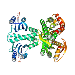
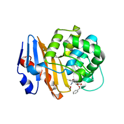
4YWV
 | | Structural insight into the substrate inhibition mechanism of NADP+-dependent succinic semialdehyde dehydrogenase from Streptococcus pyogenes | | 分子名称: | 4-oxobutanoic acid, SULFATE ION, Succinic semialdehyde dehydrogenase | | 著者 | Park, S.A, Jang, E.H, Chi, Y.M, Lee, K.S. | | 登録日 | 2015-03-21 | | 公開日 | 2015-05-06 | | 最終更新日 | 2023-11-08 | | 実験手法 | X-RAY DIFFRACTION (2.4 Å) | | 主引用文献 | Structural insight into the substrate inhibition mechanism of NADP(+)-dependent succinic semialdehyde dehydrogenase from Streptococcus pyogenes.
Biochem.Biophys.Res.Commun., 461, 2015
|
|
8SQF
 | | OXA-48 bound to inhibitor CDD-2725 | | 分子名称: | (1M)-3'-(benzyloxy)-5-hydroxy[1,1'-biphenyl]-3,4'-dicarboxylic acid, BICARBONATE ION, Beta-lactamase | | 著者 | Park, S, Judge, A, Fan, J, Sankaran, B, Prasad, B.V.V, Palzkill, T. | | 登録日 | 2023-05-04 | | 公開日 | 2024-01-03 | | 最終更新日 | 2024-01-17 | | 実験手法 | X-RAY DIFFRACTION (2.3 Å) | | 主引用文献 | Exploiting the Carboxylate-Binding Pocket of beta-Lactamase Enzymes Using a Focused DNA-Encoded Chemical Library.
J.Med.Chem., 67, 2024
|
|
5X03
 | |
2KLV
 | | Membrane-bound structure of the Pf1 major coat protein in DHPC micelle | | 分子名称: | Capsid protein G8P | | 著者 | Park, S, Son, W, Mukhopadhyay, R, Valafar, H, Opella, S.J. | | 登録日 | 2009-07-08 | | 公開日 | 2009-10-06 | | 最終更新日 | 2024-05-22 | | 実験手法 | SOLUTION NMR | | 主引用文献 | Phage-induced alignment of membrane proteins enables the measurement and structural analysis of residual dipolar couplings with dipolar waves and lambda-maps.
J.Am.Chem.Soc., 131, 2009
|
|
5Y6C
 | |
5Y6B
 | |


