Loading
PDBj
MenuPDBj@FacebookPDBj@TwitterPDBj@YouTubewwPDB FoundationwwPDB
RCSB PDBPDBeBMRBAdv. SearchSearch help
PDB: 511 results

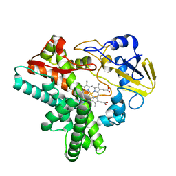
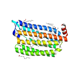
6JY9
DownloadVisualize
BU of 6jy9 by Molmil
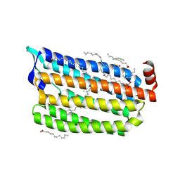
Structure of light-state marine bacterial chloride importer, NM-R3, with CW laser (ND-3%) at 95K.
Descriptor: CHLORIDE ION, Chloride pumping rhodopsin, OLEIC ACID, ...
Authors:Yun, J.H, Ohki, M, Park, S.Y, Lee, W.
Deposit date:2019-04-26
Release date:2020-03-04
Last modified:2024-10-30
Method:X-RAY DIFFRACTION (1.9 Å)
Cite:Pumping mechanism of NM-R3, a light-driven bacterial chloride importer in the rhodopsin family.
Sci Adv, 6, 2020
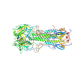
1HGD
DownloadVisualize
BU of 1hgd by Molmil
BINDING OF INFLUENZA VIRUS HEMAGGLUTININ TO ANALOGS OF ITS CELL-SURFACE RECEPTOR, SIALIC ACID: ANALYSIS BY PROTON NUCLEAR MAGNETIC RESONANCE SPECTROSCOPY AND X-RAY CRYSTALLOGRAPHY
Descriptor: 2-acetamido-2-deoxy-beta-D-glucopyranose, HEMAGGLUTININ, CHAIN HA1, ...
Authors:Sauter, N.K, Hanson, J.E, Glick, G.D, Brown, J.H, Crowther, R.L, Park, S.-J, Skehel, J.J, Wiley, D.C.
Deposit date:1991-11-01
Release date:1994-01-31
Last modified:2024-10-23
Method:X-RAY DIFFRACTION (2.7 Å)
Cite:Binding of influenza virus hemagglutinin to analogs of its cell-surface receptor, sialic acid: analysis by proton nuclear magnetic resonance spectroscopy and X-ray crystallography.
Biochemistry, 31, 1992
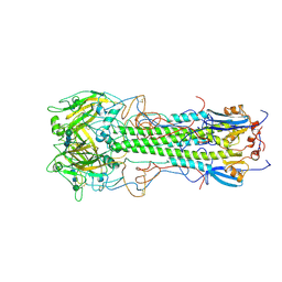
1HGF
DownloadVisualize
BU of 1hgf by Molmil
BINDING OF INFLUENZA VIRUS HEMAGGLUTININ TO ANALOGS OF ITS CELL-SURFACE RECEPTOR, SIALIC ACID: ANALYSIS BY PROTON NUCLEAR MAGNETIC RESONANCE SPECTROSCOPY AND X-RAY CRYSTALLOGRAPHY
Descriptor: 2-acetamido-2-deoxy-beta-D-glucopyranose, HEMAGGLUTININ, CHAIN HA1, ...
Authors:Sauter, N.K, Hanson, J.E, Glick, G.D, Brown, J.H, Crowther, R.L, Park, S.-J, Skehel, J.J, Wiley, D.C.
Deposit date:1991-11-01
Release date:1994-01-31
Last modified:2020-07-29
Method:X-RAY DIFFRACTION (3 Å)
Cite:Binding of influenza virus hemagglutinin to analogs of its cell-surface receptor, sialic acid: analysis by proton nuclear magnetic resonance spectroscopy and X-ray crystallography.
Biochemistry, 31, 1992
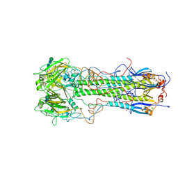
1HGE
DownloadVisualize
BU of 1hge by Molmil
BINDING OF INFLUENZA VIRUS HEMAGGLUTININ TO ANALOGS OF ITS CELL-SURFACE RECEPTOR, SIALIC ACID: ANALYSIS BY PROTON NUCLEAR MAGNETIC RESONANCE SPECTROSCOPY AND X-RAY CRYSTALLOGRAPHY
Descriptor: 2-O-methyl-5-N-acetyl-alpha-D-neuraminic acid, 2-acetamido-2-deoxy-beta-D-glucopyranose, HEMAGGLUTININ, ...
Authors:Sauter, N.K, Hanson, J.E, Glick, G.D, Brown, J.H, Crowther, R.L, Park, S.-J, Skehel, J.J, Wiley, D.C.
Deposit date:1991-11-01
Release date:1994-01-31
Last modified:2024-10-16
Method:X-RAY DIFFRACTION (2.6 Å)
Cite:Binding of influenza virus hemagglutinin to analogs of its cell-surface receptor, sialic acid: analysis by proton nuclear magnetic resonance spectroscopy and X-ray crystallography.
Biochemistry, 31, 1992
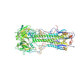
1HGJ
DownloadVisualize
BU of 1hgj by Molmil
BINDING OF INFLUENZA VIRUS HEMAGGLUTININ TO ANALOGS OF ITS CELL-SURFACE RECEPTOR, SIALIC ACID: ANALYSIS BY PROTON NUCLEAR MAGNETIC RESONANCE SPECTROSCOPY AND X-RAY CRYSTALLOGRAPHY
Descriptor: 2-acetamido-2-deoxy-beta-D-glucopyranose, HEMAGGLUTININ, CHAIN HA1, ...
Authors:Sauter, N.K, Hanson, J.E, Glick, G.D, Brown, J.H, Crowther, R.L, Park, S.-J, Skehel, J.J, Wiley, D.C.
Deposit date:1991-11-01
Release date:1994-01-31
Last modified:2020-07-29
Method:X-RAY DIFFRACTION (2.7 Å)
Cite:Binding of influenza virus hemagglutinin to analogs of its cell-surface receptor, sialic acid: analysis by proton nuclear magnetic resonance spectroscopy and X-ray crystallography.
Biochemistry, 31, 1992
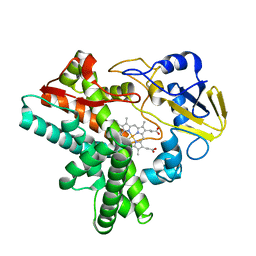
1EHF
DownloadVisualize
BU of 1ehf by Molmil
CRYSTAL STRUCTURES OF CYTOCHROME P450NOR AND ITS MUTANTS (SER286 VAL, THR) IN THE FERRIC RESTING STATE AT CRYOGENIC TEMPERATURE: A COMPARATIVE ANALYSIS WITH MONOOXYGENASE CYTOCHROME P450S
Descriptor: CYTOCHROME P450NOR, PROTOPORPHYRIN IX CONTAINING FE
Authors:Shimizu, H, Park, S.
Deposit date:2000-02-21
Release date:2000-08-21
Last modified:2024-02-07
Method:X-RAY DIFFRACTION (1.7 Å)
Cite:Crystal structures of cytochrome P450nor and its mutants (Ser286-->Val, Thr) in the ferric resting state at cryogenic temperature: a comparative analysis with monooxygenase cytochrome P450s.
J.Inorg.Biochem., 81, 2000
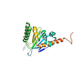
5YX3
DownloadVisualize
BU of 5yx3 by Molmil
Chalcone isomerase from the Antarctic vascular plant Deschampsia Antarctica (DaCHI1)
Descriptor: Chalcone-flavonone isomerase family protein
Authors:Lee, C.W, Park, S, Lee, J.H.
Deposit date:2017-12-01
Release date:2018-02-21
Last modified:2024-03-27
Method:X-RAY DIFFRACTION (2.7 Å)
Cite:Crystal structure and enzymatic properties of chalcone isomerase from the Antarctic vascular plant Deschampsia antarctica Desv.
PLoS ONE, 13, 2018
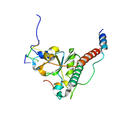
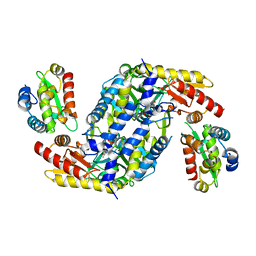
7C06
DownloadVisualize
BU of 7c06 by Molmil
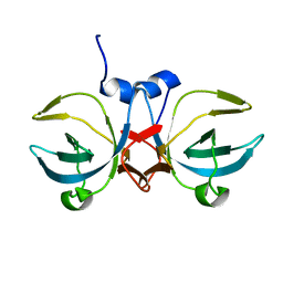
Crystal structure of yeast U2AF1 complex bound to 3' splice site RNA, 5'-UAGGU.
Descriptor: RNA (5'-R(*U*UP*AP*GP*GP*U)-3'), Splicing factor U2AF 23 kDa subunit, Splicing factor U2AF 59 kDa subunit, ...
Authors:Yoshida, H, Park, S.Y, Urano, T, Obayashi, E.
Deposit date:2020-04-30
Release date:2020-09-30
Last modified:2023-11-29
Method:X-RAY DIFFRACTION (3.02 Å)
Cite:Elucidation of the aberrant 3' splice site selection by cancer-associated mutations on the U2AF1.
Nat Commun, 11, 2020
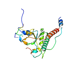
7C08
DownloadVisualize
BU of 7c08 by Molmil
Crystal structure of S34Y mutant of yeast U2AF1 complex bound to 3' splice site RNA, 5'-UAGGU.
Descriptor: RNA (5'-R(*U*UP*AP*GP*GP*U)-3'), Splicing factor U2AF 23 kDa subunit, Splicing factor U2AF 59 kDa subunit, ...
Authors:Yoshida, H, Park, S.Y, Urano, T, Obayashi, E.
Deposit date:2020-04-30
Release date:2020-09-30
Last modified:2023-11-29
Method:X-RAY DIFFRACTION (3.35 Å)
Cite:Elucidation of the aberrant 3' splice site selection by cancer-associated mutations on the U2AF1.
Nat Commun, 11, 2020
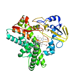
1EHG
DownloadVisualize
BU of 1ehg by Molmil
CRYSTAL STRUCTURES OF CYTOCHROME P450NOR AND ITS MUTANTS (SER286 VAL, THR) IN THE FERRIC RESTING STATE AT CRYOGENIC TEMPERATURE: A COMPARATIVE ANALYSIS WITH MONOOXYGENASE CYTOCHROME P450S
Descriptor: CYTOCHROME P450NOR, PROTOPORPHYRIN IX CONTAINING FE
Authors:Shimizu, H, Park, S.
Deposit date:2000-02-21
Release date:2000-08-21
Last modified:2024-02-07
Method:X-RAY DIFFRACTION (1.7 Å)
Cite:Crystal structures of cytochrome P450nor and its mutants (Ser286-->Val, Thr) in the ferric resting state at cryogenic temperature: a comparative analysis with monooxygenase cytochrome P450s.
J.Inorg.Biochem., 81, 2000
5YX4
DownloadVisualize
BU of 5yx4 by Molmil
Isoliquiritigenin-complexed Chalcone isomerase (S189A) from the Antarctic vascular plant Deschampsia Antarctica (DaCHI1)
Descriptor: 2',4,4'-TRIHYDROXYCHALCONE, Chalcone-flavonone isomerase family protein
Authors:Lee, C.W, Park, S, Lee, J.H.
Deposit date:2017-12-01
Release date:2018-02-14
Last modified:2024-03-27
Method:X-RAY DIFFRACTION (2.1 Å)
Cite:Crystal structure and enzymatic properties of chalcone isomerase from the Antarctic vascular plant Deschampsia antarctica Desv.
PLoS ONE, 13, 2018
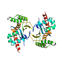
4LW4
DownloadVisualize
BU of 4lw4 by Molmil
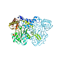
Structural changes during cysteine desulfurase CsdA and sulfur-acceptor CsdE interactions provide insight into the trans-persulfuration
Descriptor: Cysteine desulfuration protein CsdE, Cysteine sulfinate desulfinase, PYRIDOXAL-5'-PHOSPHATE
Authors:Kim, S, Park, S.Y.
Deposit date:2013-07-26
Release date:2013-08-07
Last modified:2023-11-08
Method:X-RAY DIFFRACTION (2.01 Å)
Cite:Structural changes during cysteine desulfurase CsdA and sulfur acceptor CsdE interactions provide insight into the trans-persulfuration.
J.Biol.Chem., 288, 2013
1EHE
DownloadVisualize
BU of 1ehe by Molmil
CRYSTAL STRUCTURES OF CYTOCHROME P450NOR AND ITS MUTANTS (SER286 VAL, THR) IN THE FERRIC RESTING STATE AT CRYOGENIC TEMPERATURE: A COMPARATIVE ANALYSIS WITH MONOOXYGENASE CYTOCHROME P450S
Descriptor: CYTOCHROME P450NOR, PROTOPORPHYRIN IX CONTAINING FE
Authors:Shimizu, H, Park, S.
Deposit date:2000-02-21
Release date:2000-08-21
Last modified:2024-02-07
Method:X-RAY DIFFRACTION (1.7 Å)
Cite:Crystal structures of cytochrome P450nor and its mutants (Ser286-->Val, Thr) in the ferric resting state at cryogenic temperature: a comparative analysis with monooxygenase cytochrome P450s.
J.Inorg.Biochem., 81, 2000
4LW2
DownloadVisualize
BU of 4lw2 by Molmil
Structural changes during cysteine desulfurase CsdA and sulfur-acceptor CsdE interactions provide insight into the trans-persulfuration
Descriptor: Cysteine sulfinate desulfinase, GLYCEROL, PYRIDOXAL-5'-PHOSPHATE
Authors:Kim, S, Park, S.Y.
Deposit date:2013-07-26
Release date:2013-08-07
Last modified:2023-11-08
Method:X-RAY DIFFRACTION (1.8 Å)
Cite:Structural changes during cysteine desulfurase CsdA and sulfur acceptor CsdE interactions provide insight into the trans-persulfuration.
J.Biol.Chem., 288, 2013
5H34
DownloadVisualize
BU of 5h34 by Molmil
Crystal structure of the C-terminal domain of methionyl-tRNA synthetase (MetRS-C) in Nanoarchaeum equitans
Descriptor: Methionine-tRNA ligase
Authors:Suzuki, H, Kaneko, A, Yamamoto, T, Nambo, M, Umehara, T, Yoshida, H, Park, S.Y, Tamura, K.
Deposit date:2016-10-20
Release date:2017-06-21
Last modified:2024-03-20
Method:X-RAY DIFFRACTION (1.748 Å)
Cite:Binding Properties of Split tRNA to the C-terminal Domain of Methionyl-tRNA Synthetase of Nanoarchaeum equitans.
J. Mol. Evol., 84, 2017
7VDN
DownloadVisualize
BU of 7vdn by Molmil
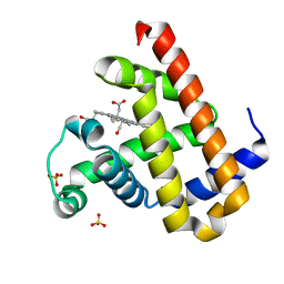
High resolution crystal structure of Sperm Whale Myoglobin in the carbonmonoxy form
Descriptor: CARBON MONOXIDE, Myoglobin, PROTOPORPHYRIN IX CONTAINING FE, ...
Authors:Shibayama, N, Sato-Tomita, A, Ishimoto, N, Park, S.Y.
Deposit date:2021-09-07
Release date:2022-09-14
Last modified:2023-11-29
Method:X-RAY DIFFRACTION (0.93 Å)
Cite:X-ray fluorescence holography of biological metal sites: Application to myoglobin.
Biochem.Biophys.Res.Commun., 635, 2022
5DSL
DownloadVisualize
BU of 5dsl by Molmil
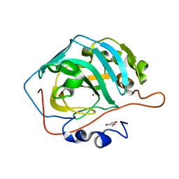
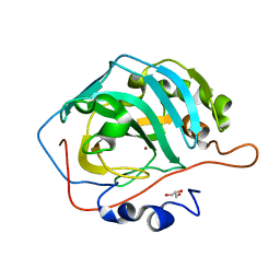
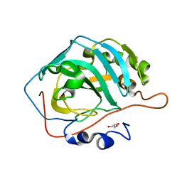
Structure of CO2 released holo-form of human carbonic anhydrase II with 10 min warming
Descriptor: Carbonic anhydrase 2, GLYCEROL, ZINC ION
Authors:Kim, C.U, Park, S.Y, McKenna, R.
Deposit date:2015-09-17
Release date:2016-05-04
Last modified:2023-11-08
Method:X-RAY DIFFRACTION (1.45 Å)
Cite:Tracking solvent and protein movement during CO2 release in carbonic anhydrase II crystals
Proc. Natl. Acad. Sci. U.S.A., 113, 2016
5DSK
DownloadVisualize
BU of 5dsk by Molmil
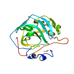
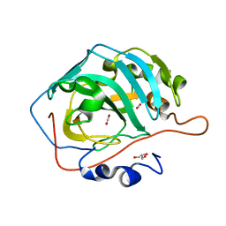
Structure of CO2 released holo-form of human carbonic anhydrase II with 3 min warming
Descriptor: Carbonic anhydrase 2, GLYCEROL, ZINC ION
Authors:Kim, C.U, Park, S.Y, McKenna, R.
Deposit date:2015-09-17
Release date:2016-05-04
Last modified:2023-11-08
Method:X-RAY DIFFRACTION (1.3 Å)
Cite:Tracking solvent and protein movement during CO2 release in carbonic anhydrase II crystals
Proc. Natl. Acad. Sci. U.S.A., 113, 2016
5DSQ
DownloadVisualize
BU of 5dsq by Molmil
Structure of CO2 released apo-form of human carbonic anhydrase II with 3 min warming
Descriptor: Carbonic anhydrase 2, GLYCEROL
Authors:Kim, C.U, Park, S.Y, McKenna, R.
Deposit date:2015-09-17
Release date:2016-05-04
Last modified:2023-11-08
Method:X-RAY DIFFRACTION (1.5 Å)
Cite:Tracking solvent and protein movement during CO2 release in carbonic anhydrase II crystals
Proc. Natl. Acad. Sci. U.S.A., 113, 2016
5DSM
DownloadVisualize
BU of 5dsm by Molmil
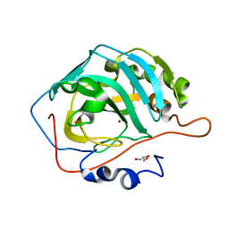
Structure of CO2 released holo-form of human carbonic anhydrase II with 25 min warming
Descriptor: Carbonic anhydrase 2, GLYCEROL, ZINC ION
Authors:Kim, C.U, Park, S.Y, McKenna, R.
Deposit date:2015-09-17
Release date:2016-05-04
Last modified:2023-11-08
Method:X-RAY DIFFRACTION (1.3 Å)
Cite:Tracking solvent and protein movement during CO2 release in carbonic anhydrase II crystals
Proc. Natl. Acad. Sci. U.S.A., 113, 2016
5DSR
DownloadVisualize
BU of 5dsr by Molmil
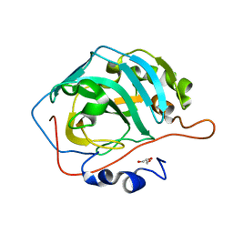
Structure of CO2 released apo-form of human carbonic anhydrase II with 10 min warming
Descriptor: Carbonic anhydrase 2, GLYCEROL
Authors:Kim, C.U, Park, S.Y, McKenna, R.
Deposit date:2015-09-17
Release date:2016-05-04
Last modified:2023-11-08
Method:X-RAY DIFFRACTION (1.3 Å)
Cite:Tracking solvent and protein movement during CO2 release in carbonic anhydrase II crystals
Proc. Natl. Acad. Sci. U.S.A., 113, 2016
5DSP
DownloadVisualize
BU of 5dsp by Molmil
Structure of CO2 released apo-form of human carbonic anhydrase II with 40 sec warming
Descriptor: Carbonic anhydrase 2, GLYCEROL
Authors:Kim, C.U, Park, S.Y, McKenna, R.
Deposit date:2015-09-17
Release date:2016-05-04
Last modified:2023-11-08
Method:X-RAY DIFFRACTION (1.4 Å)
Cite:Tracking solvent and protein movement during CO2 release in carbonic anhydrase II crystals
Proc. Natl. Acad. Sci. U.S.A., 113, 2016
5DSO
DownloadVisualize
BU of 5dso by Molmil
Structure of CO2 bound apo-form of human carbonic anhydrase II with 0 sec (no) warming
Descriptor: CARBON DIOXIDE, Carbonic anhydrase 2, GLYCEROL
Authors:Kim, C.U, Park, S.Y, McKenna, R.
Deposit date:2015-09-17
Release date:2016-05-04
Last modified:2023-11-08
Method:X-RAY DIFFRACTION (1.4 Å)
Cite:Tracking solvent and protein movement during CO2 release in carbonic anhydrase II crystals
Proc. Natl. Acad. Sci. U.S.A., 113, 2016
5ZTK
DownloadVisualize
BU of 5ztk by Molmil
Synchrotron structure of light-driven chloride pump having an NTQ motif
Descriptor: CHLORIDE ION, Chloride pumping rhodopsin, OLEIC ACID, ...
Authors:Yun, J.H, Park, J.H, Park, S.Y, Lee, W.
Deposit date:2018-05-04
Release date:2018-12-05
Last modified:2024-10-30
Method:X-RAY DIFFRACTION (1.75 Å)
Cite:Non-cryogenic structure of a chloride pump provides crucial clues to temperature-dependent channel transport efficiency
J. Biol. Chem., 294, 2019
2ZTT
DownloadVisualize
BU of 2ztt by Molmil
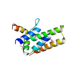
Crystal Structure of RNA polymerase PB1-PB2 subunits from Influenza A Virus
Descriptor: Polymerase basic protein 2, RNA-directed RNA polymerase catalytic subunit
Authors:Sugiyama, K, Obayashi, E, Park, S.-Y.
Deposit date:2008-10-08
Release date:2009-06-09
Last modified:2011-07-13
Method:X-RAY DIFFRACTION (2.1 Å)
Cite:Structural insight into the essential PB1-PB2 subunit contact of the influenza virus RNA polymerase
Embo J., 28, 2009

226707

PDB entries from 2024-10-30

PDB statisticsPDBj update infoContact PDBjnumon