Loading
PDBj
MenuPDBj@FacebookPDBj@TwitterPDBj@YouTubewwPDB FoundationwwPDB
RCSB PDBPDBeBMRBAdv. SearchSearch help
PDB: 552 results

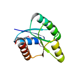
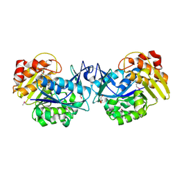
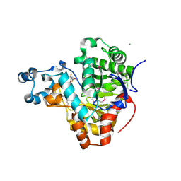
3BE3
DownloadVisualize
BU of 3be3 by Molmil
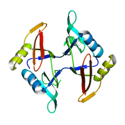
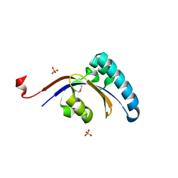
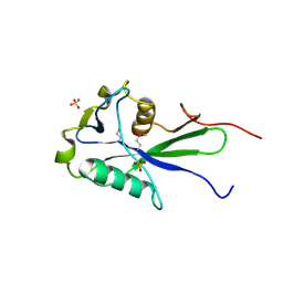
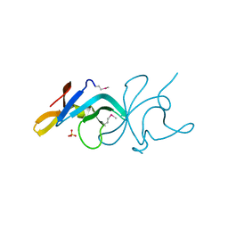
Crystal structure of a protein belonging to pfam DUF1653 from Bordetella bronchiseptica
Descriptor: SULFATE ION, Uncharacterized protein
Authors:Tyagi, R, Eswaramoorthy, S, Burley, S.K, Swaminathan, S, New York SGX Research Center for Structural Genomics (NYSGXRC)
Deposit date:2007-11-16
Release date:2007-11-27
Last modified:2024-10-30
Method:X-RAY DIFFRACTION (2.04 Å)
Cite:Crystal structure of a protein belonging to pfam DUF1653 from Bordetella bronchiseptica.
To be Published
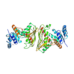
3K2G
DownloadVisualize
BU of 3k2g by Molmil
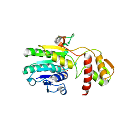
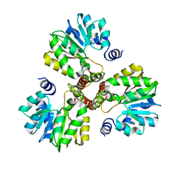
Crystal structure of a Resiniferatoxin-binding protein from Rhodobacter sphaeroides
Descriptor: (2S,3S)-1,4-DIMERCAPTOBUTANE-2,3-DIOL, MAGNESIUM ION, Resiniferatoxin-binding, ...
Authors:Kumaran, D, Burley, S.K, Swaminathan, S, New York SGX Research Center for Structural Genomics (NYSGXRC)
Deposit date:2009-09-30
Release date:2009-10-13
Last modified:2021-02-10
Method:X-RAY DIFFRACTION (1.8 Å)
Cite:Crystal structure of a Resiniferatoxin-binding protein from Rhodobacter sphaeroides
To be Published
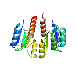
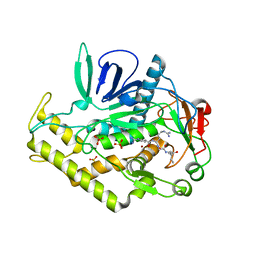
3BWI
DownloadVisualize
BU of 3bwi by Molmil
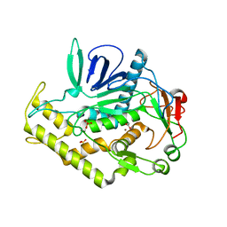
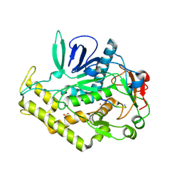
Crystal structure of the catalytic domain of botulinum neurotoxin serotype A with an acetate ion bound at the active site
Descriptor: ACETATE ION, Botulinum neurotoxin A light chain, SULFATE ION, ...
Authors:Kumaran, D, Rawat, R, Swaminathan, S.
Deposit date:2008-01-09
Release date:2008-04-22
Last modified:2023-08-30
Method:X-RAY DIFFRACTION (1.7 Å)
Cite:Structure- and Substrate-based Inhibitor Design for Clostridium botulinum Neurotoxin Serotype A
J.Biol.Chem., 283, 2008
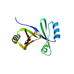
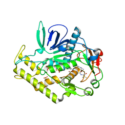
3C88
DownloadVisualize
BU of 3c88 by Molmil
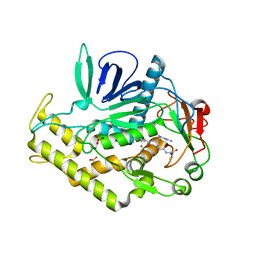
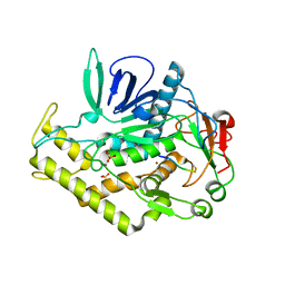
Crystal structure of the catalytic domain of botulinum neurotoxin serotype A with inhibitory peptide RRGC
Descriptor: Botulinum neurotoxin A light chain, Inhibitor peptide RRGC, SODIUM ION, ...
Authors:Kumaran, D, Swaminathan, S.
Deposit date:2008-02-11
Release date:2008-04-22
Last modified:2023-08-30
Method:X-RAY DIFFRACTION (1.6 Å)
Cite:Structure- and Substrate-based Inhibitor Design for Clostridium botulinum Neurotoxin Serotype A
J.Biol.Chem., 283, 2008
2PZZ
DownloadVisualize
BU of 2pzz by Molmil
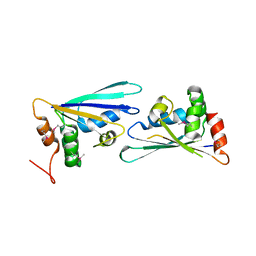
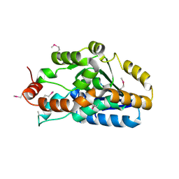
2.2 A resolution crystal structure of UPF0201 protein from Methanococcus jannaschii
Descriptor: UPF0201 protein MJ1564
Authors:Rao, K.N, Burley, S.K, Swaminathan, S, New York SGX Research Center for Structural Genomics (NYSGXRC)
Deposit date:2007-05-18
Release date:2007-05-29
Last modified:2024-10-16
Method:X-RAY DIFFRACTION (2.2 Å)
Cite:UPF201 archaeal specific family members reveal structural similarity to RNA-binding proteins but low likelihood for RNA-binding function.
Plos One, 3, 2008
3BT3
DownloadVisualize
BU of 3bt3 by Molmil
Crystal structure of a glyoxalase-related enzyme from Clostridium phytofermentans
Descriptor: Glyoxalase-related enzyme, AraC type
Authors:Rao, K.N, Burley, S.K, Swaminathan, S, New York SGX Research Center for Structural Genomics (NYSGXRC)
Deposit date:2007-12-27
Release date:2008-03-18
Last modified:2024-02-21
Method:X-RAY DIFFRACTION (2.5 Å)
Cite:Crystal structure of a glyoxalase-related enzyme from Clostridium phytofermentans.
To be Published
3KXW
DownloadVisualize
BU of 3kxw by Molmil
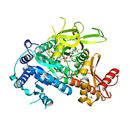
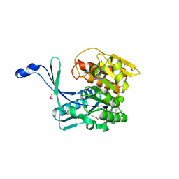
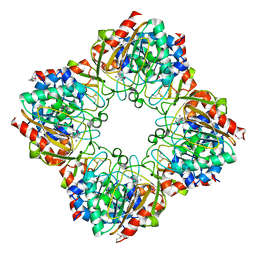
The crystal structure of fatty acid AMP ligase from Legionella pneumophila
Descriptor: 5'-O-[(S)-(dodecanoyloxy)(hydroxy)phosphoryl]adenosine, Saframycin Mx1 synthetase B
Authors:Zhang, Z, Burley, S.K, Swaminathan, S, New York SGX Research Center for Structural Genomics (NYSGXRC)
Deposit date:2009-12-04
Release date:2010-03-16
Last modified:2021-02-10
Method:X-RAY DIFFRACTION (1.851 Å)
Cite:Structural and Functional Studies of Fatty Acyl Adenylate Ligases from E. coli and L. pneumophila.
J.Mol.Biol., 406, 2011
2A9F
DownloadVisualize
BU of 2a9f by Molmil
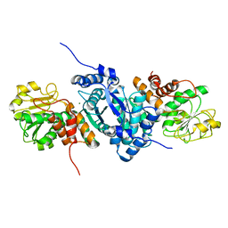
Crystal structure of a putative malic enzyme ((S)-malate:NAD+ oxidoreductase (decarboxylating))
Descriptor: MAGNESIUM ION, putative malic enzyme ((S)-malate:NAD+ oxidoreductase (decarboxylating))
Authors:Seetharaman, J, Swaminathan, S, Burley, S.K, New York SGX Research Center for Structural Genomics (NYSGXRC)
Deposit date:2005-07-11
Release date:2005-08-02
Last modified:2024-02-14
Method:X-RAY DIFFRACTION (2.5 Å)
Cite:Crystal structure of a putative malic enzyme ((S)-malate:NAD+ oxidoreductase (decarboxylating))
To be Published
3GPK
DownloadVisualize
BU of 3gpk by Molmil
Crystal Structure of PpiC-type peptidyl-prolyl cis-trans isomerase domain at 1.55A resolution.
Descriptor: PpiC-type peptidyl-prolyl cis-trans isomerase, SULFATE ION
Authors:Satyanarayana, L, Eswaramoorthy, S, Burley, S.K, Swaminathan, S, New York SGX Research Center for Structural Genomics (NYSGXRC)
Deposit date:2009-03-23
Release date:2009-03-31
Last modified:2021-02-10
Method:X-RAY DIFFRACTION (1.55 Å)
Cite:Crystal Structure of PpiC-type peptidyl-prolyl cis-trans isomerase domain at 1.55A resolution.
To be Published
3EHE
DownloadVisualize
BU of 3ehe by Molmil
Crystal structure of UDP-glucose 4 epimerase (galE-1) from Archaeoglobus fulgidus
Descriptor: NICOTINAMIDE-ADENINE-DINUCLEOTIDE, UDP-glucose 4-epimerase (GalE-1)
Authors:Agarwal, R, Burley, S.K, Swaminathan, S, New York SGX Research Center for Structural Genomics (NYSGXRC)
Deposit date:2008-09-12
Release date:2008-10-14
Last modified:2024-02-21
Method:X-RAY DIFFRACTION (1.87 Å)
Cite:Crystal structure of galE-1 from Archaeoglobus fulgidus
To be Published
2QZJ
DownloadVisualize
BU of 2qzj by Molmil
Crystal structure of a two-component response regulator from Clostridium difficile
Descriptor: Two-component response regulator
Authors:Sugadev, R, Burley, S.K, Swaminathan, S, New York SGX Research Center for Structural Genomics (NYSGXRC)
Deposit date:2007-08-16
Release date:2007-09-04
Last modified:2021-02-03
Method:X-RAY DIFFRACTION (2.89 Å)
Cite:Crystal structure of a two-component response regulator from Clostridium difficile.
To be Published
3E9M
DownloadVisualize
BU of 3e9m by Molmil
Crystal Structure of an oxidoreductase from Enterococcus faecalis
Descriptor: Oxidoreductase, Gfo/Idh/MocA family
Authors:Damodharan, L, Burley, S.K, Swaminathan, S, New York SGX Research Center for Structural Genomics (NYSGXRC)
Deposit date:2008-08-22
Release date:2008-10-14
Last modified:2021-02-10
Method:X-RAY DIFFRACTION (2.7 Å)
Cite:Crystal Structure of an oxidoreductase from Enterococcus faecalis
To be Published
3HDG
DownloadVisualize
BU of 3hdg by Molmil
Crystal structure of the N-terminal domain of an uncharacterized protein (WS1339) from Wolinella succinogenes
Descriptor: MAGNESIUM ION, uncharacterized protein
Authors:Eswaramoorthy, S, Burley, S.K, Swaminathan, S, New York SGX Research Center for Structural Genomics (NYSGXRC)
Deposit date:2009-05-07
Release date:2009-05-19
Last modified:2024-10-09
Method:X-RAY DIFFRACTION (2.27 Å)
Cite:Crystal structure of the N-terminal domain of an uncharacterized protein (WS1339) from Wolinella succinogenes
To be Published
3HDP
DownloadVisualize
BU of 3hdp by Molmil
Crystal structure of the NI(II)-bound Glyoxalase-I from Clostridium acetobutylicum
Descriptor: Glyoxalase-I, NICKEL (II) ION
Authors:Satyanarayana, L, Eswaramoorthy, S, Burley, S.K, Swaminathan, S, New York SGX Research Center for Structural Genomics (NYSGXRC)
Deposit date:2009-05-07
Release date:2009-05-26
Last modified:2023-09-06
Method:X-RAY DIFFRACTION (2.06 Å)
Cite:Crystal structure of the NI(II)-bound Glyoxalase-I from Clostridium acetobutylicum.
To be Published
3IH0
DownloadVisualize
BU of 3ih0 by Molmil
Crystal structure of an uncharacterized sugar kinase PH1459 from Pyrococcus horikoshii in complex with AMP-PNP
Descriptor: GLYCEROL, PHOSPHOAMINOPHOSPHONIC ACID-ADENYLATE ESTER, Uncharacterized sugar kinase PH1459
Authors:Kumar, G, Eswaramoorthy, S, Burley, S.K, Swaminathan, S, New York SGX Research Center for Structural Genomics (NYSGXRC)
Deposit date:2009-07-29
Release date:2009-09-08
Last modified:2023-11-22
Method:X-RAY DIFFRACTION (1.9 Å)
Cite:Crystal structure of an uncharacterized sugar kinase PH1459 from Pyrococcus horikoshii in complex with AMP-PNP
To be Published
3C8B
DownloadVisualize
BU of 3c8b by Molmil
Crystal structure of the catalytic domain of botulinum neurotoxin serotype A with inhibitory peptide RRGI
Descriptor: Botulinum neurotoxin A light chain, Inhibitor peptide RRGI, SULFATE ION, ...
Authors:Kumaran, D, Swaminathan, S.
Deposit date:2008-02-11
Release date:2008-04-22
Last modified:2024-10-30
Method:X-RAY DIFFRACTION (1.47 Å)
Cite:Structure- and Substrate-based Inhibitor Design for Clostridium botulinum Neurotoxin Serotype A
J.Biol.Chem., 283, 2008
3C89
DownloadVisualize
BU of 3c89 by Molmil
Crystal structure of the catalytic domain of botulinum neurotoxin serotype A with inhibitory peptide RRGM
Descriptor: Botulinum neurotoxin A light chain, Inhibitor peptide RRGM, SULFATE ION, ...
Authors:Kumaran, D, Swaminathan, S.
Deposit date:2008-02-11
Release date:2008-04-22
Last modified:2023-08-30
Method:X-RAY DIFFRACTION (1.58 Å)
Cite:Structure- and Substrate-based Inhibitor Design for Clostridium botulinum Neurotoxin Serotype A
J.Biol.Chem., 283, 2008
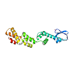
3D5L
DownloadVisualize
BU of 3d5l by Molmil
Crystal structure of regulatory protein RecX
Descriptor: Regulatory protein RecX, SULFATE ION
Authors:Agarwal, R, Burley, S.K, Swaminathan, S, New York SGX Research Center for Structural Genomics (NYSGXRC)
Deposit date:2008-05-16
Release date:2008-06-10
Last modified:2024-02-21
Method:X-RAY DIFFRACTION (2.35 Å)
Cite:Crystal structure of DNA repair regulatory protein RecX.
To be Published
3DLI
DownloadVisualize
BU of 3dli by Molmil
Crystal structure of a SAM dependent methyltransferase from Archaeoglobus fulgidus
Descriptor: methyltransferase
Authors:Agarwal, R, Burley, S.K, Swaminathan, S, New York SGX Research Center for Structural Genomics (NYSGXRC)
Deposit date:2008-06-27
Release date:2008-07-15
Last modified:2024-02-21
Method:X-RAY DIFFRACTION (2.46 Å)
Cite:Crystal structure of a SAM dependent methyltransferase from Archaeoglobus fulgidus
To be Published
3EXQ
DownloadVisualize
BU of 3exq by Molmil
Crystal structure of a NUDIX family hydrolase from Lactobacillus brevis
Descriptor: NUDIX family hydrolase, SULFATE ION
Authors:Palani, K, Burley, S.K, Swaminathan, S, New York SGX Research Center for Structural Genomics (NYSGXRC)
Deposit date:2008-10-16
Release date:2008-11-04
Last modified:2024-10-09
Method:X-RAY DIFFRACTION (2 Å)
Cite:Crystal structure of a NUDIX family hydrolase from Lactobacillus brevis
To be Published
3HP0
DownloadVisualize
BU of 3hp0 by Molmil
Crystal structure of a Putative polyketide biosynthesis enoyl-CoA hydratase (pksH) from Bacillus subtilis
Descriptor: Putative polyketide biosynthesis enoyl-CoA hydratase homolog pksH
Authors:Satyanarayana, L, Eswaramoorthy, S, Burley, S.K, Swaminathan, S, New York SGX Research Center for Structural Genomics (NYSGXRC)
Deposit date:2009-06-03
Release date:2009-06-23
Last modified:2021-02-10
Method:X-RAY DIFFRACTION (2.32 Å)
Cite:Crystal structure of a Putative polyketide biosynthesis enoyl-CoA hydratase (pksH) from Bacillus subtilis
To be Published
3H49
DownloadVisualize
BU of 3h49 by Molmil
Crystal structure of a putative Ribokinase (Apo Form) from E.coli at 1.8A resolution
Descriptor: Ribokinase
Authors:Satyanarayana, L, Eswaramoorthy, S, Burley, S.K, Swaminathan, S, New York SGX Research Center for Structural Genomics (NYSGXRC)
Deposit date:2009-04-18
Release date:2009-04-28
Last modified:2021-02-10
Method:X-RAY DIFFRACTION (1.8 Å)
Cite:Crystal structure of a putative Ribokinase (Apo Form) from E.coli at 1.8A resolution.
To be Published
3DDA
DownloadVisualize
BU of 3dda by Molmil
Crystal structure of the catalytic domain of Botulinum neurotoxin serotype a with a snap-25 peptide
Descriptor: Botulinum neurotoxin A light chain, SULFATE ION, Synaptosomal-associated protein 25, ...
Authors:Kumaran, D, Swaminathan, S.
Deposit date:2008-06-05
Release date:2008-09-16
Last modified:2024-10-30
Method:X-RAY DIFFRACTION (1.5 Å)
Cite:Substrate binding mode and its implication on drug design for botulinum neurotoxin A
Plos Pathog., 4, 2008
3DDB
DownloadVisualize
BU of 3ddb by Molmil
Crystal structure of the catalytic domain of Botulinum neurotoxin serotype a with a substrate analog peptide
Descriptor: Botulinum neurotoxin A light chain, SULFATE ION, Synaptosomal-associated protein 25, ...
Authors:Kumaran, D, Swaminathan, S.
Deposit date:2008-06-05
Release date:2008-09-16
Last modified:2023-08-30
Method:X-RAY DIFFRACTION (1.6 Å)
Cite:Substrate binding mode and its implication on drug design for botulinum neurotoxin A
Plos Pathog., 4, 2008
3FIJ
DownloadVisualize
BU of 3fij by Molmil
Crystal structure of a uncharacterized protein lin1909
Descriptor: Lin1909 protein, MANGANESE (II) ION
Authors:Sugadev, R, Burley, S.K, Swaminathan, S, New York SGX Research Center for Structural Genomics (NYSGXRC)
Deposit date:2008-12-11
Release date:2009-01-13
Last modified:2021-02-10
Method:X-RAY DIFFRACTION (2.3 Å)
Cite:Crystal structure of a uncharacterized protein lin1909
To be Published

226707

數據於2024-10-30公開中

PDB statisticsPDBj update infoContact PDBjnumon