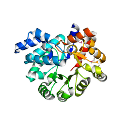
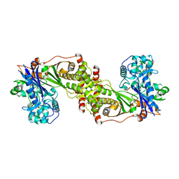
Loading
PDBj
MenuPDBj@FacebookPDBj@TwitterPDBj@YouTubewwPDB FoundationwwPDB
RCSB PDBPDBeBMRBAdv. SearchSearch help
PDB: 757 results

3NWM
DownloadVisualize
BU of 3nwm by Molmil
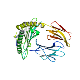
Crystal structure of a single chain construct composed of MHC class I H-2Kd, beta-2microglobulin and a peptide which is an autoantigen for type 1 diabetes
Descriptor: Peptide/beta-2microglobulin/MHC class I H-2Kd chimeric protein
Authors:Ramagopal, U.A, Samanta, D, Nathenson, S.G, Almo, S.C.
Deposit date:2010-07-09
Release date:2011-07-06
Last modified:2017-06-28
Method:X-RAY DIFFRACTION (2.7 Å)
Cite:Structural and functional characterization of a single-chain peptide-MHC molecule that modulates both naive and activated CD8+ T cells.
Proc.Natl.Acad.Sci.USA, 108, 2011
3NO1
DownloadVisualize
BU of 3no1 by Molmil
Crystal Structure of Mandelate racemase/muconate lactonizing enzyme from a Marine actinobacterium in complex with magnesium
Descriptor: MAGNESIUM ION, Mandelate racemase/muconate lactonizing enzyme
Authors:Satyanarayana, L, Burley, S.K, Swaminathan, S, New York SGX Research Center for Structural Genomics (NYSGXRC)
Deposit date:2010-06-24
Release date:2010-07-07
Last modified:2023-11-22
Method:X-RAY DIFFRACTION (2.16 Å)
Cite:Crystal Structure of Mandelate racemase/muconate lactonizing enzyme from a Marine actinobacterium in complex with magnesium
To be Published
3NPK
DownloadVisualize
BU of 3npk by Molmil
The crystal structure of geranyltranstransferase from Campylobacter jejuni
Descriptor: GLYCEROL, Geranyltranstransferase, PYROPHOSPHATE
Authors:Zhang, Z, Burley, S.K, Swaminathan, S, New York SGX Research Center for Structural Genomics (NYSGXRC)
Deposit date:2010-06-28
Release date:2010-07-14
Last modified:2023-12-27
Method:X-RAY DIFFRACTION (1.5 Å)
Cite:The crystal structure of geranyltranstransferase from Campylobacter jejuni
To be Published
3NYW
DownloadVisualize
BU of 3nyw by Molmil
Crystal Structure of a betaketoacyl-[ACP] reductase (FabG) from Bacteroides thetaiotaomicron
Descriptor: Putative oxidoreductase
Authors:Satyanarayana, L, Burley, S.K, Swaminathan, S, New York SGX Research Center for Structural Genomics (NYSGXRC)
Deposit date:2010-07-15
Release date:2010-09-15
Last modified:2021-02-10
Method:X-RAY DIFFRACTION (2.16 Å)
Cite:Crystal Structure of a betaketoacyl-[ACP] reductase (FabG) from Bacteroides thetaiotaomicron
To be Published
3NQB
DownloadVisualize
BU of 3nqb by Molmil
Crystal Structure of Adenine Deaminase from Agrobacterium tumefaciens (str. C 58)
Descriptor: Adenine deaminase 2, MANGANESE (II) ION
Authors:Bagaria, A, Kumaran, D, Burley, S.K, Swaminathan, S, New York SGX Research Center for Structural Genomics (NYSGXRC)
Deposit date:2010-06-29
Release date:2010-07-28
Last modified:2023-12-27
Method:X-RAY DIFFRACTION (2.21 Å)
Cite:Crystal Structure of Adenine Deaminase from Agrobacterium tumefaciens (str. C 58)
To be Published
3OU8
DownloadVisualize
BU of 3ou8 by Molmil
The crystal structure of adenosine deaminase from Pseudomonas aeruginosa
Descriptor: Adenosine deaminase, ZINC ION
Authors:Zhang, Z, Burley, S.K, Swaminathan, S, New York SGX Research Center for Structural Genomics (NYSGXRC)
Deposit date:2010-09-14
Release date:2010-10-13
Last modified:2021-02-10
Method:X-RAY DIFFRACTION (2.51 Å)
Cite:The crystal structure of adenosine deaminase from Pseudomonas aeruginosa
To be Published
3OR5
DownloadVisualize
BU of 3or5 by Molmil
Crystal structure of thiol:disulfide interchange protein, thioredoxin family protein from Chlorobium tepidum TLS
Descriptor: Thiol:disulfide interchange protein, thioredoxin family protein
Authors:Bagaria, A, Burley, S.K, Swaminathan, S, New York SGX Research Center for Structural Genomics (NYSGXRC)
Deposit date:2010-09-06
Release date:2010-09-22
Last modified:2021-02-10
Method:X-RAY DIFFRACTION (1.66 Å)
Cite:Crystal structure of thiol:disulfide interchange protein, thioredoxin family protein from Chlorobium tepidum TLS
TO BE PUBLISHED
3OXN
DownloadVisualize
BU of 3oxn by Molmil
The crystal structure of a putative transcriptional regulator from Vibrio parahaemolyticus
Descriptor: Putative transcriptional regulator, LysR family, SULFATE ION
Authors:Zhang, Z, Burley, S.K, Swaminathan, S, New York SGX Research Center for Structural Genomics (NYSGXRC)
Deposit date:2010-09-21
Release date:2010-10-13
Last modified:2021-02-10
Method:X-RAY DIFFRACTION (2.7 Å)
Cite:The crystal structure of a putative transcriptional regulator from Vibrio parahaemolyticus
To be Published
2GUW
DownloadVisualize
BU of 2guw by Molmil
Crystal structure of AMP Nucleosidase from Salmonella typhimurium LT2
Descriptor: AMP nucleosidase
Authors:Rao, K.N, Swaminathan, S, Burley, S.K, New York SGX Research Center for Structural Genomics (NYSGXRC)
Deposit date:2006-05-01
Release date:2006-06-06
Last modified:2024-02-14
Method:X-RAY DIFFRACTION (2.64 Å)
Cite:Crystal structure of AMP Nucleosidase from Salmonella typhimurium LT2
To be Published
3PAN
DownloadVisualize
BU of 3pan by Molmil
The crystal structure of adenosine deaminase with hypoxanthine bound from Pseudomonas aeruginosa
Descriptor: Adenosine deaminase, HYPOXANTHINE, ZINC ION
Authors:Zhang, Z, Burley, S.K, Swaminathan, S, New York SGX Research Center for Structural Genomics (NYSGXRC)
Deposit date:2010-10-19
Release date:2010-12-22
Last modified:2023-12-06
Method:X-RAY DIFFRACTION (2.627 Å)
Cite:The crystal structure of adenosine deaminase with hypoxanthine bound from Pseudomonas aeruginosa
TO BE PUBLISHED
3PZL
DownloadVisualize
BU of 3pzl by Molmil
The crystal structure of agmatine ureohydrolase of Thermoplasma volcanium
Descriptor: Agmatine ureohydrolase, MANGANESE (II) ION
Authors:Zhang, Z, Burley, S.K, Swaminathan, S, New York SGX Research Center for Structural Genomics (NYSGXRC)
Deposit date:2010-12-14
Release date:2011-01-19
Last modified:2021-02-10
Method:X-RAY DIFFRACTION (2.7 Å)
Cite:The crystal structure of agmatine ureohydrolase of Thermoplasma volcanium
To be Published
4JRW
DownloadVisualize
BU of 4jrw by Molmil
Crystal structure of Clostridium histolyticum colg collagenase PKD domain 2 at 1.6 Angstrom resolution
Descriptor: BROMIDE ION, Collagenase
Authors:Sakon, J, Philominathan, S.T.L, Gann, S, Bauer, R, Matsushita, O.
Deposit date:2013-03-22
Release date:2014-05-21
Last modified:2023-09-20
Method:X-RAY DIFFRACTION (1.6 Å)
Cite:Structures of three polycystic kidney disease-like domains from Clostridium histolyticum collagenases ColG and ColH.
Acta Crystallogr.,Sect.D, 71, 2015
1XEV
DownloadVisualize
BU of 1xev by Molmil
Crystal structure of human carbonic anhydrase II in a new crystal form
Descriptor: Carbonic anhydrase II, ZINC ION
Authors:Mazumdar, P.A, Kumaran, D, Das, A.K, Swaminathan, S.
Deposit date:2004-09-13
Release date:2005-09-20
Last modified:2023-08-23
Method:X-RAY DIFFRACTION (2.2 Å)
Cite:Crystal structure of human carbonic anhydrase II in a new crystal form
To be Published
1SGM
DownloadVisualize
BU of 1sgm by Molmil
Crystal Structure of Hypothetical Protein YXAF
Descriptor: Putative HTH-type transcriptional regulator yxaF
Authors:Seetharaman, J, Swaminathan, S, Burley, S.K, New York SGX Research Center for Structural Genomics (NYSGXRC)
Deposit date:2004-02-24
Release date:2004-08-17
Last modified:2024-02-14
Method:X-RAY DIFFRACTION (2 Å)
Cite:Crystal structure of a putative HTH-type transcriptional regulator yxaF from Bacillus subtilis.
Proteins, 63, 2006
3OC4
DownloadVisualize
BU of 3oc4 by Molmil
Crystal Structure of a pyridine nucleotide-disulfide family oxidoreductase from the Enterococcus faecalis V583
Descriptor: FLAVIN-ADENINE DINUCLEOTIDE, Oxidoreductase, pyridine nucleotide-disulfide family, ...
Authors:Kumaran, D, Baumann, K, Burley, S.K, Swaminathan, S, New York SGX Research Center for Structural Genomics (NYSGXRC)
Deposit date:2010-08-09
Release date:2010-10-06
Last modified:2021-02-10
Method:X-RAY DIFFRACTION (2.6 Å)
Cite:Crystal Structure of a pyridine nucleotide-disulfide family oxidoreductase from the Enterococcus faecalis V583
To be Published
3Q0H
DownloadVisualize
BU of 3q0h by Molmil
Structure of T-cell immunoreceptor with immunoglobulin and ITIM domains (TIGIT)
Descriptor: T cell immunoreceptor with Ig and ITIM domains
Authors:Ramagopal, U.A, Guo, H, Samanta, D, Nathenson, S.G, Almo, S.C, New York Structural Genomics Research Consortium (NYSGRC)
Deposit date:2010-12-15
Release date:2011-02-16
Last modified:2023-09-13
Method:X-RAY DIFFRACTION (1.7 Å)
Cite:Structure of T-cell immunoreceptor with immunoglobulin and ITIM domains (TIGIT)
To be published
3PU6
DownloadVisualize
BU of 3pu6 by Molmil
The crystal structure of an uncharacterized protein from Wolinella succinogenes
Descriptor: Uncharacterized protein
Authors:Zhang, Z, Burley, S.K, Swaminathan, S, New York SGX Research Center for Structural Genomics (NYSGXRC)
Deposit date:2010-12-03
Release date:2011-01-19
Last modified:2021-02-10
Method:X-RAY DIFFRACTION (2.6 Å)
Cite:The crystal structure of an uncharacterized protein from Wolinella succinogenes
TO BE PUBLISHED
2HAE
DownloadVisualize
BU of 2hae by Molmil
Crystal structure of a putative malic enzyme (malate oxidoreductase)
Descriptor: Malate oxidoreductase, NICOTINAMIDE-ADENINE-DINUCLEOTIDE, ZINC ION
Authors:Seetharaman, J, Swaminathan, S, Burley, S.K, New York SGX Research Center for Structural Genomics (NYSGXRC)
Deposit date:2006-06-12
Release date:2006-07-04
Last modified:2023-08-30
Method:X-RAY DIFFRACTION (3.13 Å)
Cite:Crystal structure of a putative malic enzyme (malate oxidoreductase)
To be Published
3QDK
DownloadVisualize
BU of 3qdk by Molmil
Structural insight on mechanism and diverse substrate selection strategy of ribulokinase
Descriptor: L-ribulose, Ribulokinase
Authors:Agarwal, R, Burley, S.K, Swaminathan, S, New York SGX Research Center for Structural Genomics (NYSGXRC)
Deposit date:2011-01-18
Release date:2011-02-09
Last modified:2024-02-21
Method:X-RAY DIFFRACTION (2.31 Å)
Cite:Structural insight into mechanism and diverse substrate selection strategy of L-ribulokinase.
Proteins, 80, 2012
3QJK
DownloadVisualize
BU of 3qjk by Molmil
Structure of Calcium Binding Protein-1 from Entamoeba histolytica in complex with Lead
Descriptor: ACETATE ION, Calcium-binding protein, LEAD (II) ION
Authors:Kumar, S, Gourinath, S.
Deposit date:2011-01-29
Release date:2012-04-11
Last modified:2023-11-01
Method:X-RAY DIFFRACTION (3.001 Å)
Cite:Structure of Calcium Binding Protein-1 from Entamoeba histolytica in complex with Lead
To be Published
3QW7
DownloadVisualize
BU of 3qw7 by Molmil
Crystal structure of the protease domain of Botulinum Neurotoxin Serotype A with a peptide inhibitor RRFC
Descriptor: Botulinum neurotoxin type A, SODIUM ION, SULFATE ION, ...
Authors:Kumaran, D, Swaminathan, S.
Deposit date:2011-02-27
Release date:2012-02-08
Last modified:2023-09-13
Method:X-RAY DIFFRACTION (1.5 Å)
Cite:Peptide inhibitors of botulinum neurotoxin serotype A: design, inhibition, cocrystal structures, structure-activity relationship and pharmacophore modeling.
Acta Crystallogr.,Sect.D, 68, 2012
2G6T
DownloadVisualize
BU of 2g6t by Molmil
Crystal structure of an uncharacterized protein from Clostridium acetobutylicum
Descriptor: Uncharacterized protein, homolog HI1244 from Haemophilus influenzae
Authors:Rao, K.N, Swaminathan, S, Burley, S.K, New York SGX Research Center for Structural Genomics (NYSGXRC)
Deposit date:2006-02-25
Release date:2006-03-14
Last modified:2021-02-03
Method:X-RAY DIFFRACTION (3 Å)
Cite:Crystal structure of an uncharacterized protein from Clostridium acetobutylicum
To be Published
3QW6
DownloadVisualize
BU of 3qw6 by Molmil
Crystal structure of the protease domain of Botulinum Neurotoxin Serotype A with a peptide inhibitor RYGC
Descriptor: Botulinum neurotoxin type A, SODIUM ION, SULFATE ION, ...
Authors:Kumaran, D, Swaminathan, S.
Deposit date:2011-02-26
Release date:2012-02-08
Last modified:2023-09-13
Method:X-RAY DIFFRACTION (1.6 Å)
Cite:Peptide inhibitors of botulinum neurotoxin serotype A: design, inhibition, cocrystal structures, structure-activity relationship and pharmacophore modeling.
Acta Crystallogr.,Sect.D, 68, 2012
1L2O
DownloadVisualize
BU of 1l2o by Molmil
SCALLOP MYOSIN S1-ADP-p-PDM IN THE ACTIN-DETACHED CONFORMATION
Descriptor: 4-[4-(2,5-DIOXO-PYRROLIDIN-1-YL)-PHENYLAMINO]-4-HYDROXY-BUTYRIC ACID, ADENOSINE-5'-DIPHOSPHATE, CALCIUM ION, ...
Authors:Himmel, M, Gourinath, S, Reshetnikova, L, Shen, Y, Szent-Gyorgyi, G, Cohen, C.
Deposit date:2002-02-22
Release date:2002-10-30
Last modified:2023-08-16
Method:X-RAY DIFFRACTION (2.8 Å)
Cite:Crystallographic findings on the internally uncoupled and near-rigor states of myosin: further insights into the mechanics of the motor.
Proc.Natl.Acad.Sci.USA, 99, 2002
3QW5
DownloadVisualize
BU of 3qw5 by Molmil
Crystal structure of the protease domain of Botulinum Neurotoxin Serotype A with a peptide inhibitor RRGF
Descriptor: Botulinum neurotoxin type A, SULFATE ION, ZINC ION, ...
Authors:Kumaran, D, Swaminathan, S.
Deposit date:2011-02-26
Release date:2012-02-08
Last modified:2023-09-13
Method:X-RAY DIFFRACTION (1.6 Å)
Cite:Peptide inhibitors of botulinum neurotoxin serotype A: design, inhibition, cocrystal structures, structure-activity relationship and pharmacophore modeling.
Acta Crystallogr.,Sect.D, 68, 2012

225681

PDB entries from 2024-10-02

PDB statisticsPDBj update infoContact PDBjnumon