Loading
PDBj
MenuPDBj@FacebookPDBj@TwitterPDBj@YouTubewwPDB FoundationwwPDB
RCSB PDBPDBeBMRBAdv. SearchSearch help
PDB: 757 results

5EY7
DownloadVisualize
BU of 5ey7 by Molmil
Crystal structure of Fructokinase from Vibrio cholerae O395 in apo form
Descriptor: Fructokinase, SODIUM ION
Authors:Paul, R, Nath, S, Sen, U.
Deposit date:2015-11-24
Release date:2016-11-30
Last modified:2023-11-08
Method:X-RAY DIFFRACTION (2.461 Å)
Cite:Crystal structure of Fructokinase from Vibrio cholerae O395 in apo form
To Be Published
5F11
DownloadVisualize
BU of 5f11 by Molmil
Crystal structure of Fructokinase from Vibrio cholerae O395 in fructose bound form
Descriptor: Fructokinase, SODIUM ION, beta-D-fructofuranose
Authors:Paul, R, Nath, S, Sen, U.
Deposit date:2015-11-28
Release date:2016-11-30
Last modified:2023-11-08
Method:X-RAY DIFFRACTION (2.3 Å)
Cite:Crystal structure of Fructokinase from Vibrio cholerae O395 in apo form
To Be Published
5EYN
DownloadVisualize
BU of 5eyn by Molmil
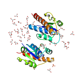
Crystal structure of Fructokinase from Vibrio cholerae O395 in fructose, ADP, Beryllium trifluoride and calcium ion bound form
Descriptor: ADENOSINE-5'-DIPHOSPHATE, BERYLLIUM TRIFLUORIDE ION, CALCIUM ION, ...
Authors:Paul, R, Nath, S, Sen, U.
Deposit date:2015-11-25
Release date:2016-11-30
Last modified:2023-11-08
Method:X-RAY DIFFRACTION (1.289 Å)
Cite:Crystal structure of Fructokinase from Vibrio cholerae O395 in apo form
To Be Published
5F0Z
DownloadVisualize
BU of 5f0z by Molmil
Crystal structure of Fructokinase from Vibrio cholerae O395 in fructose, ADP and calcium ion bound form
Descriptor: ADENOSINE-5'-DIPHOSPHATE, CALCIUM ION, Fructokinase, ...
Authors:Paul, R, Nath, S, Sen, U.
Deposit date:2015-11-28
Release date:2016-11-30
Last modified:2023-11-08
Method:X-RAY DIFFRACTION (1.75 Å)
Cite:Crystal structure of Fructokinase from Vibrio cholerae O395 in apo form
To Be Published
5Z3M
DownloadVisualize
BU of 5z3m by Molmil
Crystal structure of Low Molecular Weight Phosphotyrosine phosphatase (VcLMWPTP-2) from Vibrio choleraeO395
Descriptor: 3[N-MORPHOLINO]PROPANE SULFONIC ACID, GLYCEROL, Phosphotyrosine protein phosphatase, ...
Authors:Chatterjee, S, Nath, S, Sen, U.
Deposit date:2018-01-08
Release date:2018-09-19
Last modified:2023-11-22
Method:X-RAY DIFFRACTION (2.6 Å)
Cite:Vibrio cholerae LMWPTP-2 display unique surface charge and grooves around the active site: Indicative of distinctive substrate specificity and scope to design specific inhibitor.
Biochim Biophys Acta Proteins Proteom, 1867, 2019
4DVD
DownloadVisualize
BU of 4dvd by Molmil
Crystal structure of the disulphide linked knotted homodimer of Psu
Descriptor: Polarity suppression protein
Authors:Banerjee, R, Nath, S, Sen, U.
Deposit date:2012-02-23
Release date:2012-11-21
Last modified:2023-11-08
Method:X-RAY DIFFRACTION (3 Å)
Cite:The first structure of polarity suppression protein, Psu from enterobacteria phage P4, reveals a novel fold and a knotted dimer
J.Biol.Chem., 287, 2012
5B74
DownloadVisualize
BU of 5b74 by Molmil
Crystal structure of conjoined Pyrococcus furiosus L-asparaginase with peptide
Descriptor: CHLORIDE ION, CITRATE ANION, L-asparaginase, ...
Authors:Sharma, P, Tomar, R, Nath, S.K, Kundu, B, Ashish, F.
Deposit date:2016-06-05
Release date:2017-06-14
Last modified:2023-11-08
Method:X-RAY DIFFRACTION (2.039 Å)
Cite:Heat induces end to end repetitive association in P. furiosus L-asparaginase which enables its thermophilic property.
Sci Rep, 10, 2020
5B5U
DownloadVisualize
BU of 5b5u by Molmil
Crystal structure of truncated Pyrococcus furiosus L-asparaginase with peptide
Descriptor: ASPARTIC ACID, CHLORIDE ION, GLYCEROL, ...
Authors:Sharma, P, Tomar, R, Nath, S.K, Kundu, B, Ashish, F.
Deposit date:2016-05-23
Release date:2017-05-31
Last modified:2023-11-08
Method:X-RAY DIFFRACTION (2.61 Å)
Cite:Heat induces end to end repetitive association in P. furiosus L-asparaginase which enables its thermophilic property.
Sci Rep, 10, 2020
3HKF
DownloadVisualize
BU of 3hkf by Molmil
Murine unglycosylated IgG Fc fragment
Descriptor: CHLORIDE ION, Igh protein, MAGNESIUM ION
Authors:Feige, M.J, Nath, S, Catharino, S.R, Weinfurtner, D, Steinbacher, S, Buchner, J.
Deposit date:2009-05-23
Release date:2009-11-10
Last modified:2023-11-01
Method:X-RAY DIFFRACTION (2.5 Å)
Cite:Structure of the murine unglycosylated IgG1 Fc fragment
J.Mol.Biol., 391, 2009
5D7V
DownloadVisualize
BU of 5d7v by Molmil
Crystal structure of PTK6 kinase domain
Descriptor: GLYCEROL, PHOSPHATE ION, Protein-tyrosine kinase 6
Authors:Thakur, M.K, Birudukota, S, Swaminathan, S, Tyagi, R, Gosu, R.
Deposit date:2015-08-14
Release date:2016-08-17
Last modified:2023-11-08
Method:X-RAY DIFFRACTION (2.33 Å)
Cite:Crystal structure of the kinase domain of human protein tyrosine kinase 6 (PTK6) at 2.33 angstrom resolution
Biochem.Biophys.Res.Commun., 478, 2016
8I11
DownloadVisualize
BU of 8i11 by Molmil
Crystal structure of LOV1 domain of phototropin from Klebsormidium nitens
Descriptor: FLAVIN MONONUCLEOTIDE, Phototropin
Authors:Gautam, A.K, Sharma, S, Gourinath, S, Kateriya, S.
Deposit date:2023-01-12
Release date:2024-01-24
Method:X-RAY DIFFRACTION (1.855 Å)
Cite:Crystal structure of LOV1 domain of phototropin from klebsormidium nitens
To Be Published
8IL9
DownloadVisualize
BU of 8il9 by Molmil
Crystal structure of the LOV1 Q122N mutant of Klebsormidium nitens phototropin
Descriptor: FLAVIN MONONUCLEOTIDE, Phototropin
Authors:Gautam, A.K, Sharma, S, Gourinath, S, Kateriya, S.
Deposit date:2023-03-03
Release date:2024-03-06
Method:X-RAY DIFFRACTION (2.16 Å)
Cite:Crystal structure of LOV1 Q122N mutant of phototropin from klebsormidium nitens
To Be Published
4IM9
DownloadVisualize
BU of 4im9 by Molmil
Cystal structure of DnaG primase C-terminal domain from Vibrio cholerae
Descriptor: DNA primase
Authors:Abdul Rehman, S.A, Tarique, K.F, Gourinath, S.
Deposit date:2013-01-02
Release date:2014-05-07
Method:X-RAY DIFFRACTION (2.46 Å)
Cite:Cystal structure of DnaG primase C-terminal domain from Vibrio cholerae
To be Published
4IL5
DownloadVisualize
BU of 4il5 by Molmil
Crystal structure of O-Acetyl Serine Sulfhydrylase from Entamoeba histolytica in complex with isoleucine
Descriptor: Cysteine synthase, ISOLEUCINE, SULFATE ION
Authors:Raj, I, Gourinath, S.
Deposit date:2012-12-29
Release date:2013-12-04
Last modified:2024-03-20
Method:X-RAY DIFFRACTION (2.03 Å)
Cite:Molecular basis of ligand recognition by OASS from E. histolytica: insights from structural and molecular dynamics simulation studies
Biochim.Biophys.Acta, 1830, 2013
3EB2
DownloadVisualize
BU of 3eb2 by Molmil
Crystal structure of Dihydrodipicolinate Synthase from Rhodopseudomonas palustris at 2.0A resolution
Descriptor: Putative dihydrodipicolinate synthetase, TRIETHYLENE GLYCOL
Authors:Satyanarayana, L, Eswaramoorthy, S, Burley, S.K, Swaminathan, S, New York SGX Research Center for Structural Genomics (NYSGXRC)
Deposit date:2008-08-26
Release date:2008-09-02
Last modified:2024-02-21
Method:X-RAY DIFFRACTION (2.04 Å)
Cite:Crystal structure of Dihydrodipicolinate Synthase from Rhodopseudomonas palustris at 2.0A resolution
To be Published
2ISN
DownloadVisualize
BU of 2isn by Molmil
Crystal structure of a phosphatase from a pathogenic strain Toxoplasma gondii
Descriptor: NYSGXRC-8828z, phosphatase, PRASEODYMIUM ION, ...
Authors:Agarwal, R, Burley, S.K, Swaminathan, S, New York SGX Research Center for Structural Genomics (NYSGXRC)
Deposit date:2006-10-18
Release date:2006-10-31
Last modified:2021-02-03
Method:X-RAY DIFFRACTION (1.9 Å)
Cite:Structural genomics of protein phosphatases.
J.STRUCT.FUNCT.GENOM., 8, 2007
4S1I
DownloadVisualize
BU of 4s1i by Molmil
Pyridoxal Kinase of Entamoeba histolytica with PLP
Descriptor: MAGNESIUM ION, PYRIDOXAL-5'-PHOSPHATE, Pyridoxal kinase
Authors:Tarique, K.F, Devi, S, Abdul Rehman, S.A, Gourinath, S.
Deposit date:2015-01-14
Release date:2015-02-04
Last modified:2023-09-20
Method:X-RAY DIFFRACTION (1.6 Å)
Cite:Characterization and functional insights into the Entamoeba histolytica pyridoxal kinase, an enzyme essential for its survival.
J.Struct.Biol., 212, 2020
4S1M
DownloadVisualize
BU of 4s1m by Molmil
Crystal Structure of Pyridoxal Kinase from Entamoeba histolytica
Descriptor: MAGNESIUM ION, Pyridoxal kinase
Authors:Tarique, K.F, Devi, S, Abdul Rehman, S.A, Gourinath, S.
Deposit date:2015-01-14
Release date:2015-01-28
Last modified:2024-04-03
Method:X-RAY DIFFRACTION (1.64 Å)
Cite:Characterization and functional insights into the Entamoeba histolytica pyridoxal kinase, an enzyme essential for its survival.
J.Struct.Biol., 212, 2020
3E9M
DownloadVisualize
BU of 3e9m by Molmil
Crystal Structure of an oxidoreductase from Enterococcus faecalis
Descriptor: Oxidoreductase, Gfo/Idh/MocA family
Authors:Damodharan, L, Burley, S.K, Swaminathan, S, New York SGX Research Center for Structural Genomics (NYSGXRC)
Deposit date:2008-08-22
Release date:2008-10-14
Last modified:2021-02-10
Method:X-RAY DIFFRACTION (2.7 Å)
Cite:Crystal Structure of an oxidoreductase from Enterococcus faecalis
To be Published
3E3V
DownloadVisualize
BU of 3e3v by Molmil
Crystal structure of RecX from Lactobacillus salivarius
Descriptor: Regulatory protein recX
Authors:Agarwal, R, Burley, S.K, Swaminathan, S, New York SGX Research Center for Structural Genomics (NYSGXRC)
Deposit date:2008-08-08
Release date:2008-09-02
Last modified:2024-02-21
Method:X-RAY DIFFRACTION (2.04 Å)
Cite:Crystal structure of RecX
To be Published
2I6E
DownloadVisualize
BU of 2i6e by Molmil
Crystal structure of protein DR0370 from Deinococcus radiodurans, Pfam DUF178
Descriptor: Hypothetical protein, SULFATE ION
Authors:Tyagi, R, Kumaran, D, Burley, S.K, Swaminathan, S, New York SGX Research Center for Structural Genomics (NYSGXRC)
Deposit date:2006-08-28
Release date:2006-09-05
Last modified:2021-02-03
Method:X-RAY DIFFRACTION (2.5 Å)
Cite:X-ray structures of two proteins belonging to Pfam DUF178 revealed unexpected structural similarity to the DUF191 Pfam family.
Bmc Struct.Biol., 7, 2007
2I76
DownloadVisualize
BU of 2i76 by Molmil
Crystal structure of protein TM1727 from Thermotoga maritima
Descriptor: Hypothetical protein, NADPH DIHYDRO-NICOTINAMIDE-ADENINE-DINUCLEOTIDE PHOSPHATE
Authors:Madegowda, M, Eswaramoorthy, S, Seetharaman, J, Burley, S.K, Swaminathan, S, New York SGX Research Center for Structural Genomics (NYSGXRC)
Deposit date:2006-08-30
Release date:2006-10-03
Last modified:2021-02-03
Method:X-RAY DIFFRACTION (3 Å)
Cite:Crystal structure of hypothetical protein TM1727 from Thermatoga maritima
TO BE PUBLISHED
2I3O
DownloadVisualize
BU of 2i3o by Molmil
Crystal structure of gamma-glutamyl transferase related protein from Thermoplasma acidophilum
Descriptor: Gamma-glutamyltransferase related protein
Authors:Rao, K.N, Eswaramoorthy, S, Burley, S.K, Swaminathan, S, New York SGX Research Center for Structural Genomics (NYSGXRC)
Deposit date:2006-08-19
Release date:2006-08-29
Last modified:2021-02-03
Method:X-RAY DIFFRACTION (2.03 Å)
Cite:Crystal structure of gamma-glutamyl transferase related protein from Thermoplasma acidophilum
To be Published
3EUW
DownloadVisualize
BU of 3euw by Molmil
Crystal Structure of a Myo-inositol dehydrogenase from Corynebacterium glutamicum ATCC 13032
Descriptor: Myo-inositol dehydrogenase, SODIUM ION
Authors:Kumaran, D, Mahmood, A, Burley, S.K, Swaminathan, S, New York SGX Research Center for Structural Genomics (NYSGXRC)
Deposit date:2008-10-11
Release date:2008-10-21
Last modified:2023-12-27
Method:X-RAY DIFFRACTION (2.3 Å)
Cite:Crystal Structure of a Myo-inositol dehydrogenase from Corynebacterium glutamicum ATCC 13032
To be Published
4RKI
DownloadVisualize
BU of 4rki by Molmil
Crystal structure of sliding beta clamp from Helicobacter pylori
Descriptor: DNA polymerase III subunit beta
Authors:Satyawali, P, Tarique, K.F, Abdul Rehman, S.A, Gourinath, S.
Deposit date:2014-10-13
Release date:2016-01-13
Last modified:2023-09-20
Method:X-RAY DIFFRACTION (2.05 Å)
Cite:Structural insight into beta-Clamp and its interaction with DNA Ligase in Helicobacter pylori.
Sci Rep, 6, 2016

222926

PDB entries from 2024-07-24

PDB statisticsPDBj update infoContact PDBjnumon