Loading
PDBj
MenuPDBj@FacebookPDBj@TwitterPDBj@YouTubewwPDB FoundationwwPDB
RCSB PDBPDBeBMRBAdv. SearchSearch help
PDB: 762 results

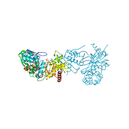
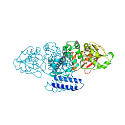
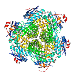
3MSR
DownloadVisualize
BU of 3msr by Molmil
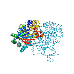
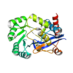
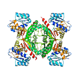
The crystal structure of an amidohydrolase from Mycoplasma synoviae
Descriptor: GLYCEROL, PHOSPHATE ION, amidohydrolases
Authors:Zhang, Z, Burley, S.K, Swaminathan, S, New York SGX Research Center for Structural Genomics (NYSGXRC)
Deposit date:2010-04-29
Release date:2010-05-12
Last modified:2021-02-10
Method:X-RAY DIFFRACTION (2.162 Å)
Cite:The crystal structure of an amidohydrolase from Mycoplasma synoviae
To be Published
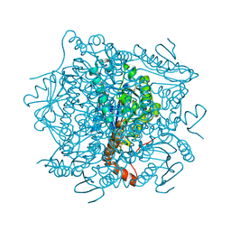
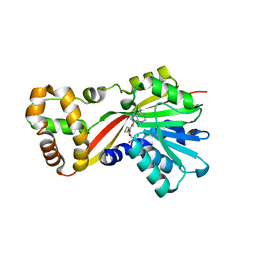
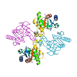
4K3W
DownloadVisualize
BU of 4k3w by Molmil
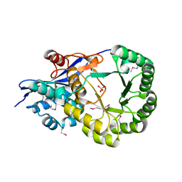
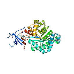
Crystal structure of an enoyl-CoA hydratase/isomerase from Marinobacter aquaeolei
Descriptor: Enoyl-CoA hydratase/isomerase
Authors:Eswaramoorthy, S, Chamala, S, Evans, B, Foti, F, Gizzi, A, Hillerich, B, Kar, A, Lafleur, J, Seidel, R, Villigas, G, Zencheck, W, Al Obaidi, N, Almo, S.C, Swaminathan, S, New York Structural Genomics Research Consortium (NYSGRC)
Deposit date:2013-04-11
Release date:2013-04-24
Last modified:2024-10-09
Method:X-RAY DIFFRACTION (2.31 Å)
Cite:Crystal structure of an enoyl-CoA hydratase/isomerase from Marinobacter aquaeolei
To be Published
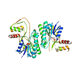
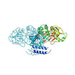
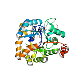
3KZB
DownloadVisualize
BU of 3kzb by Molmil
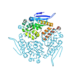
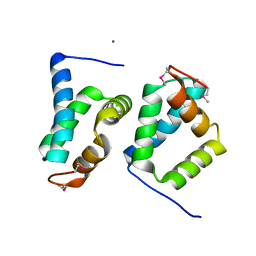
Crystal structure of xylulokinase from Chromobacterium violaceum
Descriptor: ADENOSINE-5'-DIPHOSPHATE, Xylulokinase
Authors:Zhang, Z, Burley, S.K, Swaminathan, S, New York SGX Research Center for Structural Genomics (NYSGXRC)
Deposit date:2009-12-08
Release date:2010-03-09
Last modified:2023-11-22
Method:X-RAY DIFFRACTION (2.705 Å)
Cite:Crystal structure of xylulokinase from Chromobacterium violaceum
To be Published
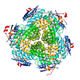
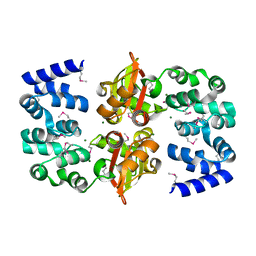
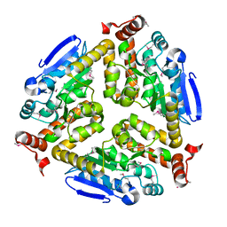
4JCS
DownloadVisualize
BU of 4jcs by Molmil
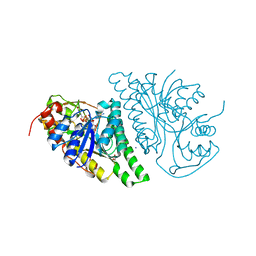
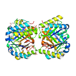
Crystal structure of Enoyl-CoA hydratase/isomerase from Cupriavidus metallidurans CH34
Descriptor: Enoyl-CoA hydratase/isomerase
Authors:Eswaramoorthy, S, Chamala, S, Chamala, B, Foti, F, Gizzi, A, Hillerich, B, Kar, A, Lafleur, J, Seidel, R, Villigas, G, Zencheck, W, Al Obaidi, N, Almo, S.C, Swaminathan, S, New York Structural Genomics Research Consortium (NYSGRC)
Deposit date:2013-02-22
Release date:2013-03-13
Method:X-RAY DIFFRACTION (1.77 Å)
Cite:Crystal structure of Enoyl-CoA hydratase/isomerase from Cupriavidus metallidurans CH34
To be Published
3M2P
DownloadVisualize
BU of 3m2p by Molmil
The crystal structure of UDP-N-acetylglucosamine 4-epimerase from Bacillus cereus
Descriptor: UDP-N-acetylglucosamine 4-epimerase, URIDINE-5'-DIPHOSPHATE
Authors:Zhang, Z, Burley, S.K, Swaminathan, S, New York SGX Research Center for Structural Genomics (NYSGXRC)
Deposit date:2010-03-08
Release date:2010-04-07
Last modified:2021-02-10
Method:X-RAY DIFFRACTION (2.95 Å)
Cite:The crystal structure of UDP-N-acetylglucosamine 4-epimerase from Bacillus cereus
To be Published
3MSY
DownloadVisualize
BU of 3msy by Molmil
Crystal Structure of Mandelate racemase/muconate lactonizing enzyme from a Marine actinobacterium
Descriptor: Mandelate racemase/muconate lactonizing enzyme
Authors:Satyanarayana, L, Burley, S.K, Swaminathan, S, New York SGX Research Center for Structural Genomics (NYSGXRC)
Deposit date:2010-04-29
Release date:2010-06-30
Last modified:2024-10-30
Method:X-RAY DIFFRACTION (2.5 Å)
Cite:Crystal Structure of Mandelate racemase/muconate lactonizing enzyme from a Marine actinobacterium
To be Published
3NAS
DownloadVisualize
BU of 3nas by Molmil
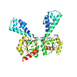
The crystal structure of beta-phosphoglucomutase from Bacillus subtilis
Descriptor: beta-phosphoglucomutase
Authors:Zhang, Z, Burley, S.K, Swaminathan, S, New York SGX Research Center for Structural Genomics (NYSGXRC)
Deposit date:2010-06-02
Release date:2010-07-28
Last modified:2021-02-10
Method:X-RAY DIFFRACTION (3 Å)
Cite:The crystal structure of beta-phosphoglucomutase from Bacillus subtilis
To be Published
2NN4
DownloadVisualize
BU of 2nn4 by Molmil
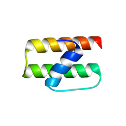
Crystal structure of Bacillus subtilis yqgQ, Pfam DUF910
Descriptor: Hypothetical protein yqgQ
Authors:Damodharan, L, Eswaramoorthy, S, Burley, S.K, Swaminathan, S, New York SGX Research Center for Structural Genomics (NYSGXRC)
Deposit date:2006-10-23
Release date:2006-10-31
Last modified:2023-12-27
Method:X-RAY DIFFRACTION (2.1 Å)
Cite:Structure of YqgQ protein from Bacillus subtilis, a conserved hypothetical protein.
Acta Crystallogr.,Sect.F, 66, 2010
3QJ1
DownloadVisualize
BU of 3qj1 by Molmil
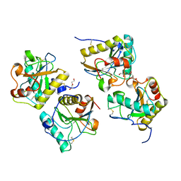
Crystal structure of camel peptidoglycan recognition protein, PGRP-S with a trapped diethylene glycol in the ligand diffusion channel at 3.2 A resolution
Descriptor: DI(HYDROXYETHYL)ETHER, GLYCEROL, L(+)-TARTARIC ACID, ...
Authors:Sharma, P, Yamini, S, Sinha, M, Kaur, P, Sharma, S, Singh, T.P.
Deposit date:2011-01-28
Release date:2011-02-16
Last modified:2024-10-30
Method:X-RAY DIFFRACTION (3.2 Å)
Cite:Crystal structure of camel peptidoglycan recognition protein, PGRP-S with a trapped diethylene glycol in the ligand diffusion channel at 3.2 A resolution
To be Published
2OOD
DownloadVisualize
BU of 2ood by Molmil
Crystal structure of guanine deaminase from Bradyrhizobium japonicum
Descriptor: Blr3880 protein, GUANINE, ZINC ION
Authors:Agarwal, R, Burley, S.K, Swaminathan, S, New York SGX Research Center for Structural Genomics (NYSGXRC)
Deposit date:2007-01-25
Release date:2007-02-20
Last modified:2024-10-09
Method:X-RAY DIFFRACTION (2.62 Å)
Cite:Crystal structure of guanine deaminase from Bradyrhizobium japonicum
To be Published
2OPJ
DownloadVisualize
BU of 2opj by Molmil
Crystal structure of O-succinylbenzoate synthase
Descriptor: O-succinylbenzoate-CoA synthase
Authors:Sugadev, R, Burley, S.K, Swaminathan, S, New York SGX Research Center for Structural Genomics (NYSGXRC)
Deposit date:2007-01-29
Release date:2007-02-20
Last modified:2023-12-27
Method:X-RAY DIFFRACTION (1.6 Å)
Cite:Crystal structure of O-succinylbenzoate synthase
To be Published
2OQH
DownloadVisualize
BU of 2oqh by Molmil
Crystal structure of an isomerase from Streptomyces coelicolor A3(2)
Descriptor: Putative isomerase, SULFATE ION
Authors:Agarwal, R, Burley, S.K, Swaminathan, S, New York SGX Research Center for Structural Genomics (NYSGXRC)
Deposit date:2007-01-31
Release date:2007-02-20
Last modified:2024-10-09
Method:X-RAY DIFFRACTION (1.98 Å)
Cite:Crystal structure of an isomerase from Streptomyces coelicolor
To be Published
4JOT
DownloadVisualize
BU of 4jot by Molmil
Crystal structure of enoyl-CoA hydrotase from Deinococcus radiodurans R1
Descriptor: Enoyl-CoA hydratase, putative, GLYCEROL
Authors:Eswaramoorthy, S, Chamala, S, Evans, B, Foti, F, Gizzi, A, Hillerich, B, Kar, A, Lafleur, J, Seidel, R, Villigas, G, Zencheck, W, Al Obaidi, N, Almo, S.C, Swaminathan, S, New York Structural Genomics Research Consortium (NYSGRC)
Deposit date:2013-03-18
Release date:2013-04-17
Last modified:2024-10-30
Method:X-RAY DIFFRACTION (1.94 Å)
Cite:Crystal structure of enoyl-CoA hydrotase from Deinococcus radiodurans R1
To be Published
4LIS
DownloadVisualize
BU of 4lis by Molmil
Crystal Structure of UDP-galactose-4-epimerase from Aspergillus nidulans
Descriptor: GLYCEROL, IODIDE ION, NICOTINAMIDE-ADENINE-DINUCLEOTIDE, ...
Authors:Dalrymple, S.A, Ko, J, Sheoran, I, Kaminskyj, S.G.W, Sanders, D.A.R.
Deposit date:2013-07-03
Release date:2013-10-23
Last modified:2023-09-20
Method:X-RAY DIFFRACTION (2.8 Å)
Cite:Elucidation of Substrate Specificity in Aspergillus nidulans UDP-Galactose-4-Epimerase.
Plos One, 8, 2013
3NND
DownloadVisualize
BU of 3nnd by Molmil
The crystal structure of ABC transporter from Rhodopseudomonas palustris
Descriptor: Possible substrate binding protein of ABC transporter system
Authors:Zhang, Z, Burley, S.K, Swaminathan, S, New York SGX Research Center for Structural Genomics (NYSGXRC)
Deposit date:2010-06-23
Release date:2010-10-13
Last modified:2024-10-16
Method:X-RAY DIFFRACTION (2.8 Å)
Cite:The crystal structure of ABC transporter from Rhodopseudomonas palustris
To be Published
2OOF
DownloadVisualize
BU of 2oof by Molmil
The crystal structure of 4-imidazolone-5-propanoate amidohydrolase from environmental sample
Descriptor: 4-imidazolone-5-propanoate amidohydrolase, FE (III) ION
Authors:Tyagi, R, Eswaramoorthy, S, Burley, S.K, Swaminathan, S, New York SGX Research Center for Structural Genomics (NYSGXRC)
Deposit date:2007-01-25
Release date:2007-02-20
Last modified:2023-12-27
Method:X-RAY DIFFRACTION (2.2 Å)
Cite:The crystal structure of 4-imidazolone-5-propanoate amidohydrolase from environmental sample
To be Published
2OY9
DownloadVisualize
BU of 2oy9 by Molmil
Crystal structure of an unknown conserved protein- Pfam: UPF0223
Descriptor: MAGNESIUM ION, UPF0223 protein BH2638
Authors:Sugadev, R, Burley, S.K, Swaminathan, S, New York SGX Research Center for Structural Genomics (NYSGXRC)
Deposit date:2007-02-21
Release date:2007-03-20
Last modified:2024-10-30
Method:X-RAY DIFFRACTION (1.6 Å)
Cite:Crystal structure of an unknown conserved protein - Pfam: UPF0223
To be Published
2P0L
DownloadVisualize
BU of 2p0l by Molmil
Crystal structure of a Lipoate-protein ligase A
Descriptor: Lipoate-protein ligase A
Authors:Sugadev, R, Burley, S.K, Swaminathan, S, New York SGX Research Center for Structural Genomics (NYSGXRC)
Deposit date:2007-02-28
Release date:2007-03-20
Last modified:2024-10-09
Method:X-RAY DIFFRACTION (2.04 Å)
Cite:Crystal structure of a Lipoate-protein ligase A
To be Published
4LKB
DownloadVisualize
BU of 4lkb by Molmil
Crystal structure of a putative 4-Oxalocrotonate Tautomerase from Nostoc sp. PCC 7120
Descriptor: GLYCEROL, SULFATE ION, hypothetical protein alr4568/putative 4-Oxalocrotonate Tautomerase
Authors:Kumaran, D, Chamala, S, Evans, B, Foti, R, Gizzi, A, Hillerich, B, Kar, A, Lafleur, J, Seidel, R, Villigas, G, Zencheck, W, Al Obaidi, N, Almo, S.C, Swaminathan, S, New York Structural Genomics Research Consortium (NYSGRC)
Deposit date:2013-07-07
Release date:2013-07-24
Method:X-RAY DIFFRACTION (2.16 Å)
Cite:Crystal structure of a putative 4-Oxalocrotonate Tautomerase from Nostoc sp. PCC 7120
To be Published
5W7P
DownloadVisualize
BU of 5w7p by Molmil
Crystal structure of OxaC
Descriptor: OxaC, S-ADENOSYLMETHIONINE
Authors:Newmister, S.A, Romminger, S, Schmidt, J.J, Williams, R.M, Smith, J.L, Berlinck, R.G.S, Sherman, D.H.
Deposit date:2017-06-20
Release date:2018-06-27
Last modified:2024-03-13
Method:X-RAY DIFFRACTION (2.4 Å)
Cite:Unveiling sequential late-stage methyltransferase reactions in the meleagrin/oxaline biosynthetic pathway.
Org. Biomol. Chem., 16, 2018
5W7K
DownloadVisualize
BU of 5w7k by Molmil
Crystal structure of OxaG
Descriptor: CHLORIDE ION, OxaG, S-ADENOSYL-L-HOMOCYSTEINE
Authors:Newmister, S.A, Romminger, S, Schmidt, J.J, Williams, R.M, Smith, J.L, Berlinck, R.G.S, Sherman, D.H.
Deposit date:2017-06-20
Release date:2018-06-27
Last modified:2024-03-13
Method:X-RAY DIFFRACTION (1.994 Å)
Cite:Unveiling sequential late-stage methyltransferase reactions in the meleagrin/oxaline biosynthetic pathway.
Org. Biomol. Chem., 16, 2018
5W7S
DownloadVisualize
BU of 5w7s by Molmil
Crystal structure of OxaC in complex with sinefungin and meleagrin
Descriptor: (3E,7aR,12aS)-6-hydroxy-3-[(1H-imidazol-4-yl)methylidene]-12-methoxy-7a-(2-methylbut-3-en-2-yl)-7a,12-dihydro-1H,5H-imidazo[1',2':1,2]pyrido[2,3-b]indole-2,5(3H)-dione, OxaC, SINEFUNGIN
Authors:Newmister, S.A, Romminger, S, Schmidt, J.J, Williams, R.M, Smith, J.L, Berlinck, R.G.S, Sherman, D.H.
Deposit date:2017-06-20
Release date:2018-06-27
Last modified:2023-10-04
Method:X-RAY DIFFRACTION (2.948 Å)
Cite:Unveiling sequential late-stage methyltransferase reactions in the meleagrin/oxaline biosynthetic pathway.
Org. Biomol. Chem., 16, 2018
2OUX
DownloadVisualize
BU of 2oux by Molmil
Crystal structure of the soluble part of a magnesium transporter
Descriptor: MAGNESIUM ION, Magnesium transporter
Authors:Sugadev, R, Burley, S.K, Swaminathan, S, New York SGX Research Center for Structural Genomics (NYSGXRC)
Deposit date:2007-02-12
Release date:2007-03-20
Last modified:2024-10-30
Method:X-RAY DIFFRACTION (2.16 Å)
Cite:Structural studies on cytosolic domain of magnesium transporter MgtE from Enterococcus faecalis.
Proteins, 78, 2010
3NO1
DownloadVisualize
BU of 3no1 by Molmil
Crystal Structure of Mandelate racemase/muconate lactonizing enzyme from a Marine actinobacterium in complex with magnesium
Descriptor: MAGNESIUM ION, Mandelate racemase/muconate lactonizing enzyme
Authors:Satyanarayana, L, Burley, S.K, Swaminathan, S, New York SGX Research Center for Structural Genomics (NYSGXRC)
Deposit date:2010-06-24
Release date:2010-07-07
Last modified:2023-11-22
Method:X-RAY DIFFRACTION (2.16 Å)
Cite:Crystal Structure of Mandelate racemase/muconate lactonizing enzyme from a Marine actinobacterium in complex with magnesium
To be Published
2PB9
DownloadVisualize
BU of 2pb9 by Molmil
Crystal structure of C-terminal domain of phosphomethylpyrimidine kinase
Descriptor: PHOSPHATE ION, Phosphomethylpyrimidine kinase
Authors:Eswaramoorthy, S, Burley, S.K, Swaminathan, S, New York SGX Research Center for Structural Genomics (NYSGXRC)
Deposit date:2007-03-28
Release date:2007-04-10
Last modified:2024-10-30
Method:X-RAY DIFFRACTION (2.7 Å)
Cite:Crystal structure of C-terminal domain of phosphomethylpyrimidine kinase
To be Published

226707

PDB entries from 2024-10-30

PDB statisticsPDBj update infoContact PDBjnumon