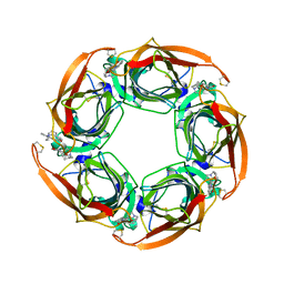
6B8L
 
 | |
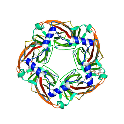
6CQ6
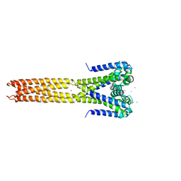
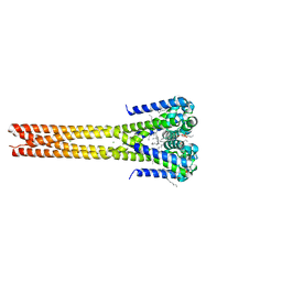
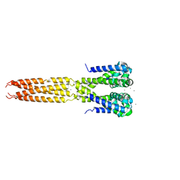
 | | K2P2.1(TREK-1) apo structure | | 分子名称: | CADMIUM ION, DECANE, POTASSIUM ION, ... | | 著者 | Lolicato, M, Minor, D.L. | | 登録日 | 2018-03-14 | | 公開日 | 2018-03-28 | | 最終更新日 | 2023-10-04 | | 実験手法 | X-RAY DIFFRACTION (3.1 Å) | | 主引用文献 | K2P2.1 (TREK-1)-activator complexes reveal a cryptic selectivity filter binding site.
Nature, 547, 2017
|
|
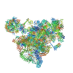
7AJT
 | | Cryo-EM structure of the 90S-exosome super-complex (state Pre-A1-exosome) | | 分子名称: | 13 kDa ribonucleoprotein-associated protein, 18S rRNA, 40S ribosomal protein S1-A, ... | | 著者 | Cheng, J, Lau, B, Flemming, D, Venuta, G.L, Berninghausen, O, Beckmann, R, Hurt, E. | | 登録日 | 2020-09-29 | | 公開日 | 2020-12-30 | | 最終更新日 | 2021-02-03 | | 実験手法 | ELECTRON MICROSCOPY (4.6 Å) | | 主引用文献 | Structure of the Maturing 90S Pre-ribosome in Association with the RNA Exosome.
Mol.Cell, 81, 2021
|
|
4MQA
 | | Human beta-tryptase co-crystal structure with {(1,1,3,3-tetramethyldisiloxane-1,3-diyl)bis[5-(methylsulfanyl)benzene-3,1-diyl]}bis({4-[3-(aminomethyl)phenyl]piperidin-1-yl}methanone) | | 分子名称: | SULFATE ION, TETRAETHYLENE GLYCOL, Tryptase alpha/beta-1, ... | | 著者 | White, A, Lakshminarasimhan, D, Suto, R. | | 登録日 | 2013-09-16 | | 公開日 | 2015-03-18 | | 実験手法 | X-RAY DIFFRACTION (2.25 Å) | | 主引用文献 | Target-directed self-assembly of homodimeric drugs
To be Published
|
|
7AJU
 | | Cryo-EM structure of the 90S-exosome super-complex (state Post-A1-exosome) | | 分子名称: | 13 kDa ribonucleoprotein-associated protein, 18S rRNA, 40S ribosomal protein S1-A, ... | | 著者 | Cheng, J, Lau, B, Flemming, D, Venuta, G.L, Berninghausen, O, Beckmann, R, Hurt, E. | | 登録日 | 2020-09-29 | | 公開日 | 2020-12-30 | | 最終更新日 | 2024-05-01 | | 実験手法 | ELECTRON MICROSCOPY (3.8 Å) | | 主引用文献 | Structure of the Maturing 90S Pre-ribosome in Association with the RNA Exosome.
Mol.Cell, 81, 2021
|
|
6AOD
 | | FXIa antibody complex | | 分子名称: | COBALT (II) ION, Coagulation factor XI, FXIa Antibody FAB Heavy Chain, ... | | 著者 | Lolicato, M, Minor, D.L. | | 登録日 | 2017-08-15 | | 公開日 | 2018-03-14 | | 最終更新日 | 2023-10-04 | | 実験手法 | X-RAY DIFFRACTION (1.8 Å) | | 主引用文献 | Structural Basis for Activity and Specificity of an Anticoagulant Anti-FXIa Monoclonal Antibody and a Reversal Agent.
Structure, 26, 2018
|
|
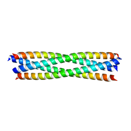
2OVC
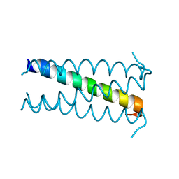 | |
4LTQ
 | | Bacterial sodium channel in low calcium, P42 space group | | 分子名称: | Ion transport protein | | 著者 | Shaya, D, Findeisen, F, Abderemane-Ali, F, Arrigoni, C, Wong, S, Reddy Nurva, S, Loussouarn, G, Minor, D.L. | | 登録日 | 2013-07-23 | | 公開日 | 2013-10-23 | | 最終更新日 | 2023-09-20 | | 実験手法 | X-RAY DIFFRACTION (5.5 Å) | | 主引用文献 | Structure of a prokaryotic sodium channel pore reveals essential gating elements and an outer ion binding site common to eukaryotic channels.
J.Mol.Biol., 426, 2014
|
|
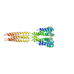
4LTO
 | | Bacterial sodium channel in high calcium, I222 space group | | 分子名称: | CALCIUM ION, Ion transport protein, NICKEL (II) ION | | 著者 | Shaya, D, Findeisen, F, Abderemane-Ali, F, Arrigoni, C, Wong, S, Reddy Nurva, S, Loussouarn, G, Minor, D.L. | | 登録日 | 2013-07-23 | | 公開日 | 2013-10-23 | | 最終更新日 | 2023-09-20 | | 実験手法 | X-RAY DIFFRACTION (3.46 Å) | | 主引用文献 | Structure of a prokaryotic sodium channel pore reveals essential gating elements and an outer ion binding site common to eukaryotic channels.
J.Mol.Biol., 426, 2014
|
|
7X4L
 | | Crystal structure of Bacteroides thetaiotaomicron glutamate decarboxylase mutant Y303F-PLP complex | | 分子名称: | Glutamate decarboxylase, PYRIDOXAL-5'-PHOSPHATE | | 著者 | Liu, S, Guoming, D, Yulu, W, Boting, W, Xin, F. | | 登録日 | 2022-03-02 | | 公開日 | 2023-05-03 | | 最終更新日 | 2023-11-29 | | 実験手法 | X-RAY DIFFRACTION (2.59 Å) | | 主引用文献 | Coordinated regulation of Bacteroides thetaiotaomicron glutamate decarboxylase activity by multiple elements under different pH.
Food Chem, 403, 2023
|
|
4LTR
 | | Bacterial sodium channel, His245Gly mutant, I222 space group | | 分子名称: | Ion transport protein | | 著者 | Shaya, D, Findeisen, F, Abderemane-Ali, F, Arrigoni, C, Wong, S, Reddy Nurva, S, Loussouarn, G, Minor, D.L. | | 登録日 | 2013-07-23 | | 公開日 | 2013-10-23 | | 最終更新日 | 2023-09-20 | | 実験手法 | X-RAY DIFFRACTION (5.8 Å) | | 主引用文献 | Structure of a prokaryotic sodium channel pore reveals essential gating elements and an outer ion binding site common to eukaryotic channels.
J.Mol.Biol., 426, 2014
|
|
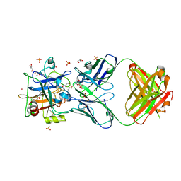
3GEQ
 | | Structural basis for the chemical rescue of Src kinase activity | | 分子名称: | 1-TERT-BUTYL-3-(4-CHLORO-PHENYL)-1H-PYRAZOLO[3,4-D]PYRIMIDIN-4-YLAMINE, Proto-oncogene tyrosine-protein kinase Src | | 著者 | Muratore, K.E, Seeliger, M.A, Wang, Z, Fomina, D, Neiswinger, J, Havranek, J.J, Baker, D, Kuriyan, J, Cole, P.A. | | 登録日 | 2009-02-25 | | 公開日 | 2009-04-28 | | 最終更新日 | 2023-09-06 | | 実験手法 | X-RAY DIFFRACTION (2.2 Å) | | 主引用文献 | Comparative analysis of mutant tyrosine kinase chemical rescue.
Biochemistry, 48, 2009
|
|
5HK7
 | | Bacterial sodium channel pore, 2.95 Angstrom resolution | | 分子名称: | 2-{[(S)-(2-aminoethoxy)(hydroxy)phosphoryl]oxy}ethyl heptadecanoate, CHLORIDE ION, Ion transport protein, ... | | 著者 | Shaya, D, Findeisen, F, Rohaim, A, Minor, D.L. | | 登録日 | 2016-01-14 | | 公開日 | 2016-03-09 | | 最終更新日 | 2023-09-27 | | 実験手法 | X-RAY DIFFRACTION (2.95 Å) | | 主引用文献 | Unfolding of a Temperature-Sensitive Domain Controls Voltage-Gated Channel Activation.
Cell, 164, 2016
|
|
4LTP
 | | Bacterial sodium channel in high calcium, I222 space group, crystal 2 | | 分子名称: | CALCIUM ION, Ion transport protein | | 著者 | Shaya, D, Findeisen, F, Abderemane-Ali, F, Arrigoni, C, Wong, S, Reddy Nurva, S, Loussouarn, G, Minor, D.L. | | 登録日 | 2013-07-23 | | 公開日 | 2013-10-23 | | 最終更新日 | 2023-09-20 | | 実験手法 | X-RAY DIFFRACTION (3.8 Å) | | 主引用文献 | Structure of a prokaryotic sodium channel pore reveals essential gating elements and an outer ion binding site common to eukaryotic channels.
J.Mol.Biol., 426, 2014
|
|
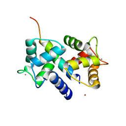
5HJ8
 | |
5HLN
 | | X-RAY CRYSTAL STRUCTURE OF GSK3B IN COMPLEX WITH CHIR99021 | | 分子名称: | 2-(N-MORPHOLINO)-ETHANESULFONIC ACID, CHIR99021, Glycogen synthase kinase-3 beta, ... | | 著者 | White, A, Lakshminarasimhan, D, Nadupalli, A, Suto, R.K. | | 登録日 | 2016-01-15 | | 公開日 | 2016-05-25 | | 最終更新日 | 2016-07-27 | | 実験手法 | X-RAY DIFFRACTION (3.1 Å) | | 主引用文献 | Inhibitors of Glycogen Synthase Kinase 3 with Exquisite Kinome-Wide Selectivity and Their Functional Effects.
Acs Chem.Biol., 11, 2016
|
|
3V0H
 | | Crystal structure of Ciona intestinalis voltage sensor-containing phosphatase (Ci-VSP), residues 241-576(C363S), complexed with D-MYO-inositol-1,4,5-triphosphate | | 分子名称: | D-MYO-INOSITOL-1,4,5-TRIPHOSPHATE, Voltage-sensor containing phosphatase | | 著者 | Liu, L, Kohout, S.C, Xu, Q, Muller, S, Kimberlin, C, Isacoff, E.Y, Minor, D.L. | | 登録日 | 2011-12-08 | | 公開日 | 2012-05-09 | | 最終更新日 | 2023-09-13 | | 実験手法 | X-RAY DIFFRACTION (1.85 Å) | | 主引用文献 | A glutamate switch controls voltage-sensitive phosphatase function.
Nat.Struct.Mol.Biol., 19, 2012
|
|
3V0G
 | | Crystal structure of Ciona intestinalis voltage sensor-containing phosphatase (Ci-VSP), residues 241-576(C363S), form III | | 分子名称: | PHOSPHATE ION, Voltage-sensor containing phosphatase | | 著者 | Liu, L, Kohout, S.C, Xu, Q, Muller, S, Kimberlin, C, Isacoff, E.Y, Minor, D.L. | | 登録日 | 2011-12-08 | | 公開日 | 2012-05-09 | | 最終更新日 | 2023-09-13 | | 実験手法 | X-RAY DIFFRACTION (1.6 Å) | | 主引用文献 | A glutamate switch controls voltage-sensitive phosphatase function.
Nat.Struct.Mol.Biol., 19, 2012
|
|
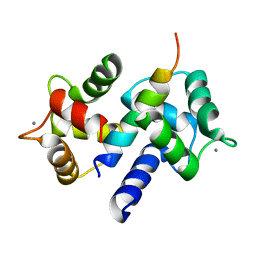
3E7K
 | |
3DVE
 | | Crystal Structure of Ca2+/CaM-CaV2.2 IQ domain complex | | 分子名称: | CALCIUM ION, Calmodulin, NICKEL (II) ION, ... | | 著者 | Kim, E.Y, Rumpf, C.H, Fujiwara, Y, Cooley, E.S, Van Petegem, F, Minor, D.L. | | 登録日 | 2008-07-18 | | 公開日 | 2008-11-04 | | 最終更新日 | 2024-02-21 | | 実験手法 | X-RAY DIFFRACTION (2.35 Å) | | 主引用文献 | Structures of Ca(V)2 Ca(2+)/CaM-IQ Domain Complexes Reveal Binding Modes that Underlie Calcium-Dependent Inactivation and Facilitation.
Structure, 16, 2008
|
|
3DVM
 | | Crystal Structure of Ca2+/CaM-CaV2.1 IQ domain complex | | 分子名称: | CALCIUM ION, Calmodulin, Voltage-dependent P/Q-type calcium channel subunit alpha-1A | | 著者 | Kim, E.Y, Rumpf, C.H, Fujiwara, Y, Cooley, E.S, Van Petegem, F, Minor, D.L. | | 登録日 | 2008-07-18 | | 公開日 | 2008-11-04 | | 最終更新日 | 2024-02-21 | | 実験手法 | X-RAY DIFFRACTION (2.6 Å) | | 主引用文献 | Structures of Ca(V)2 Ca(2+)/CaM-IQ Domain Complexes Reveal Binding Modes that Underlie Calcium-Dependent Inactivation and Facilitation.
Structure, 16, 2008
|
|
3DVK
 | | Crystal Structure of Ca2+/CaM-CaV2.3 IQ domain complex | | 分子名称: | CALCIUM ION, Calmodulin, Voltage-dependent R-type calcium channel subunit alpha-1E | | 著者 | Kim, E.Y, Rumpf, C.H, Fujiwara, Y, Cooley, E.S, Van Petegem, F, Minor, D.L. | | 登録日 | 2008-07-18 | | 公開日 | 2008-11-04 | | 最終更新日 | 2024-02-21 | | 実験手法 | X-RAY DIFFRACTION (2.3 Å) | | 主引用文献 | Structures of Ca(V)2 Ca(2+)/CaM-IQ Domain Complexes Reveal Binding Modes that Underlie Calcium-Dependent Inactivation and Facilitation.
Structure, 16, 2008
|
|
3DVJ
 | | Crystal Structure of Ca2+/CaM-CaV2.2 IQ domain (without cloning artifact, HM to TV) complex | | 分子名称: | CALCIUM ION, Calmodulin, Voltage-dependent N-type calcium channel subunit alpha-1B | | 著者 | Kim, E.Y, Rumpf, C.H, Fujiwara, Y, Cooley, E.S, Van Petegem, F, Minor, D.L. | | 登録日 | 2008-07-18 | | 公開日 | 2008-11-04 | | 最終更新日 | 2024-02-21 | | 実験手法 | X-RAY DIFFRACTION (2.8 Å) | | 主引用文献 | Structures of Ca(V)2 Ca(2+)/CaM-IQ Domain Complexes Reveal Binding Modes that Underlie Calcium-Dependent Inactivation and Facilitation.
Structure, 16, 2008
|
|
2XYS
 | | Crystal structure of Aplysia californica AChBP in complex with strychnine | | 分子名称: | SOLUBLE ACETYLCHOLINE RECEPTOR, STRYCHNINE | | 著者 | Brams, M, Pandya, A, Kuzmin, D, van Elk, R, Krijnen, L, Yakel, J.L, Tsetlin, V, Smit, A.B, Ulens, C. | | 登録日 | 2010-11-19 | | 公開日 | 2011-03-23 | | 最終更新日 | 2023-12-20 | | 実験手法 | X-RAY DIFFRACTION (1.909 Å) | | 主引用文献 | A Structural and Mutagenic Blueprint for Molecular Recognition of Strychnine and D-Tubocurarine by Different Cys-Loop Receptors.
Plos Biol., 9, 2011
|
|
2XYT
 | | Crystal structure of Aplysia californica AChBP in complex with d- tubocurarine | | 分子名称: | D-TUBOCURARINE, SOLUBLE ACETYLCHOLINE RECEPTOR | | 著者 | Brams, M, Pandya, A, Kuzmin, D, van Elk, R, Krijnen, L, Yakel, J.L, Tsetlin, V, Smit, A.B, Ulens, C. | | 登録日 | 2010-11-19 | | 公開日 | 2011-03-23 | | 最終更新日 | 2023-12-20 | | 実験手法 | X-RAY DIFFRACTION (2.05 Å) | | 主引用文献 | A Structural and Mutagenic Blueprint for Molecular Recognition of Strychnine and D-Tubocurarine by Different Cys-Loop Receptors.
Plos Biol., 9, 2011
|
|


