Loading
PDBj
MenuPDBj@FacebookPDBj@TwitterPDBj@YouTubewwPDB FoundationwwPDB
RCSB PDBPDBeBMRBAdv. SearchSearch help
PDB: 298 results

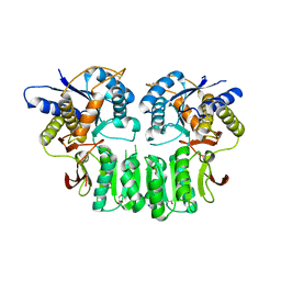
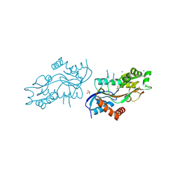
4ZDM
DownloadVisualize
BU of 4zdm by Molmil
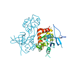
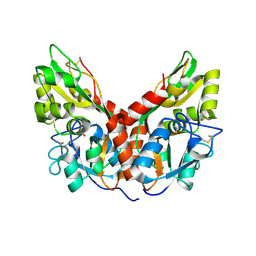
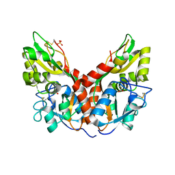
Pleurobrachia bachei iGluR3 LBD Glycine Complex
Descriptor: GLYCINE, Glutamate receptor kainate-like protein, SODIUM ION, ...
Authors:Grey, R.J, Mayer, M.L.
Deposit date:2015-04-17
Release date:2015-10-21
Last modified:2024-10-16
Method:X-RAY DIFFRACTION (1.5 Å)
Cite:Glycine activated ion channel subunits encoded by ctenophore glutamate receptor genes.
Proc.Natl.Acad.Sci.USA, 112, 2015
5UN2
DownloadVisualize
BU of 5un2 by Molmil
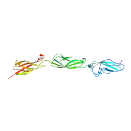
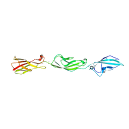
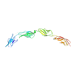
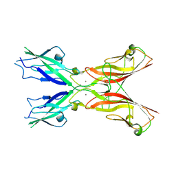
Crystal Structure of Mouse Cadherin-23 EC19-21 with non-syndromic deafness (DFNB12) associated mutation R2029W
Descriptor: CALCIUM ION, Cadherin-23, POTASSIUM ION
Authors:Jaiganesh, A, Sotomayor, M.
Deposit date:2017-01-30
Release date:2018-02-07
Last modified:2023-10-04
Method:X-RAY DIFFRACTION (2.96 Å)
Cite:Zooming in on Cadherin-23: Structural Diversity and Potential Mechanisms of Inherited Deafness.
Structure, 26, 2018
5UZ8
DownloadVisualize
BU of 5uz8 by Molmil
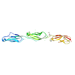
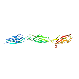
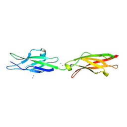
Crystal Structure of Mouse Cadherin-23 EC22-24
Descriptor: CALCIUM ION, CHLORIDE ION, Cadherin-23, ...
Authors:Patel, A, Jaiganesh, A, Sotomayor, M.
Deposit date:2017-02-25
Release date:2018-02-28
Last modified:2023-10-04
Method:X-RAY DIFFRACTION (1.85 Å)
Cite:Zooming in on Cadherin-23: Structural Diversity and Potential Mechanisms of Inherited Deafness.
Structure, 26, 2018
5TPK
DownloadVisualize
BU of 5tpk by Molmil
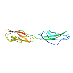
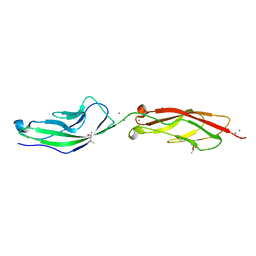
Crystal Structure of Mouse Protocadherin-15 EC7-8 V875A
Descriptor: CALCIUM ION, Protocadherin-15
Authors:Chen, C, Sotomayor, M.
Deposit date:2016-10-20
Release date:2017-10-25
Last modified:2023-10-04
Method:X-RAY DIFFRACTION (2 Å)
Cite:Structural determinants of protocadherin-15 mechanics and function in hearing and balance perception.
Proc.Natl.Acad.Sci.USA, 2020
5CYX
DownloadVisualize
BU of 5cyx by Molmil
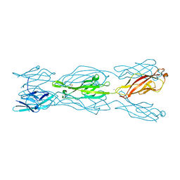
Crystal Structure of Mouse Protocadherin-24 EC1-3
Descriptor: CALCIUM ION, CHLORIDE ION, Protein Cdhr2
Authors:Johnson, Z.R, Sotomayor, M.
Deposit date:2015-07-31
Release date:2016-11-02
Last modified:2024-10-16
Method:X-RAY DIFFRACTION (2.1 Å)
Cite:Crystal Structure of Mouse Protocadherin-24 EC1-3
To Be Published
5ULU
DownloadVisualize
BU of 5ulu by Molmil
Crystal Structure of Mouse Cadherin-23 EC19-21 (S2087P) with non-syndromic deafness (DFNB12) associated mutation D2148N
Descriptor: CALCIUM ION, Cadherin-23
Authors:Jaiganesh, A, Sotomayor, M.
Deposit date:2017-01-25
Release date:2018-01-31
Last modified:2023-10-04
Method:X-RAY DIFFRACTION (2.85 Å)
Cite:Zooming in on Cadherin-23: Structural Diversity and Potential Mechanisms of Inherited Deafness.
Structure, 26, 2018
4KCC
DownloadVisualize
BU of 4kcc by Molmil
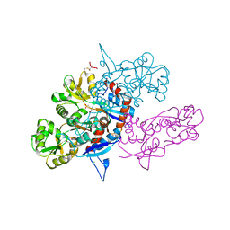
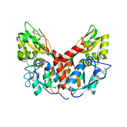
Crystal Structure of the NMDA Receptor GluN1 Ligand Binding Domain Apo State
Descriptor: Glutamate receptor ionotropic, NMDA 1, PHOSPHATE ION
Authors:Berger, A.J, Lau, A.Y, Mayer, M.L.
Deposit date:2013-04-24
Release date:2013-07-31
Last modified:2023-09-20
Method:X-RAY DIFFRACTION (1.894 Å)
Cite:Conformational Analysis of NMDA Receptor GluN1, GluN2, and GluN3 Ligand-Binding Domains Reveals Subtype-Specific Characteristics.
Structure, 21, 2013
3U93
DownloadVisualize
BU of 3u93 by Molmil
Crystal structure of the GluK3 ligand binding domain complex with glutamate and zinc: P2221 form
Descriptor: CHLORIDE ION, GLUTAMIC ACID, Glutamate receptor, ...
Authors:Kumar, J, Mayer, M.L.
Deposit date:2011-10-17
Release date:2012-09-26
Last modified:2024-11-06
Method:X-RAY DIFFRACTION (1.881 Å)
Cite:Zinc Potentiates GluK3 Glutamate Receptor Function by Stabilizing the Ligand Binding Domain Dimer Interface.
Neuron, 76, 2012
2YMB
DownloadVisualize
BU of 2ymb by Molmil
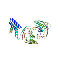
Structures of MITD1
Descriptor: CHARGED MULTIVESICULAR BODY PROTEIN 1A, MIT DOMAIN-CONTAINING PROTEIN 1
Authors:Hadders, M.A, Agromayor, M, Obita, T, Perisic, O, Caballe, A, Kloc, M, Lamers, M.H, Williams, R.L, Martin-Serrano, J.
Deposit date:2012-10-08
Release date:2012-10-24
Last modified:2023-12-20
Method:X-RAY DIFFRACTION (3.404 Å)
Cite:Escrt-III Binding Protein Mitd1 is Involved in Cytokinesis and Has an Unanticipated Pld Fold that Binds Membranes.
Proc.Natl.Acad.Sci.USA, 109, 2012
5W1D
DownloadVisualize
BU of 5w1d by Molmil
Crystal Structure of Mouse Protocadherin-15 EC4-7
Descriptor: CALCIUM ION, POTASSIUM ION, Protocadherin-15
Authors:Klanseck, C.F, Neel, B.L, Sotomayor, M.
Deposit date:2017-06-02
Release date:2018-06-06
Last modified:2023-10-04
Method:X-RAY DIFFRACTION (3.35 Å)
Cite:Structural determinants of protocadherin-15 mechanics and function in hearing and balance perception.
Proc.Natl.Acad.Sci.USA, 2020
5W4T
DownloadVisualize
BU of 5w4t by Molmil
Crystal Structure of Fish Cadherin-23 EC1-3
Descriptor: (4R)-2-METHYLPENTANE-2,4-DIOL, (4S)-2-METHYL-2,4-PENTANEDIOL, CALCIUM ION, ...
Authors:De-la-Torre, P, Sotomayor, M.
Deposit date:2017-06-12
Release date:2018-06-20
Last modified:2023-10-04
Method:X-RAY DIFFRACTION (2.65 Å)
Cite:Zooming in on Cadherin-23: Structural Diversity and Potential Mechanisms of Inherited Deafness.
Structure, 26, 2018
4YKP
DownloadVisualize
BU of 4ykp by Molmil
Mnemiopsis leidyi ML032222a iGluR LBD serine complex
Descriptor: GLYCINE, MAGNESIUM ION, ML032222a iGluR, ...
Authors:Alberstein, R.G, Mayer, M.L.
Deposit date:2015-03-04
Release date:2015-10-21
Last modified:2024-10-16
Method:X-RAY DIFFRACTION (1.46 Å)
Cite:Glycine activated ion channel subunits encoded by ctenophore glutamate receptor genes.
Proc.Natl.Acad.Sci.USA, 112, 2015
5WJM
DownloadVisualize
BU of 5wjm by Molmil
Crystal Structure of Mouse Cadherin-23 EC17-18
Descriptor: (4S)-2-METHYL-2,4-PENTANEDIOL, CALCIUM ION, Cadherin-23, ...
Authors:De-la-Torre, P, Sotomayor, M.
Deposit date:2017-07-23
Release date:2018-07-04
Last modified:2023-10-04
Method:X-RAY DIFFRACTION (2.9 Å)
Cite:Zooming in on Cadherin-23: Structural Diversity and Potential Mechanisms of Inherited Deafness.
Structure, 26, 2018
5ULY
DownloadVisualize
BU of 5uly by Molmil
Crystal Structure of Human Protocadherin-15 EC2-3
Descriptor: CALCIUM ION, Protocadherin-15
Authors:Wimalasena, L.N, Sotomayor, M.
Deposit date:2017-01-25
Release date:2018-08-08
Last modified:2023-10-04
Method:X-RAY DIFFRACTION (2.64 Å)
Cite:Structural determinants of protocadherin-15 mechanics and function in hearing and balance perception.
Proc.Natl.Acad.Sci.USA, 2020
4YKI
DownloadVisualize
BU of 4yki by Molmil
Mnemiopsis leidyi ML032222a iGluR LBD glycine complex
Descriptor: GLYCINE, MAGNESIUM ION, ML032222a iGluR, ...
Authors:Alberstein, R.G, Mayer, M.L.
Deposit date:2015-03-04
Release date:2015-10-21
Last modified:2024-10-30
Method:X-RAY DIFFRACTION (1.21 Å)
Cite:Glycine activated ion channel subunits encoded by ctenophore glutamate receptor genes.
Proc.Natl.Acad.Sci.USA, 112, 2015
5WJ8
DownloadVisualize
BU of 5wj8 by Molmil
Crystal Structure of Human Cadherin-23 EC13-14
Descriptor: CALCIUM ION, CHLORIDE ION, Cadherin-23, ...
Authors:Velez-Cortes, F, Conghui, C, De-la-Torre, P, Sotomayor, M.
Deposit date:2017-07-21
Release date:2018-07-04
Last modified:2023-11-15
Method:X-RAY DIFFRACTION (1.86 Å)
Cite:Zooming in on Cadherin-23: Structural Diversity and Potential Mechanisms of Inherited Deafness.
Structure, 26, 2018
4YKJ
DownloadVisualize
BU of 4ykj by Molmil
Mnemiopsis leidyi ML032222a iGluR LBD complex with Alanine
Descriptor: ALANINE, GLYCINE, ML032222a iGluR
Authors:Alberstein, R.G, Mayer, M.L.
Deposit date:2015-03-04
Release date:2015-10-21
Last modified:2024-11-06
Method:X-RAY DIFFRACTION (1.401 Å)
Cite:Glycine activated ion channel subunits encoded by ctenophore glutamate receptor genes.
Proc.Natl.Acad.Sci.USA, 112, 2015
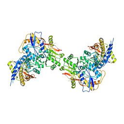
3EBB
DownloadVisualize
BU of 3ebb by Molmil
PLAP/P97 complex
Descriptor: MAGNESIUM ION, PHOSPHOLIPASE A2-ACTIVATING PROTEIN, TRANSITIONAL ENDOPLASMIC RETICULUM ATPASE (TER ATP
Authors:Walker, J.R, Qiu, L, Akutsu, M, Slessarev, Y, Amaya, M.F, Li, Y, Bountra, C, Weigelt, J, Arrowsmith, C.H, Edwards, A.M, Bochkarev, A, Dhe-Paganon, S, Structural Genomics Consortium (SGC)
Deposit date:2008-08-27
Release date:2009-02-24
Last modified:2024-10-09
Method:X-RAY DIFFRACTION (1.9 Å)
Cite:Structure and function of the PLAA/Ufd3-p97/Cdc48 complex.
J.Biol.Chem., 285, 2010
3U92
DownloadVisualize
BU of 3u92 by Molmil
Crystal structure of the GluK3 ligand binding domain complex with kainate and zinc: P2221 form
Descriptor: 3-(CARBOXYMETHYL)-4-ISOPROPENYLPROLINE, CHLORIDE ION, Glutamate receptor, ...
Authors:Kumar, J, Mayer, M.L.
Deposit date:2011-10-17
Release date:2012-09-26
Last modified:2024-10-30
Method:X-RAY DIFFRACTION (1.9 Å)
Cite:Zinc Potentiates GluK3 Glutamate Receptor Function by Stabilizing the Ligand Binding Domain Dimer Interface.
Neuron, 76, 2012
3OLZ
DownloadVisualize
BU of 3olz by Molmil
Crystal structure of the GluK3 (GluR7) ATD dimer at 2.75 Angstrom resolution
Descriptor: 2-acetamido-2-deoxy-beta-D-glucopyranose, CHLORIDE ION, Glutamate receptor, ...
Authors:Kumar, J, Mayer, M.L.
Deposit date:2010-08-26
Release date:2010-11-03
Last modified:2024-10-30
Method:X-RAY DIFFRACTION (2.75 Å)
Cite:Crystal Structures of the Glutamate Receptor Ion Channel GluK3 and GluK5 Amino-Terminal Domains.
J.Mol.Biol., 404, 2010
3OM0
DownloadVisualize
BU of 3om0 by Molmil
Crystal structure of the GluK5 (KA2) ATD crystallographic dimer at 1.4 Angstrom resolution
Descriptor: 2-acetamido-2-deoxy-beta-D-glucopyranose, ACETATE ION, GLYCEROL, ...
Authors:Kumar, J, Mayer, M.L.
Deposit date:2010-08-26
Release date:2010-11-03
Last modified:2024-04-03
Method:X-RAY DIFFRACTION (1.401 Å)
Cite:Crystal Structures of the Glutamate Receptor Ion Channel GluK3 and GluK5 Amino-Terminal Domains.
J.Mol.Biol., 404, 2010
3OM1
DownloadVisualize
BU of 3om1 by Molmil
Crystal structure of the GluK5 (KA2) ATD dimer at 1.7 Angstrom Resolution
Descriptor: 2-acetamido-2-deoxy-beta-D-glucopyranose, 2-acetamido-2-deoxy-beta-D-glucopyranose-(1-4)-2-acetamido-2-deoxy-beta-D-glucopyranose, GLYCEROL, ...
Authors:Kumar, J, Mayer, M.L.
Deposit date:2010-08-26
Release date:2010-11-03
Last modified:2023-09-06
Method:X-RAY DIFFRACTION (1.677 Å)
Cite:Crystal Structures of the Glutamate Receptor Ion Channel GluK3 and GluK5 Amino-Terminal Domains.
J.Mol.Biol., 404, 2010
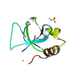
3QJ6
DownloadVisualize
BU of 3qj6 by Molmil
The crystal structure of PWWP domain of human Hepatoma-derived growth factor 2 in complex with H3K79me3 peptide
Descriptor: H3K79me3, Hepatoma-derived growth factor-related protein 2, SULFATE ION, ...
Authors:Zeng, H, Amaya, M.F, Tempel, W, Walker, J.R, Mackenzie, F, Bountra, C, Weigelt, J, Arrowsmith, C.H, Edwards, A.M, Min, J, Wu, H, Structural Genomics Consortium (SGC)
Deposit date:2011-01-28
Release date:2011-02-23
Last modified:2023-09-13
Method:X-RAY DIFFRACTION (2.3 Å)
Cite:Structural and Histone Binding Ability Characterizations of Human PWWP Domains.
Plos One, 6, 2011
3QLV
DownloadVisualize
BU of 3qlv by Molmil
Crystal structure of the GluK2/GluK5 (GluR6/KA2) ATD tetramer assembly
Descriptor: Glutamate receptor, ionotropic kainate 2, ionotropic kainate 5
Authors:Kumar, J, Mayer, M.L.
Deposit date:2011-02-03
Release date:2011-08-03
Last modified:2023-09-13
Method:X-RAY DIFFRACTION (3.94 Å)
Cite:Structure and assembly mechanism for heteromeric kainate receptors.
Neuron, 71, 2011
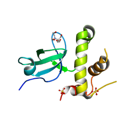
3QKJ
DownloadVisualize
BU of 3qkj by Molmil
The PWWP domain of human DNA (CYTOSINE-5-)-METHYLTRANSFERASE 3 BETA in complex with a bis-tris molecule
Descriptor: 2-[BIS-(2-HYDROXY-ETHYL)-AMINO]-2-HYDROXYMETHYL-PROPANE-1,3-DIOL, DNA cytosine-5 methyltransferase 3 beta isoform 6 variant, SULFATE ION
Authors:Zeng, H, Amaya, M.F, Mackenzie, F, Weigelt, J, Sundstrom, M, Arrowsmith, C.H, Edwards, A.M, Botchkarev, A, Min, J, Wu, H, Structural Genomics Consortium (SGC)
Deposit date:2011-02-01
Release date:2011-03-09
Last modified:2024-02-21
Method:X-RAY DIFFRACTION (2.04 Å)
Cite:Structural and Histone Binding Ability Characterizations of Human PWWP Domains.
Plos One, 6, 2011

227111

PDB entries from 2024-11-06

PDB statisticsPDBj update infoContact PDBjnumon