4M0I
 
 | |
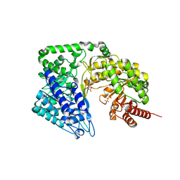
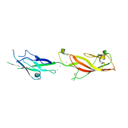
7Z3J
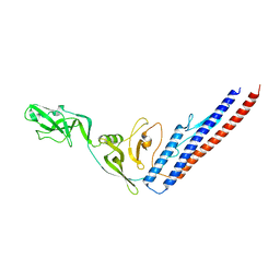
 | | Structure of crystallisable rat Phospholipase C gamma 1 in complex with inositol 1,4,5-trisphosphate | | 分子名称: | 1-phosphatidylinositol 4,5-bisphosphate phosphodiesterase gamma-1, CALCIUM ION, D-MYO-INOSITOL-1,4,5-TRIPHOSPHATE, ... | | 著者 | Pinotsis, N, Bunney, T.D, Katan, M. | | 登録日 | 2022-03-02 | | 公開日 | 2022-07-20 | | 最終更新日 | 2024-01-31 | | 実験手法 | X-RAY DIFFRACTION (2 Å) | | 主引用文献 | Characterization of the membrane interactions of phospholipase C gamma reveals key features of the active enzyme.
Sci Adv, 8, 2022
|
|
6E8F
 | |
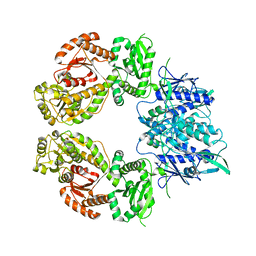
4ZTB
 | | Crystal structure of nsP2 protease from Chikungunya virus in P212121 space group at 2.59 A (4molecules/ASU). | | 分子名称: | GLYCEROL, Protease nsP2 | | 著者 | Narwal, M, Pratap, S, Singh, H, Kumar, P, Tomar, S. | | 登録日 | 2015-05-14 | | 公開日 | 2016-06-15 | | 最終更新日 | 2024-03-20 | | 実験手法 | X-RAY DIFFRACTION (2.59 Å) | | 主引用文献 | Crystal structure of chikungunya virus nsP2 cysteine protease reveals a putative flexible loop blocking its active site.
Int.J.Biol.Macromol., 116, 2018
|
|
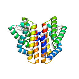
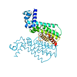
4ZTY
 | | Neurospora crassa cobalamin-independent methionine synthase complexed with Cd2+ | | 分子名称: | 2-AMINO-2-HYDROXYMETHYL-PROPANE-1,3-DIOL, CADMIUM ION, Cobalamin-Independent Methionine synthase, ... | | 著者 | Wheatley, R.W, Ng, K.K, Kapoor, M. | | 登録日 | 2015-05-15 | | 公開日 | 2015-12-09 | | 最終更新日 | 2024-03-06 | | 実験手法 | X-RAY DIFFRACTION (1.88 Å) | | 主引用文献 | Fungal cobalamin-independent methionine synthase: Insights from the model organism, Neurospora crassa.
Arch.Biochem.Biophys., 590, 2015
|
|
7ZEP
 | | Human SLFN11 E209A dimer | | 分子名称: | Schlafen family member 11, ZINC ION | | 著者 | Metzner, F.J, Kugler, M, Wenzl, S.J, Lammens, K. | | 登録日 | 2022-03-31 | | 公開日 | 2022-09-28 | | 最終更新日 | 2024-07-24 | | 実験手法 | ELECTRON MICROSCOPY (3.2 Å) | | 主引用文献 | Mechanistic understanding of human SLFN11.
Nat Commun, 13, 2022
|
|
1IUL
 | | The structure of cell-free ID.343 from Thermus thermophilus | | 分子名称: | hypothetical protein TT1466 | | 著者 | Wada, T, Shirouzu, M, Park, S.-Y, Tame, J.R, Kuramitsu, S, Yokoyama, S, RIKEN Structural Genomics/Proteomics Initiative (RSGI) | | 登録日 | 2002-03-05 | | 公開日 | 2003-07-15 | | 最終更新日 | 2023-12-27 | | 実験手法 | X-RAY DIFFRACTION (2 Å) | | 主引用文献 | Structure of a conserved CoA-binding protein synthesized by a cell-free system.
Acta Crystallogr.,Sect.D, 59, 2003
|
|
2MF9
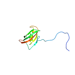 | | Solution structure of the N-terminal domain of human FKBP38 (FKBP38NTD) | | 分子名称: | Peptidyl-prolyl cis-trans isomerase FKBP8 | | 著者 | Kang, C, Ye, H, Simon, B, Sattler, M, Yoon, H.S. | | 登録日 | 2013-10-08 | | 公開日 | 2013-11-06 | | 最終更新日 | 2024-05-01 | | 実験手法 | SOLUTION NMR | | 主引用文献 | Functional role of the flexible N-terminal extension of FKBP38 in catalysis.
Sci Rep, 3, 2013
|
|
3LJF
 | | The X-ray structure of iron superoxide dismutase from Pseudoalteromonas haloplanktis (crystal form II) | | 分子名称: | FE (III) ION, alpha-D-glucopyranose-(1-1)-alpha-D-glucopyranose, iron superoxide dismutase | | 著者 | Merlino, A, Russo Krauss, I, Rossi, B, Conte, M, Vergara, A, Sica, F. | | 登録日 | 2010-01-26 | | 公開日 | 2010-09-08 | | 最終更新日 | 2023-09-06 | | 実験手法 | X-RAY DIFFRACTION (2.1 Å) | | 主引用文献 | Structure and flexibility in cold-adapted iron superoxide dismutases: the case of the enzyme isolated from Pseudoalteromonas haloplanktis.
J.Struct.Biol., 172, 2010
|
|
4ZVB
 | |
6EB5
 | |
4M3E
 | | Rapid and efficient design of new inhibitors of Mycobacterium tuberculosis transcriptional repressor EthR using fragment growing, merging and linking approaches | | 分子名称: | 4-(2-{[(propylsulfonyl)amino]methyl}-1,3-thiazol-4-yl)-N-(3,3,3-trifluoropropyl)benzamide, HTH-type transcriptional regulator EthR | | 著者 | Villemagne, B, Flipo, M, Blondiaux, N, Crauste, C, Malaquin, S, Leroux, F, Piveteau, C, Villeret, V, Brodin, P, Villoutreix, B, Sperandio, O, Wohlkonig, A, Wintjens, R, Deprez, B, Baulard, A, Willand, N. | | 登録日 | 2013-08-06 | | 公開日 | 2014-06-25 | | 最終更新日 | 2024-02-28 | | 実験手法 | X-RAY DIFFRACTION (2.109 Å) | | 主引用文献 | Ligand Efficiency Driven Design of New Inhibitors of Mycobacterium tuberculosis Transcriptional Repressor EthR Using Fragment Growing, Merging, and Linking Approaches.
J.Med.Chem., 57, 2014
|
|
1IWT
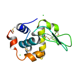 | | Crystal Structure Analysis of Human lysozyme at 113K. | | 分子名称: | CHLORIDE ION, LYSOZYME C | | 著者 | Joti, Y, Nakasako, M, Kidera, A, Go, N. | | 登録日 | 2002-06-03 | | 公開日 | 2002-09-04 | | 最終更新日 | 2023-12-27 | | 実験手法 | X-RAY DIFFRACTION (1.4 Å) | | 主引用文献 | Nonlinear temperature dependence of the crystal structure of lysozyme: correlation between coordinate shifts and thermal factors.
Acta Crystallogr.,Sect.D, 58, 2002
|
|
4ZV9
 | | 2.00 Angstrom resolution crystal structure of an uncharacterized protein from Escherichia coli O157:H7 str. Sakai | | 分子名称: | DI(HYDROXYETHYL)ETHER, GLYCEROL, PHOSPHATE ION, ... | | 著者 | Halavaty, A.S, Wawrzak, Z, Filippova, E.V, Kiryukhina, O, Endres, M, Joachimiak, A, Anderson, W.F, Midwest Center for Structural Genomics (MCSG) | | 登録日 | 2015-05-18 | | 公開日 | 2015-06-17 | | 実験手法 | X-RAY DIFFRACTION (2 Å) | | 主引用文献 | 2.00 Angstrom resolution crystal structure of an uncharacterized protein from Escherichia coli O157:H7 str. Sakai
To Be Published
|
|
4LXU
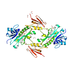 | | dTdp-Fuc3N and 5-N-Formyl-THF | | 分子名称: | (3R,4S,5R,6R)-4-amino-3,5-dihydroxy-6-methyloxan-2-yl][hydroxy-[[(2R,3S,5R)-3-hydroxy-5-(5-methyl-2,4-dioxopyrimidin-1-yl)oxolan-2-yl]methoxy]phosphoryl] hydrogen phosphate, 1,2-ETHANEDIOL, 3[N-MORPHOLINO]PROPANE SULFONIC ACID, ... | | 著者 | Thoden, J.B, Goneau, M.-F, Gilbert, M, Holden, H.M. | | 登録日 | 2013-07-30 | | 公開日 | 2013-08-14 | | 最終更新日 | 2023-09-20 | | 実験手法 | X-RAY DIFFRACTION (1.4 Å) | | 主引用文献 | Structure of a sugar N-formyltransferase from Campylobacter jejuni.
Biochemistry, 52, 2013
|
|
6IUE
 | | DNA helical wire containing Hg(II) | | 分子名称: | DNA (5'-D(*TP*TP*TP*GP*C)-3'), MERCURY (II) ION | | 著者 | Ono, A, Kanazawa, H, Ito, H, Goto, M, Nakamura, K, Saneyoshi, H, Kondo, J. | | 登録日 | 2018-11-28 | | 公開日 | 2019-10-16 | | 最終更新日 | 2024-03-27 | | 実験手法 | X-RAY DIFFRACTION (1.901 Å) | | 主引用文献 | A Novel DNA Helical Wire Containing HgII-Mediated T:T and T:G Pairs.
Angew.Chem.Int.Ed.Engl., 58, 2019
|
|
7ZCE
 | |
1IYB
 | |
6ECJ
 | |
4M5A
 | | Crystal structure of the complex of Ribosome inactivating protein from Momordica balsamina inhibited by asymmetric dimethyl arginine at 1.70 A resolution | | 分子名称: | 2-acetamido-2-deoxy-beta-D-glucopyranose, NG,NG-DIMETHYL-L-ARGININE, rRNA N-glycosidase | | 著者 | Yamini, S, Pandey, S, Kushwaha, G.S, Sinha, M, Bhushan, A, Kaur, P, Sharma, S, Singh, T.P. | | 登録日 | 2013-08-08 | | 公開日 | 2013-08-28 | | 最終更新日 | 2023-12-06 | | 実験手法 | X-RAY DIFFRACTION (1.7 Å) | | 主引用文献 | Crystal structure of the complex of Ribosome inactivating protein from Momordica balsamina inhibited by asymmetric dimethyl arginine at 1.70 A resolution
To be Published
|
|
6ISL
 | | SnoaL-like cyclase XimE with its product xiamenmycin B | | 分子名称: | (2R,3S)-2-methyl-2-(4-methylpent-3-enyl)-3-oxidanyl-3,4-dihydrochromene-6-carboxylic acid, XimE, SnoaL-like domain protein | | 著者 | He, B, Zhou, T, Bu, X, Weng, J, Xu, J, Lin, S, Zheng, J, Zhao, Y, Xu, M. | | 登録日 | 2018-11-16 | | 公開日 | 2019-11-20 | | 最終更新日 | 2024-03-27 | | 実験手法 | X-RAY DIFFRACTION (1.77 Å) | | 主引用文献 | Enzymatic Pyran Formation Involved in Xiamenmycin Biosynthesis
Acs Catalysis, 9, 2019
|
|
1IO1
 | | CRYSTAL STRUCTURE OF F41 FRAGMENT OF FLAGELLIN | | 分子名称: | PHASE 1 FLAGELLIN | | 著者 | Samatey, F.A, Imada, K, Nagashima, S, Vondervisz, F, Kumasaka, T, Yamamoto, M, Namba, K. | | 登録日 | 2000-12-28 | | 公開日 | 2001-04-04 | | 最終更新日 | 2023-12-27 | | 実験手法 | X-RAY DIFFRACTION (2 Å) | | 主引用文献 | Structure of the bacterial flagellar protofilament and implications for a switch for supercoiling
Nature, 410, 2001
|
|
1IOQ
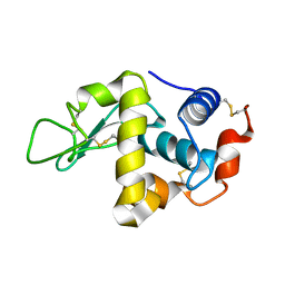 | | STABILIZATION OF HEN EGG WHITE LYSOZYME BY A CAVITY-FILLING MUTATION | | 分子名称: | LYSOZYME C | | 著者 | Ohmura, T, Ueda, T, Ootsuka, K, Saito, M, Imoto, T. | | 登録日 | 2001-03-28 | | 公開日 | 2001-04-11 | | 最終更新日 | 2023-12-27 | | 実験手法 | X-RAY DIFFRACTION (1.79 Å) | | 主引用文献 | Stabilization of hen egg white lysozyme by a cavity-filling mutation.
Protein Sci., 10, 2001
|
|
4M6D
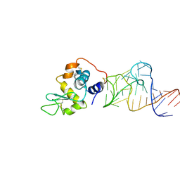 | | Crystal structure of the aptamer minF-lysozyme complex. | | 分子名称: | Lysozyme C, aptamer | | 著者 | Malashkevich, V.N, Padlan, F.C, Toro, R, Girvin, M, Almo, S.C, New York Structural Genomics Research Consortium (NYSGRC) | | 登録日 | 2013-08-09 | | 公開日 | 2013-12-11 | | 最終更新日 | 2023-09-20 | | 実験手法 | X-RAY DIFFRACTION (2.68 Å) | | 主引用文献 | Crystal structure of the aptamer minF-lysozyme complex.
To be Published
|
|
1IOS
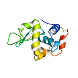 | | STABILIZATION OF HEN EGG WHITE LYSOZYME BY A CAVITY-FILLING MUTATION | | 分子名称: | LYSOZYME C | | 著者 | Ohmura, T, Ueda, T, Ootsuka, K, Saito, M, Imoto, T. | | 登録日 | 2001-03-28 | | 公開日 | 2001-04-11 | | 最終更新日 | 2023-12-27 | | 実験手法 | X-RAY DIFFRACTION (1.76 Å) | | 主引用文献 | Stabilization of hen egg white lysozyme by a cavity-filling mutation.
Protein Sci., 10, 2001
|
|


