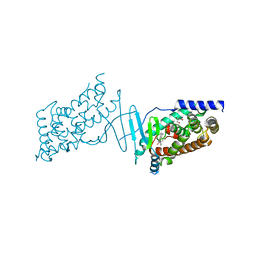
7PXD
 
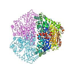
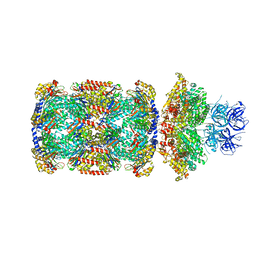 | | Substrate-engaged mycobacterial Proteasome-associated ATPase in complex with open-gate 20S CP - composite map (state B) | | 分子名称: | AAA ATPase forming ring-shaped complexes, ADENOSINE-5'-TRIPHOSPHATE, MAGNESIUM ION, ... | | 著者 | Jomaa, A, Kavalchuk, M, Weber-Ban, E. | | 登録日 | 2021-10-08 | | 公開日 | 2022-01-19 | | 最終更新日 | 2024-07-17 | | 実験手法 | ELECTRON MICROSCOPY (4 Å) | | 主引用文献 | Structural basis of prokaryotic ubiquitin-like protein engagement and translocation by the mycobacterial Mpa-proteasome complex.
Nat Commun, 13, 2022
|
|
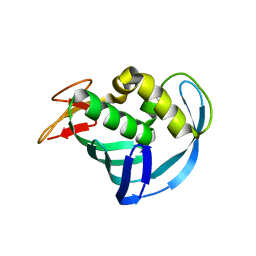
7PT2
 | |
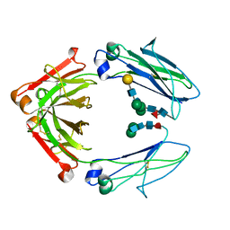
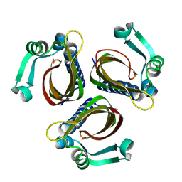
1A0F
 | | CRYSTAL STRUCTURE OF GLUTATHIONE S-TRANSFERASE FROM ESCHERICHIA COLI COMPLEXED WITH GLUTATHIONESULFONIC ACID | | 分子名称: | GLUTATHIONE S-TRANSFERASE, GLUTATHIONE SULFONIC ACID | | 著者 | Nishida, M, Harada, S, Noguchi, S, Inoue, H, Takahashi, K, Satow, Y. | | 登録日 | 1997-11-29 | | 公開日 | 1999-01-13 | | 最終更新日 | 2024-02-07 | | 実験手法 | X-RAY DIFFRACTION (2.1 Å) | | 主引用文献 | Three-dimensional structure of Escherichia coli glutathione S-transferase complexed with glutathione sulfonate: catalytic roles of Cys10 and His106.
J.Mol.Biol., 281, 1998
|
|
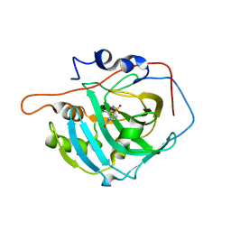
7PT1
 | |
8BMV
 | | Ligand binding domain of the P. Putida receptor McpH in complex with Uric acid | | 分子名称: | Methyl-accepting chemotaxis protein McpH, URIC ACID | | 著者 | Gavira, J.A, Krell, T, Fernandez, M, Martinez-Rodriguez, S. | | 登録日 | 2022-11-11 | | 公開日 | 2024-07-24 | | 実験手法 | X-RAY DIFFRACTION (1.95 Å) | | 主引用文献 | Ubiquitous purine sensor modulates diverse signal transduction pathways in bacteria.
Nat Commun, 15, 2024
|
|
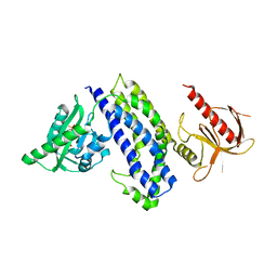
5HY9
 | | Glycosylated, disulfide-linked Knob-into-Hole Fc fragment | | 分子名称: | 2-acetamido-2-deoxy-beta-D-glucopyranose-(1-2)-beta-D-mannopyranose-(1-3)-beta-D-mannopyranose-(1-4)-2-acetamido-2-deoxy-beta-D-glucopyranose-(1-4)-[alpha-L-fucopyranose-(1-6)]2-acetamido-2-deoxy-beta-D-glucopyranose, Ig gamma-1 chain C region, beta-D-galactopyranose-(1-4)-2-acetamido-2-deoxy-beta-D-glucopyranose-(1-2)-beta-D-mannopyranose-(1-6)-[2-acetamido-2-deoxy-beta-D-glucopyranose-(1-2)-beta-D-mannopyranose-(1-3)]beta-D-mannopyranose-(1-4)-2-acetamido-2-deoxy-beta-D-glucopyranose-(1-4)-[alpha-L-fucopyranose-(1-6)]2-acetamido-2-deoxy-beta-D-glucopyranose | | 著者 | Kuglstatter, A, Stihle, M, Benz, J. | | 登録日 | 2016-02-01 | | 公開日 | 2017-02-01 | | 最終更新日 | 2024-01-10 | | 実験手法 | X-RAY DIFFRACTION (2.7 Å) | | 主引用文献 | Structural differences between glycosylated, disulfide-linked heterodimeric Knob-into-Hole Fc fragment and its homodimeric Knob-Knob and Hole-Hole side products.
Protein Eng. Des. Sel., 30, 2017
|
|
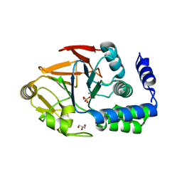
7PT3
 | |
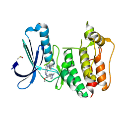
1IWQ
 | | Crystal Structure of MARCKS calmodulin binding domain peptide complexed with Ca2+/Calmodulin | | 分子名称: | CALCIUM ION, CALMODULIN, MARCKS | | 著者 | Yamauchi, E, Nakatsu, T, Matsubara, M, Kato, H, Taniguchi, H, RIKEN Structural Genomics/Proteomics Initiative (RSGI) | | 登録日 | 2002-05-31 | | 公開日 | 2003-03-11 | | 最終更新日 | 2023-10-25 | | 実験手法 | X-RAY DIFFRACTION (2 Å) | | 主引用文献 | Crystal structure of a MARCKS peptide containing the calmodulin-binding domain in complex with Ca(2+)-calmodulin
NAT.STRUCT.BIOL., 10, 2003
|
|
5HN3
 | |
5EG9
 | | The cap binding site of influenza virus protein PB2 as a drug target | | 分子名称: | Polymerase basic protein 2 | | 著者 | Severin, C, Rocha de Moura, T, Liu, Y, Li, K, Zheng, X, Luo, M. | | 登録日 | 2015-10-26 | | 公開日 | 2016-02-10 | | 最終更新日 | 2023-09-27 | | 実験手法 | X-RAY DIFFRACTION (2.3 Å) | | 主引用文献 | The cap-binding site of influenza virus protein PB2 as a drug target.
Acta Crystallogr D Struct Biol, 72, 2016
|
|
8CDZ
 | | human carbonic anhydrase I complexed with 4-(3-butylureido)benzenesulfonamide | | 分子名称: | 1-butyl-3-(4-sulfamoylphenyl)urea, Carbonic anhydrase 1, DIMETHYL SULFOXIDE, ... | | 著者 | Angeli, A, Ferraroni, M. | | 登録日 | 2023-02-01 | | 公開日 | 2024-02-21 | | 最終更新日 | 2024-09-04 | | 実験手法 | X-RAY DIFFRACTION (1.44 Å) | | 主引用文献 | Ureidobenzenesulfonamides as Selective Carbonic Anhydrase I, IX, and XII Inhibitors.
Molecules, 28, 2023
|
|
5HZJ
 | | Crystal structure of photoinhibitable Intersectin1 containing wildtype LOV2 domain | | 分子名称: | FLAVIN MONONUCLEOTIDE, Intersectin-1,NPH1-1,Intersectin-1 | | 著者 | Tarnawski, M, Dagliyan, O, Chu, P.H, Shirvanyants, D, Dokholyan, N.V, Hahn, K.M, Schlichting, I. | | 登録日 | 2016-02-02 | | 公開日 | 2016-12-21 | | 最終更新日 | 2024-01-10 | | 実験手法 | X-RAY DIFFRACTION (2.6 Å) | | 主引用文献 | Engineering extrinsic disorder to control protein activity in living cells.
Science, 354, 2016
|
|
6OBS
 | | PP1 Y134K | | 分子名称: | GLYCEROL, MANGANESE (II) ION, PHOSPHATE ION, ... | | 著者 | Choy, M.S, Moon, T.M, Bray, J.A, Archuleta, T.L, Shi, W, Peti, W, Page, R. | | 登録日 | 2019-03-21 | | 公開日 | 2019-09-18 | | 最終更新日 | 2023-10-11 | | 実験手法 | X-RAY DIFFRACTION (1.803 Å) | | 主引用文献 | SDS22 selectively recognizes and traps metal-deficient inactive PP1.
Proc.Natl.Acad.Sci.USA, 116, 2019
|
|
8CF9
 | | Crystal structure of the human PXR ligand-binding domain in complex with sclareol | | 分子名称: | GLYCEROL, Nuclear receptor subfamily 1 group I member 2, sclareol | | 著者 | Carivenc, C, Derosa, Q, Grimaldi, M, Boulahtouf, A, Balaguer, P, Bourguet, W. | | 登録日 | 2023-02-03 | | 公開日 | 2024-02-21 | | 実験手法 | X-RAY DIFFRACTION (2 Å) | | 主引用文献 | Crystal structure of the human PXR ligand-binding domain in complex with sclareol
To Be Published
|
|
7PT4
 | | Actinobacterial 2-hydroxyacyl-CoA lyase (AcHACL) structure in complex with a covalently bound reaction intermediate as well as products formyl-CoA and acetone | | 分子名称: | 2-hydroxyacyl-CoA lyase, 3-[(4-AMINO-2-METHYLPYRIMIDIN-5-YL)METHYL]-2-{(1R,11R,15S,17R)-19-[(2R,3S,4R,5R)-5-(6-AMINO-9H-PURIN-9-YL)-4-HYDROXY-3-(PHOSPHONOOXY)TETRAHYDROFURAN-2-YL]-1,11,15,17-TETRAHYDROXY-12,12-DIMETHYL-15,17-DIOXIDO-6,10-DIOXO-14,16,18-TRIOXA-2-THIA-5,9-DIAZA-15,17-DIPHOSPHANONADEC-1-YL}-5-(2-{[(R)-HYDROXY(PHOSPHONOOXY)PHOSPHORYL]OXY}ETHYL)-4-METHYL-1,3-THIAZOL-3-IUM, ACETONE, ... | | 著者 | Zahn, M, Rohwerder, T. | | 登録日 | 2021-09-25 | | 公開日 | 2022-02-02 | | 最終更新日 | 2024-01-31 | | 実験手法 | X-RAY DIFFRACTION (1.64 Å) | | 主引用文献 | Mechanistic details of the actinobacterial lyase-catalyzed degradation reaction of 2-hydroxyisobutyryl-CoA.
J.Biol.Chem., 298, 2022
|
|
5EHH
 | | Structure of human DPP3 in complex with endomorphin-2. | | 分子名称: | Dipeptidyl peptidase 3, Endomorphin-2, MAGNESIUM ION, ... | | 著者 | Kumar, P, Reithofer, V, Reisinger, M, Pavkov-Keller, T, Wallner, S, Macheroux, P, Gruber, K. | | 登録日 | 2015-10-28 | | 公開日 | 2016-04-13 | | 最終更新日 | 2024-01-10 | | 実験手法 | X-RAY DIFFRACTION (2.38 Å) | | 主引用文献 | Substrate complexes of human dipeptidyl peptidase III reveal the mechanism of enzyme inhibition.
Sci Rep, 6, 2016
|
|
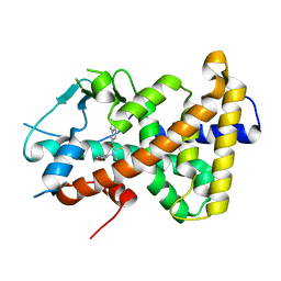
5HOC
 | | p73 homo-tetramerization domain mutant II | | 分子名称: | Tumor protein p73 | | 著者 | Coutandin, D, Krojer, T, Salah, E, Mathea, S, Sumyk, M, Knapp, S, Dotsch, V. | | 登録日 | 2016-01-19 | | 公開日 | 2016-10-19 | | 最終更新日 | 2024-01-10 | | 実験手法 | X-RAY DIFFRACTION (1.36007786 Å) | | 主引用文献 | Mechanism of TAp73 inhibition by Delta Np63 and structural basis of p63/p73 hetero-tetramerization.
Cell Death Differ., 23, 2016
|
|
5EHB
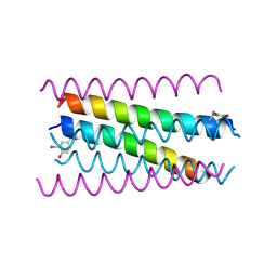 | | A de novo designed hexameric coiled-coil peptide with iodotyrosine | | 分子名称: | pHiosYI | | 著者 | Lizatovic, R, Aurelius, O, Stenstrom, O, Drakenberg, T, Akke, M, Logan, D.T, Andre, I. | | 登録日 | 2015-10-28 | | 公開日 | 2016-06-15 | | 最終更新日 | 2018-01-17 | | 実験手法 | X-RAY DIFFRACTION (3.19 Å) | | 主引用文献 | A De Novo Designed Coiled-Coil Peptide with a Reversible pH-Induced Oligomerization Switch.
Structure, 24, 2016
|
|
1IDP
 | |
5EHO
 | | Rapid Discovery of Pyrido[3,4-d]pyrimidine Inhibitors of Monopolar Spindle kinase 1 (MPS1) Using a Structure-Based Hydridization Approach | | 分子名称: | DIMETHYL SULFOXIDE, Dual specificity protein kinase TTK, ~{N}8-cyclohexyl-~{N}2-[2-methoxy-4-(1-methylpyrazol-4-yl)phenyl]pyrido[3,4-d]pyrimidine-2,8-diamine | | 著者 | Innocenti, P, Woodward, H.L, Solanki, S, Naud, N, Westwood, I.M, Cronin, N, Hayes, A, Roberts, J, Henley, A.T, Baker, R, Faisal, A, Mak, G, Box, G, Valenti, M, De Haven Brandon, A, O'Fee, L, Saville, J, Schmitt, J, Burke, R, van Montfort, R.L.M, Raymaud, F.I, Eccles, S.A, Linardopoulos, S, Blagg, J, Hoelder, S. | | 登録日 | 2015-10-28 | | 公開日 | 2016-11-09 | | 最終更新日 | 2024-05-08 | | 実験手法 | X-RAY DIFFRACTION (2.18 Å) | | 主引用文献 | Rapid Discovery of Pyrido[3,4-d]pyrimidine Inhibitors of Monopolar Spindle kinase 1 (MPS1) Using a Structure-Based Hydridization Approach
To Be Published
|
|
2LVU
 | | Solution structure of Miz-1 zinc finger 10 | | 分子名称: | ZINC ION, Zinc finger and BTB domain-containing protein 17 | | 著者 | Bedard, M, Maltais, L, Beaulieu, M, Bernard, D, Lavigne, P. | | 登録日 | 2012-07-11 | | 公開日 | 2012-07-25 | | 最終更新日 | 2024-05-01 | | 実験手法 | SOLUTION NMR | | 主引用文献 | NMR structure note: solution structure of human Miz-1 zinc fingers 8 to 10.
J.Biomol.Nmr, 54, 2012
|
|
8CH8
 | | Crystal structure of the human PXR ligand-binding domain in complex with liranaftate | | 分子名称: | Nuclear receptor subfamily 1 group I member 2, ~{O}-(5,6,7,8-tetrahydronaphthalen-2-yl) ~{N}-(6-methoxypyridin-2-yl)-~{N}-methyl-carbamothioate | | 著者 | Carivenc, C, Derosa, Q, Grimaldi, M, Boulahtouf, A, Balaguer, P, Bourguet, W. | | 登録日 | 2023-02-07 | | 公開日 | 2024-02-21 | | 実験手法 | X-RAY DIFFRACTION (2.15 Å) | | 主引用文献 | Crystal structure of the human PXR ligand-binding domain in complex with liranaftate
To Be Published
|
|
8CHY
 | | Crystal structure of an 8-repeat consensus TPR superhelix with Zinc. | | 分子名称: | (4S)-2-METHYL-2,4-PENTANEDIOL, ACETATE ION, CHLORIDE ION, ... | | 著者 | Liutkus, M, Rojas, A.L, Cortajarena, A.L. | | 登録日 | 2023-02-08 | | 公開日 | 2024-02-21 | | 最終更新日 | 2024-04-24 | | 実験手法 | X-RAY DIFFRACTION (2 Å) | | 主引用文献 | Diverse crystalline protein scaffolds through metal-dependent polymorphism.
Protein Sci., 33, 2024
|
|
1A90
 | | RECOMBINANT MUTANT CHICKEN EGG WHITE CYSTATIN, NMR, 31 STRUCTURES | | 分子名称: | CYSTATIN | | 著者 | Dieckmann, T, Mitschang, L, Hofmann, M, Kos, J, Turk, V, Auerswald, E.A, Jaenicke, R, Oschkinat, H. | | 登録日 | 1998-04-14 | | 公開日 | 1998-06-17 | | 最終更新日 | 2022-02-16 | | 実験手法 | SOLUTION NMR | | 主引用文献 | The structures of native phosphorylated chicken cystatin and of a recombinant unphosphorylated variant in solution.
J.Mol.Biol., 234, 1993
|
|
8CI8
 | |


