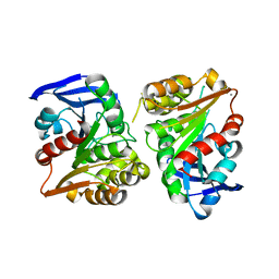
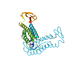
2YVI
 
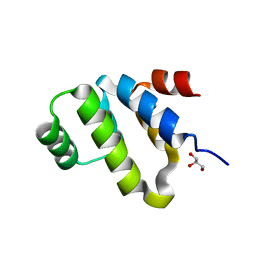 | | Crystal structure of a death domain of human ankryn protein | | 分子名称: | Ankyrin-1, GLYCEROL | | 著者 | Ihsanawati, Bessho, Y, Chen, L, Liu, Z.J, Wang, B.C, Shirouzu, M, Yokoyama, S, RIKEN Structural Genomics/Proteomics Initiative (RSGI) | | 登録日 | 2007-04-12 | | 公開日 | 2008-04-15 | | 最終更新日 | 2024-03-13 | | 実験手法 | X-RAY DIFFRACTION (1.92 Å) | | 主引用文献 | Crystal structure of a death domain of human ankryn protein
To be Published
|
|
1SL8
 | | Calcium-loaded apo-aequorin from Aequorea victoria | | 分子名称: | Aequorin 1, CALCIUM ION | | 著者 | Deng, L, Markova, S.V, Vysotski, E.S, Liu, Z.J, Lee, J, Rose, J, Wang, B.C, Southeast Collaboratory for Structural Genomics (SECSG) | | 登録日 | 2004-03-05 | | 公開日 | 2004-12-28 | | 最終更新日 | 2023-11-29 | | 実験手法 | X-RAY DIFFRACTION (1.7 Å) | | 主引用文献 | All three Ca2+-binding loops of photoproteins bind calcium ions: The crystal structures of calcium-loaded apo-aequorin and apo-obelin.
Protein Sci., 14, 2005
|
|
1SL7
 | | Crystal structure of calcium-loaded apo-obelin from Obelia longissima | | 分子名称: | CALCIUM ION, Obelin | | 著者 | Deng, L, Markova, S.V, Vysotski, E.S, Liu, Z.J, Lee, J, Rose, J, Wang, B.C, Southeast Collaboratory for Structural Genomics (SECSG) | | 登録日 | 2004-03-05 | | 公開日 | 2004-12-28 | | 最終更新日 | 2023-08-23 | | 実験手法 | X-RAY DIFFRACTION (2.2 Å) | | 主引用文献 | All three Ca2+-binding loops of photoproteins bind calcium ions: The crystal structures of calcium-loaded apo-aequorin and apo-obelin.
Protein Sci., 14, 2005
|
|
2PW6
 | | Crystal structure of uncharacterized protein JW3007 from Escherichia coli K12 | | 分子名称: | Uncharacterized protein ygiD, ZINC ION | | 著者 | Newton, M.G, Takagi, Y, Ebihara, A, Shinkai, A, Kuramitsu, S, Yokayama, S, Li, Y, Chen, L, Zhu, J, Ruble, J, Liu, Z.J, Rose, J.P, Wang, B.-C, Southeast Collaboratory for Structural Genomics (SECSG), RIKEN Structural Genomics/Proteomics Initiative (RSGI) | | 登録日 | 2007-05-10 | | 公開日 | 2007-06-12 | | 最終更新日 | 2018-01-24 | | 実験手法 | X-RAY DIFFRACTION (2.27 Å) | | 主引用文献 | Crystal structure of uncharacterized protein JW3007 from Escherichia coli K12.
To be Published
|
|
2QVO
 | | Crystal structure of AF1382 from Archaeoglobus fulgidus | | 分子名称: | Uncharacterized protein AF_1382 | | 著者 | Zhu, J, Zhao, M, Fu, Z.-Q, Yang, H, Chang, J, Hao, X, Chen, L, Liu, Z.J, Rose, J.P, Wang, B.C, Southeast Collaboratory for Structural Genomics (SECSG) | | 登録日 | 2007-08-08 | | 公開日 | 2007-09-04 | | 最終更新日 | 2024-02-21 | | 実験手法 | X-RAY DIFFRACTION (1.85 Å) | | 主引用文献 | Crystal structure of AF1382 from Archaeoglobus fulgidus.
To be Published
|
|
3CQ2
 | | Structure of the DTDP-4-Keto-L-Rhamnose Reductase related protein (other form) from Thermus Thermophilus HB8 | | 分子名称: | Putative uncharacterized protein TTHB138 | | 著者 | Jeyakanthan, J, Kanaujia, S.P, Sekar, K, Satoh, S, Kitamura, Y, Ebihara, A, Chen, L, Liu, Z.J, Wang, B.C, Yokoyama, S, Kuramitsu, S, RIKEN Structural Genomics/Proteomics Initiative (RSGI) | | 登録日 | 2008-04-02 | | 公開日 | 2009-04-07 | | 最終更新日 | 2023-11-15 | | 実験手法 | X-RAY DIFFRACTION (1.9 Å) | | 主引用文献 | Structure of the DTDP-4-Keto-L-Rhamnose Reductase related protein from Thermus Thermophilus HB8
To be Published
|
|
1LKM
 | | Crystal structure of Desulfovibrio vulgaris rubrerythrin all-iron(III) form | | 分子名称: | FE (III) ION, Rubrerythrin all-iron(III) form | | 著者 | Jin, S, Kurtz Jr, D.M, Liu, Z.J, Rose, J, Wang, B.C. | | 登録日 | 2002-04-25 | | 公開日 | 2002-09-18 | | 最終更新日 | 2023-08-16 | | 実験手法 | X-RAY DIFFRACTION (1.69 Å) | | 主引用文献 | X-ray Crystal Structures of Reduced Rubrerythrin and its Azide Adduct: A Structure-Based Mechanism for a Non-Heme DiIron Peroxidase
J.Am.Chem.Soc., 124, 2002
|
|
5TGZ
 | | Crystal Structure of the Human Cannabinoid Receptor CB1 | | 分子名称: | (2R)-2,3-dihydroxypropyl (9Z)-octadec-9-enoate, 4-[4-[2-(2,4-dichlorophenyl)-4-methyl-5-(piperidin-1-ylcarbamoyl)pyrazol-3-yl]phenyl]but-3-ynyl nitrate, Cannabinoid receptor 1,Flavodoxin,Cannabinoid receptor 1, ... | | 著者 | Hua, T, Vemuri, K, Pu, M, Qu, L, Han, G.W, Wu, Y, Zhao, S, Shui, W, Li, S, Korde, A, Laprairie, R.B, Stahl, E.L, Ho, J.H, Zvonok, N, Zhou, H, Kufareva, I, Wu, B, Zhao, Q, Hanson, M.A, Bohn, L.M, Makriyannis, A, Stevens, R.C, Liu, Z.J. | | 登録日 | 2016-09-28 | | 公開日 | 2016-11-02 | | 最終更新日 | 2023-11-08 | | 実験手法 | X-RAY DIFFRACTION (2.8 Å) | | 主引用文献 | Crystal Structure of the Human Cannabinoid Receptor CB1.
Cell, 167, 2016
|
|
1LKO
 | | Crystal structure of Desulfovibrio vulgaris rubrerythrin all-iron(II) form | | 分子名称: | FE (II) ION, Rubrerythrin all-iron(II) form | | 著者 | Jin, S, Kurtz Jr, D.M, Liu, Z.J, Rose, J, Wang, B.C. | | 登録日 | 2002-04-25 | | 公開日 | 2002-09-18 | | 最終更新日 | 2024-02-14 | | 実験手法 | X-RAY DIFFRACTION (1.63 Å) | | 主引用文献 | X-ray Crystal Structures of Reduced Rubrerythrin and its Azide Adduct: A Structure-Based Mechanism for a Non-Heme DiIron Peroxidase
J.Am.Chem.Soc., 124, 2002
|
|
3ECR
 | | Structure of human porphobilinogen deaminase | | 分子名称: | 3-[5-{[3-(2-carboxyethyl)-4-(carboxymethyl)-5-methyl-1H-pyrrol-2-yl]methyl}-4-(carboxymethyl)-1H-pyrrol-3-yl]propanoic acid, Porphobilinogen deaminase | | 著者 | Song, G, Li, Y, Cheng, C, Zhao, Y, Gao, A, Zhang, R, Joachimiak, A, Shaw, N, Liu, Z.J. | | 登録日 | 2008-09-01 | | 公開日 | 2008-09-30 | | 最終更新日 | 2024-03-20 | | 実験手法 | X-RAY DIFFRACTION (2.182 Å) | | 主引用文献 | Structural insight into acute intermittent porphyria.
Faseb J., 23, 2009
|
|
1LKP
 | | Crystal structure of Desulfovibrio vulgaris rubrerythrin all-iron(II) form, azide adduct | | 分子名称: | AZIDE ION, FE (II) ION, Rubrerythrin | | 著者 | Jin, S, Kurtz Jr, D.M, Liu, Z.J, Rose, J, Wang, B.C. | | 登録日 | 2002-04-25 | | 公開日 | 2002-09-18 | | 最終更新日 | 2024-04-03 | | 実験手法 | X-RAY DIFFRACTION (1.64 Å) | | 主引用文献 | X-ray Crystal Structures of Reduced Rubrerythrin and its Azide Adduct: A Structure-Based Mechanism for a Non-Heme Diiron Peroxidase
J.Am.Chem.Soc., 124, 2002
|
|
3FCX
 | | Crystal structure of human esterase D | | 分子名称: | CALCIUM ION, MAGNESIUM ION, S-formylglutathione hydrolase | | 著者 | Wu, D, Li, Y, Song, G, Zhang, D, Shaw, N, Liu, Z.J. | | 登録日 | 2008-11-24 | | 公開日 | 2008-12-09 | | 最終更新日 | 2023-11-01 | | 実験手法 | X-RAY DIFFRACTION (1.5 Å) | | 主引用文献 | Crystal structure of human esterase D: a potential genetic marker of retinoblastoma
Faseb J., 23, 2009
|
|
3FHA
 | | Structure of endo-beta-N-acetylglucosaminidase A | | 分子名称: | CALCIUM ION, Endo-beta-N-acetylglucosaminidase, GLYCEROL, ... | | 著者 | Yin, J, Li, L, Shaw, N, Li, Y, Song, J.K, Zhang, W, Xia, C, Zhang, R, Joachimiak, A, Zhang, H.C, Wang, L.X, Wang, P, Liu, Z.J. | | 登録日 | 2008-12-09 | | 公開日 | 2009-04-28 | | 最終更新日 | 2021-11-10 | | 実験手法 | X-RAY DIFFRACTION (2 Å) | | 主引用文献 | Structural basis and catalytic mechanism for the dual functional endo-beta-N-acetylglucosaminidase A.
Plos One, 4, 2009
|
|
3LU1
 | | Crystal Structure Analysis of WbgU: a UDP-GalNAc 4-epimerase | | 分子名称: | GLYCINE, NICOTINAMIDE-ADENINE-DINUCLEOTIDE, SODIUM ION, ... | | 著者 | Bhatt, V.S, Guo, C.Y, Zhao, G, Yi, W, Liu, Z.J, Wang, P.G. | | 登録日 | 2010-02-16 | | 公開日 | 2010-07-21 | | 最終更新日 | 2023-09-06 | | 実験手法 | X-RAY DIFFRACTION (2.5 Å) | | 主引用文献 | Altered architecture of substrate binding region defines the unique specificity of UDP-GalNAc 4-epimerases.
Protein Sci., 20, 2011
|
|
3GED
 | | Fingerprint and Structural Analysis of a Apo SCOR enzyme from Clostridium thermocellum | | 分子名称: | GLYCEROL, SODIUM ION, Short-chain dehydrogenase/reductase SDR, ... | | 著者 | Huether, R, Liu, Z.J, Xu, H, Wang, B.C, Pletnev, V, Mao, Q, Umland, T, Duax, W. | | 登録日 | 2009-02-25 | | 公開日 | 2009-03-17 | | 最終更新日 | 2023-09-06 | | 実験手法 | X-RAY DIFFRACTION (1.698 Å) | | 主引用文献 | Sequence fingerprint and structural analysis of the SCOR enzyme A3DFK9 from Clostridium thermocellum.
Proteins, 78, 2010
|
|
3GEG
 | | Fingerprint and Structural Analysis of a SCOR enzyme with its bound cofactor from Clostridium thermocellum | | 分子名称: | GLYCEROL, NICOTINAMIDE-ADENINE-DINUCLEOTIDE, SODIUM ION, ... | | 著者 | Huether, R, Liu, Z.J, Xu, H, Wang, B.C, Pletnev, V, Mao, Q, Umland, T, Duax, W. | | 登録日 | 2009-02-25 | | 公開日 | 2009-03-17 | | 最終更新日 | 2023-09-06 | | 実験手法 | X-RAY DIFFRACTION (2.102 Å) | | 主引用文献 | Sequence fingerprint and structural analysis of the SCOR enzyme A3DFK9 from Clostridium thermocellum.
Proteins, 78, 2010
|
|
5H1Y
 | |
1QYB
 | | X-ray crystal structure of Desulfovibrio vulgaris rubrerythrin with zinc substituted into the [Fe(SCys)4] site and alternative diiron site structures | | 分子名称: | FE (III) ION, Rubrerythrin, SULFATE ION, ... | | 著者 | Jin, S, Kurtz, D.M, Liu, Z.J, Rose, J, Wang, B.C. | | 登録日 | 2003-09-10 | | 公開日 | 2004-03-30 | | 最終更新日 | 2023-08-23 | | 実験手法 | X-RAY DIFFRACTION (1.75 Å) | | 主引用文献 | X-ray Crystal Structure of Desulfovibrio vulgaris Rubrerythrin with Zinc Substituted into the [Fe(SCys)(4)] Site and Alternative Diiron Site Structures.
Biochemistry, 43, 2004
|
|
1TOV
 | | Structural genomics of Caenorhabditis elegans: CAP-GLY domain of F53F4.3 | | 分子名称: | Hypothetical protein F53F4.3 in chromosome V, SULFATE ION | | 著者 | Li, S, Finley, J, Liu, Z.J, Qiu, S.H, Luan, C.H, Carson, M, Tsao, J, Johnson, D, Lin, G, Zhao, J, Thomas, W, Nagy, L.A, Sha, B, Delucas, L.J, Richardson, D, Richardson, J, Wang, B.C, Luo, M, Southeast Collaboratory for Structural Genomics (SECSG) | | 登録日 | 2004-06-15 | | 公開日 | 2004-07-27 | | 最終更新日 | 2024-02-14 | | 実験手法 | X-RAY DIFFRACTION (1.77 Å) | | 主引用文献 | Crystal Structure of the Cytoskeleton-Associated Protein Glycine-Rich (CAP-Gly) Domain
J.Biol.Chem., 277, 2002
|
|
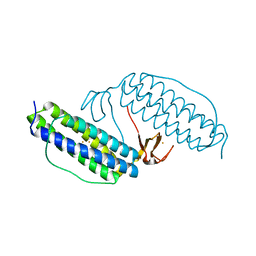
3U3S
 | | The S-SAD phased crystal structure of the ecto-domain of Death Receptor 6 (DR6) | | 分子名称: | Tumor necrosis factor receptor superfamily member 21 | | 著者 | Ru, H, Zhao, L.X, Ding, W, Jiao, L.Y, Shaw, N, Zhang, L.G, Hung, L.W, Matsugaki, N, Wakatsuki, S, Liu, Z.J. | | 登録日 | 2011-10-06 | | 公開日 | 2012-05-02 | | 最終更新日 | 2013-07-10 | | 実験手法 | X-RAY DIFFRACTION (2.7 Å) | | 主引用文献 | S-SAD phasing study of death receptor 6 and its solution conformation revealed by SAXS.
Acta Crystallogr.,Sect.D, 68, 2012
|
|
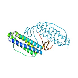
3U3P
 | | The S-SAD phased crystal structure of the ecto-domain of Death Receptor 6 (DR6) | | 分子名称: | Tumor necrosis factor receptor superfamily member 21 | | 著者 | Ru, H, Zhao, L.X, Ding, W, Jiao, L.Y, Shaw, N, Zhang, L.G, Hung, L.W, Matsugaki, N, Wakatsuki, S, Liu, Z.J. | | 登録日 | 2011-10-06 | | 公開日 | 2012-05-02 | | 最終更新日 | 2013-07-10 | | 実験手法 | X-RAY DIFFRACTION (2.09 Å) | | 主引用文献 | S-SAD phasing study of death receptor 6 and its solution conformation revealed by SAXS
Acta Crystallogr.,Sect.D, 68, 2012
|
|
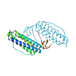
3U3V
 | | The S-SAD phased crystal structure of the ecto-domain of Death Receptor 6 (DR6) | | 分子名称: | Tumor necrosis factor receptor superfamily member 21 | | 著者 | Ru, H, Zhao, L.X, Ding, W, Jiao, L.Y, Shaw, N, Zhang, L.G, Hung, L.W, Matsugaki, N, Wakatsuki, S, Liu, Z.J. | | 登録日 | 2011-10-06 | | 公開日 | 2012-05-02 | | 最終更新日 | 2012-07-11 | | 実験手法 | X-RAY DIFFRACTION (2.96 Å) | | 主引用文献 | S-SAD phasing study of death receptor 6 and its solution conformation revealed by SAXS
Acta Crystallogr.,Sect.D, 68, 2012
|
|
1VK1
 | | Conserved hypothetical protein from Pyrococcus furiosus Pfu-392566-001 | | 分子名称: | Conserved hypothetical protein, DI(HYDROXYETHYL)ETHER, PHOSPHATE ION, ... | | 著者 | Shah, A, Liu, Z.J, Tempel, W, Chen, L, Lee, D, Yang, H, Chang, J, Zhao, M, Ng, J, Rose, J, Brereton, P.S, Izumi, M, Jenney Jr, F.E, Poole II, F.L, Shah, C, Sugar, F.J, Adams, M.W.W, Richardson, D.C, Richardson, J.S, Wang, B.C, Southeast Collaboratory for Structural Genomics (SECSG) | | 登録日 | 2004-04-13 | | 公開日 | 2004-08-10 | | 最終更新日 | 2023-12-27 | | 実験手法 | X-RAY DIFFRACTION (1.2 Å) | | 主引用文献 | (NZ)CH...O contacts assist crystallization of a ParB-like nuclease.
Bmc Struct.Biol., 7, 2007
|
|
3UT7
 | | Structural view of a non Pfam singleton and crystal packing analysis | | 分子名称: | Putative uncharacterized protein, SULFATE ION | | 著者 | Cheng, C, Shaw, N, Zhang, X, Zhang, M, Ding, W, Wang, B.C, Liu, Z.J. | | 登録日 | 2011-11-25 | | 公開日 | 2012-03-28 | | 最終更新日 | 2024-03-20 | | 実験手法 | X-RAY DIFFRACTION (3.01 Å) | | 主引用文献 | Structural view of a non pfam singleton and crystal packing analysis.
Plos One, 7, 2012
|
|
3UT8
 | | Structural view of a non Pfam singleton and crystal packing analysis | | 分子名称: | IODIDE ION, Putative uncharacterized protein | | 著者 | Cheng, C, Shaw, N, Zhang, X, Zhang, M, Ding, W, Wang, B.C, Liu, Z.J. | | 登録日 | 2011-11-25 | | 公開日 | 2012-03-28 | | 最終更新日 | 2024-03-20 | | 実験手法 | X-RAY DIFFRACTION (2.168 Å) | | 主引用文献 | Structural view of a non pfam singleton and crystal packing analysis.
Plos One, 7, 2012
|
|


