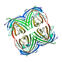
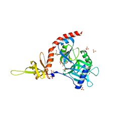
1KPC
 
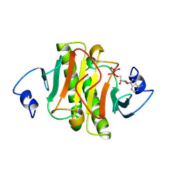
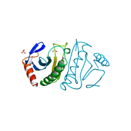
 | | PKCI-1-APO+ZINC | | 分子名称: | HUMAN PROTEIN KINASE C INTERACTING PROTEIN 1 (ZINC PROTEIN) | | 著者 | Lima, C.D, Klein, M.G, Weinstein, I.B, Hendrickson, W.A. | | 登録日 | 1996-01-06 | | 公開日 | 1996-07-11 | | 最終更新日 | 2024-02-14 | | 実験手法 | X-RAY DIFFRACTION (2.2 Å) | | 主引用文献 | Three-dimensional structure of human protein kinase C interacting protein 1, a member of the HIT family of proteins.
Proc.Natl.Acad.Sci.USA, 93, 1996
|
|
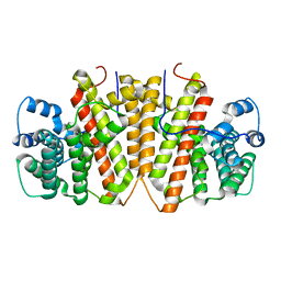
1RI6
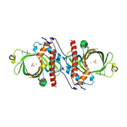
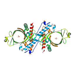
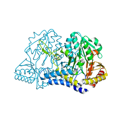
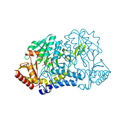
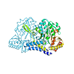
 | | Structure of a putative isomerase from E. coli | | 分子名称: | putative isomerase ybhE | | 著者 | Lima, C.D, Kniewel, R, Solorzano, V, Wu, J, Burley, S.K, New York SGX Research Center for Structural Genomics (NYSGXRC) | | 登録日 | 2003-11-16 | | 公開日 | 2003-11-25 | | 最終更新日 | 2024-02-14 | | 実験手法 | X-RAY DIFFRACTION (2 Å) | | 主引用文献 | Structure of a putative 7-bladed propeller isomerase
To be Published
|
|
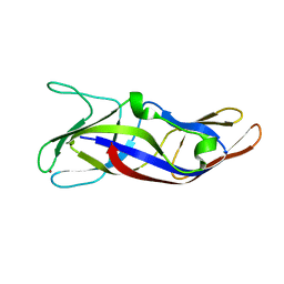
1KPE
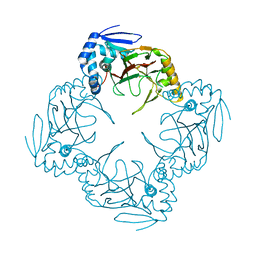
 | | PKCI-TRANSITION STATE ANALOG | | 分子名称: | ADENOSINE-5'-DITUNGSTATE, PROTEIN KINASE C INTERACTING PROTEIN | | 著者 | Lima, C.D, Klein, M.G, Hendrickson, W.A. | | 登録日 | 1997-09-25 | | 公開日 | 1998-03-25 | | 最終更新日 | 2024-02-14 | | 実験手法 | X-RAY DIFFRACTION (1.8 Å) | | 主引用文献 | Structure-based analysis of catalysis and substrate definition in the HIT protein family.
Science, 278, 1997
|
|
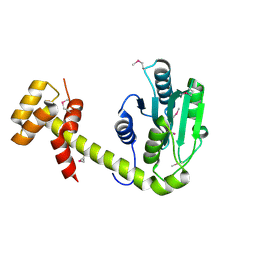
1D8H
 | | X-RAY CRYSTAL STRUCTURE OF YEAST RNA TRIPHOSPHATASE IN COMPLEX WITH SULFATE AND MANGANESE IONS. | | 分子名称: | MANGANESE (II) ION, SULFATE ION, mRNA TRIPHOSPHATASE CET1 | | 著者 | Lima, C.D, Wang, L.K, Shuman, S. | | 登録日 | 1999-10-24 | | 公開日 | 1999-11-29 | | 最終更新日 | 2024-02-07 | | 実験手法 | X-RAY DIFFRACTION (2 Å) | | 主引用文献 | Structure and mechanism of yeast RNA triphosphatase: an essential component of the mRNA capping apparatus.
Cell(Cambridge,Mass.), 99, 1999
|
|
1D8I
 | |
1JR7
 | |
1JF9
 | |
1KMK
 | |
1KMJ
 | |
2FIT
 | | FHIT (FRAGILE HISTIDINE TRIAD PROTEIN) | | 分子名称: | FRAGILE HISTIDINE PROTEIN, SULFATE ION, beta-D-fructofuranose | | 著者 | Lima, C.D, D'Amico, K.L, Naday, I, Rosenbaum, G, Westbrook, E.M, Hendrickson, W.A. | | 登録日 | 1997-05-17 | | 公開日 | 1997-11-19 | | 最終更新日 | 2020-07-29 | | 実験手法 | X-RAY DIFFRACTION (1.9 Å) | | 主引用文献 | MAD analysis of FHIT, a putative human tumor suppressor from the HIT protein family.
Structure, 5, 1997
|
|
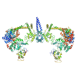
8D0B
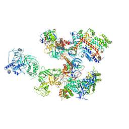 | | Human CST-DNA polymerase alpha/primase preinitiation complex bound to 4xTEL-foldback template | | 分子名称: | CST complex subunit CTC1, CST complex subunit STN1, CST complex subunit TEN1, ... | | 著者 | He, Q, Lin, X, Chavez, B.L, Agrawal, S, Lusk, B.L, Lim, C. | | 登録日 | 2022-05-26 | | 公開日 | 2022-06-22 | | 最終更新日 | 2024-02-14 | | 実験手法 | ELECTRON MICROSCOPY (3.43 Å) | | 主引用文献 | Structures of the human CST-Pol alpha-primase complex bound to telomere templates.
Nature, 608, 2022
|
|
8D9D
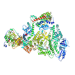 | | Human DNA polymerase-alpha/primase elongation complex II bound to primer/template | | 分子名称: | 2'-DEOXYADENOSINE 5'-TRIPHOSPHATE, DNA (5'-D(*AP*TP*GP*GP*TP*CP*GP*TP*GP*CP*CP*GP*CP*CP*AP*AP*TP*AP*A)-3'), DNA polymerase alpha catalytic subunit, ... | | 著者 | He, Q, Baranovskiy, A, Lim, C, Tahirov, T. | | 登録日 | 2022-06-09 | | 公開日 | 2023-04-19 | | 最終更新日 | 2024-06-12 | | 実験手法 | ELECTRON MICROSCOPY (3.59 Å) | | 主引用文献 | Structures of human primosome elongation complexes.
Nat.Struct.Mol.Biol., 30, 2023
|
|
8D96
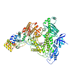 | | Human DNA polymerase alpha/primase elongation complex I bound to primer/template | | 分子名称: | 2'-DEOXYADENOSINE 5'-TRIPHOSPHATE, DNA (5'-D(*AP*TP*AP*AP*TP*GP*GP*TP*CP*GP*TP*GP*CP*CP*GP*CP*CP*AP*AP*TP*AP*A)-3'), DNA polymerase alpha catalytic subunit, ... | | 著者 | He, Q, Baranovskiy, A, Lim, C, Tahirov, T. | | 登録日 | 2022-06-09 | | 公開日 | 2023-04-19 | | 最終更新日 | 2024-06-12 | | 実験手法 | ELECTRON MICROSCOPY (3.35 Å) | | 主引用文献 | Structures of human primosome elongation complexes.
Nat.Struct.Mol.Biol., 30, 2023
|
|
8D0K
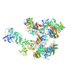 | | Human CST-DNA polymerase alpha/primase preinitiation complex bound to 4xTEL-foldback template - PRIM2C advanced PIC | | 分子名称: | CST complex subunit CTC1, CST complex subunit STN1, CST complex subunit TEN1, ... | | 著者 | He, Q, Lin, X, Chavez, B.L, Agrawal, S, Lusk, B.L, Lim, C. | | 登録日 | 2022-05-26 | | 公開日 | 2022-06-22 | | 最終更新日 | 2024-02-14 | | 実験手法 | ELECTRON MICROSCOPY (4.27 Å) | | 主引用文献 | Structures of the human CST-Pol alpha-primase complex bound to telomere templates.
Nature, 608, 2022
|
|
6CIU
 | | Structure of a Thr-rich interface in an Azami Green tetramer | | 分子名称: | Azami-Green | | 著者 | Oi, C, Lim, C.S, Knecht, K.M, Xiong, Y, Regan, L. | | 登録日 | 2018-02-25 | | 公開日 | 2018-09-26 | | 最終更新日 | 2023-11-15 | | 実験手法 | X-RAY DIFFRACTION (1.7 Å) | | 主引用文献 | A threonine zipper that mediates protein-protein interactions: Structure and prediction.
Protein Sci., 27, 2018
|
|
8GY0
 | | Agrocybe pediades linalool sunthase (Ap.LS) | | 分子名称: | Terpene synthase | | 著者 | Rehka, T, Sharma, D, Lin, F, Lim, C, Choong, Y.K, Chacko, J, Zhang, C. | | 登録日 | 2022-09-21 | | 公開日 | 2023-04-12 | | 最終更新日 | 2024-05-08 | | 実験手法 | X-RAY DIFFRACTION (1.988 Å) | | 主引用文献 | Structural Understanding of Fungal Terpene Synthases for the Formation of Linear or Cyclic Terpene Products.
Acs Catalysis, 13, 2023
|
|
5ZLQ
 | | Crystal Structure of C1orf123 | | 分子名称: | UPF0587 protein C1orf123, ZINC ION | | 著者 | Furukawa, Y, Lim, C.T, Tosha, T. | | 登録日 | 2018-03-29 | | 公開日 | 2018-10-10 | | 最終更新日 | 2024-03-27 | | 実験手法 | X-RAY DIFFRACTION (2 Å) | | 主引用文献 | Identification of a novel zinc-binding protein, C1orf123, as an interactor with a heavy metal-associated domain
PLoS ONE, 13, 2018
|
|
6UPS
 | |
2XA8
 | | Crystal structure of the Fab domain of omalizumab at 2.41A | | 分子名称: | OMALIZUMAB HEAVY CHAIN, OMALIZUMAB LIGHT CHAIN | | 著者 | Huang, C.H, Hung, F.H.A, Lim, C, Chang, T.W, Ma, C. | | 登録日 | 2010-03-30 | | 公開日 | 2011-05-11 | | 最終更新日 | 2023-12-20 | | 実験手法 | X-RAY DIFFRACTION (2.42 Å) | | 主引用文献 | Structural and Physical Basis for Anti-IgE Therapy.
Sci Rep, 5, 2015
|
|
4LKX
 | | Humanized antibody 4B12 Fab complexed with a CemX segment | | 分子名称: | CemX segment, Fab fragment heavy chain, Fab fragment light chain | | 著者 | Chu, H.M, Wright, J, Chan, Y.H, Lin, C.J, Chang, T.W, Lim, C. | | 登録日 | 2013-07-09 | | 公開日 | 2014-01-29 | | 最終更新日 | 2023-11-08 | | 実験手法 | X-RAY DIFFRACTION (1.92 Å) | | 主引用文献 | Two potential therapeutic antibodies bind to a peptide segment of membrane-bound IgE in different conformations.
Nat Commun, 5, 2014
|
|
1W4Z
 | | Structure of actinorhodin polyketide (actIII) Reductase | | 分子名称: | FORMIC ACID, KETOACYL REDUCTASE, NADP NICOTINAMIDE-ADENINE-DINUCLEOTIDE PHOSPHATE | | 著者 | Hadfield, A.T, Limpkin, C, Teartasin, W, Simpson, T.J, Crosby, J, Crump, M.P. | | 登録日 | 2004-08-03 | | 公開日 | 2004-12-10 | | 最終更新日 | 2023-12-13 | | 実験手法 | X-RAY DIFFRACTION (2.5 Å) | | 主引用文献 | The Crystal Structure of the Actiii Actinorhodin Polyketide Reductase; Proposed Mechanism for Acp and Polyketide Binding
Structure, 12, 2004
|
|
2GRO
 | | Crystal Structure of human RanGAP1-Ubc9-N85Q | | 分子名称: | Ran GTPase-activating protein 1, Ubiquitin-conjugating enzyme E2 I | | 著者 | Yunus, A.A, Lima, C.D. | | 登録日 | 2006-04-24 | | 公開日 | 2006-05-30 | | 最終更新日 | 2024-02-14 | | 実験手法 | X-RAY DIFFRACTION (1.7 Å) | | 主引用文献 | Lysine activation and functional analysis of E2-mediated conjugation in the SUMO pathway.
Nat.Struct.Mol.Biol., 13, 2006
|
|
2GRN
 | | Crystal Structure of human RanGAP1-Ubc9 | | 分子名称: | Ran GTPase-activating protein 1, Ubiquitin-conjugating enzyme E2 I | | 著者 | Yunus, A.A, Lima, C.D. | | 登録日 | 2006-04-24 | | 公開日 | 2006-05-30 | | 最終更新日 | 2024-02-14 | | 実験手法 | X-RAY DIFFRACTION (1.8 Å) | | 主引用文献 | Lysine activation and functional analysis of E2-mediated conjugation in the SUMO pathway.
Nat.Struct.Mol.Biol., 13, 2006
|
|
4PZ7
 | | PCE1 guanylyltransferase | | 分子名称: | GLYCEROL, SULFATE ION, mRNA-capping enzyme subunit alpha | | 著者 | Doamekpor, S.K, Lima, C.D. | | 登録日 | 2014-03-28 | | 公開日 | 2014-06-25 | | 最終更新日 | 2023-09-20 | | 実験手法 | X-RAY DIFFRACTION (2.109 Å) | | 主引用文献 | How an mRNA capping enzyme reads distinct RNA polymerase II and Spt5 CTD phosphorylation codes.
Genes Dev., 28, 2014
|
|
7S7B
 | |


