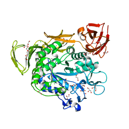
3NMV
 
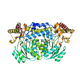
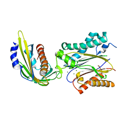 | | Crystal structure of pyrabactin-bound abscisic acid receptor PYL2 mutant A93F in complex with type 2C protein phosphatase ABI2 | | 分子名称: | 4-bromo-N-(pyridin-2-ylmethyl)naphthalene-1-sulfonamide, Abscisic acid receptor PYL2, MAGNESIUM ION, ... | | 著者 | Zhou, X.E, Melcher, K, Ng, L.-M, Soon, F.-F, Xu, Y, Suino-Powell, K.M, Kovach, A, Li, J, Yong, E.-L, Xu, H.E. | | 登録日 | 2010-06-22 | | 公開日 | 2010-08-25 | | 最終更新日 | 2023-09-06 | | 実験手法 | X-RAY DIFFRACTION (2.1 Å) | | 主引用文献 | Identification and mechanism of ABA receptor antagonism.
Nat.Struct.Mol.Biol., 17, 2010
|
|
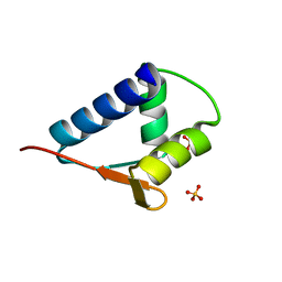
2JXO
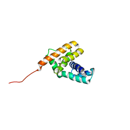
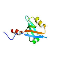 | | Structure of the second PDZ domain of NHERF-1 | | 分子名称: | Ezrin-radixin-moesin-binding phosphoprotein 50 | | 著者 | Cheng, H, Li, J, Dai, Z, Bu, Z, Roder, H. | | 登録日 | 2007-11-27 | | 公開日 | 2008-12-09 | | 最終更新日 | 2024-05-29 | | 実験手法 | SOLUTION NMR | | 主引用文献 | Autoinhibitory Interactions between the PDZ2 and C-terminal Domains in the Scaffolding Protein NHERF1
Structure, 17, 2009
|
|
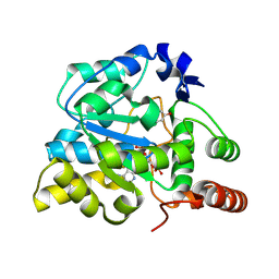
3K40
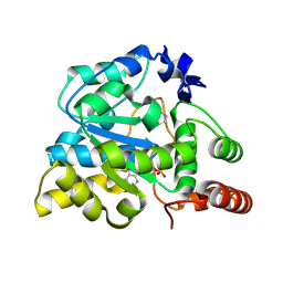
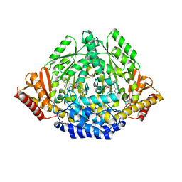 | | Crystal structure of Drosophila 3,4-dihydroxyphenylalanine decarboxylase | | 分子名称: | Aromatic-L-amino-acid decarboxylase, GLYCEROL | | 著者 | Han, Q, Ding, H, Robinson, H, Christensen, B.M, Li, J. | | 登録日 | 2009-10-05 | | 公開日 | 2010-02-02 | | 最終更新日 | 2023-11-22 | | 実験手法 | X-RAY DIFFRACTION (1.75 Å) | | 主引用文献 | Crystal structure and substrate specificity of Drosophila 3,4-dihydroxyphenylalanine decarboxylase
Plos One, 5, 2010
|
|
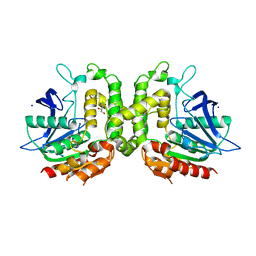
6JI3
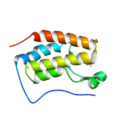 | | BRD4-BD1 bound with ligand 103 | | 分子名称: | (3~{R})-4-cyclopropyl-1,3-dimethyl-6-(1~{H}-pyrrol-2-yl)-3~{H}-quinoxalin-2-one, Bromodomain-containing protein 4 | | 著者 | Cao, D.Y, Li, Y.L, Du, Z.Y, Li, J, Xiong, B. | | 登録日 | 2019-02-20 | | 公開日 | 2020-02-26 | | 最終更新日 | 2023-11-22 | | 実験手法 | X-RAY DIFFRACTION (2.2 Å) | | 主引用文献 | brd4-bd1 bound with ligand 103
To Be Published
|
|
2HUF
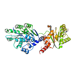
 | | Crystal structure of Aedes aegypti alanine glyoxylate aminotransferase | | 分子名称: | 1-BUTANOL, Alanine glyoxylate aminotransferase | | 著者 | Han, Q, Robinson, H, Gao, Y.G, Vogelaar, N, Wilson, S.R, Rizzi, M, Li, J. | | 登録日 | 2006-07-26 | | 公開日 | 2006-09-26 | | 最終更新日 | 2023-11-15 | | 実験手法 | X-RAY DIFFRACTION (1.75 Å) | | 主引用文献 | Crystal Structures of Aedes aegypti Alanine Glyoxylate Aminotransferase.
J.Biol.Chem., 281, 2006
|
|
2JR0
 | | Solution structure of NusB from Aquifex Aeolicus | | 分子名称: | N utilization substance protein B homolog | | 著者 | Das, R, Loss, S, Li, J, Tarasov, S, Wingfield, P, Waugh, D.S, Byrd, R.A, Altieri, A.S. | | 登録日 | 2007-06-18 | | 公開日 | 2008-02-19 | | 最終更新日 | 2024-05-08 | | 実験手法 | SOLUTION NMR | | 主引用文献 | Structural biophysics of the NusB:NusE antitermination complex.
J.Mol.Biol., 376, 2008
|
|
4GF6
 | | crystal structure of GFP with cuprum bound at the Incorporated metal Chelating Amino Acid PYZ151 | | 分子名称: | CALCIUM ION, COPPER (II) ION, green fluorescent protein | | 著者 | Dong, J, Liu, X, Li, J, Wang, J, Gong, W. | | 登録日 | 2012-08-03 | | 公開日 | 2012-08-29 | | 最終更新日 | 2023-11-15 | | 実験手法 | X-RAY DIFFRACTION (1.1 Å) | | 主引用文献 | Genetic incorporation of a metal-chelating amino Acid as a probe for protein electron transfer.
Angew.Chem.Int.Ed.Engl., 51, 2012
|
|
4G5R
 | | Structure of LGN GL4/Galphai3 complex | | 分子名称: | CITRIC ACID, G-protein-signaling modulator 2, GUANOSINE-5'-DIPHOSPHATE, ... | | 著者 | Jia, M, Li, J, Zhu, J, Wen, W, Zhang, M, Wang, W. | | 登録日 | 2012-07-18 | | 公開日 | 2012-09-05 | | 最終更新日 | 2024-03-20 | | 実験手法 | X-RAY DIFFRACTION (3.481 Å) | | 主引用文献 | Crystal Structures of the scaffolding protein LGN reveal the general mechanism by which GoLoco binding motifs inhibit the release of GDP from Galphai subunits in G-coupled heterotrimeric proteins
To be Published
|
|
4G5O
 | | Structure of LGN GL4/Galphai3(Q147L) complex | | 分子名称: | CITRIC ACID, G-protein-signaling modulator 2, GUANOSINE-5'-DIPHOSPHATE, ... | | 著者 | Jia, M, Li, J, Zhu, J, Wen, W, Zhang, M, Wang, W. | | 登録日 | 2012-07-18 | | 公開日 | 2012-09-05 | | 最終更新日 | 2024-03-20 | | 実験手法 | X-RAY DIFFRACTION (2.9 Å) | | 主引用文献 | Crystal Structures of the scaffolding protein LGN reveal the general mechanism by which GoLoco binding motifs inhibit the release of GDP from Galphai subunits in G-coupled heterotrimeric proteins
To be Published
|
|
4GES
 | | crystal structure of GFP-TYR151PYZ with an unnatural amino acid incorporation | | 分子名称: | Green fluorescent protein | | 著者 | Dong, J, Liu, X, Li, J, Wang, J, Gong, W. | | 登録日 | 2012-08-02 | | 公開日 | 2012-08-29 | | 最終更新日 | 2023-11-15 | | 実験手法 | X-RAY DIFFRACTION (1.23 Å) | | 主引用文献 | Genetic incorporation of a metal-chelating amino Acid as a probe for protein electron transfer.
Angew.Chem.Int.Ed.Engl., 51, 2012
|
|
4G5Q
 | | Structure of LGN GL4/Galphai1 complex | | 分子名称: | CITRIC ACID, G-protein-signaling modulator 2, GUANOSINE-5'-DIPHOSPHATE, ... | | 著者 | Jia, M, Li, J, Zhu, J, Wen, W, Zhang, M, Wang, W. | | 登録日 | 2012-07-18 | | 公開日 | 2012-09-05 | | 最終更新日 | 2024-03-20 | | 実験手法 | X-RAY DIFFRACTION (2.9 Å) | | 主引用文献 | Crystal Structures of the scaffolding protein LGN reveal the general mechanism by which GoLoco binding motifs inhibit the release of GDP from Galphai subunits in G-coupled heterotrimeric proteins
To be Published
|
|
6KOE
 | | X-ray Structure of the proton-pumping cytochrome aa3-600 menaquinol oxidase from Bacillus subtilis | | 分子名称: | 2-HEPTYL-4-HYDROXY QUINOLINE N-OXIDE, AA3-600 quinol oxidase subunit I, AA3-600 quinol oxidase subunit IIII, ... | | 著者 | Xu, J, Ding, Z, Liu, B, Li, J, Gennis, R.B, Zhu, J. | | 登録日 | 2019-08-09 | | 公開日 | 2020-01-15 | | 最終更新日 | 2023-11-22 | | 実験手法 | X-RAY DIFFRACTION (3.75 Å) | | 主引用文献 | Structure of the cytochromeaa3-600 heme-copper menaquinol oxidase bound to inhibitor HQNO shows TM0 is part of the quinol binding site.
Proc.Natl.Acad.Sci.USA, 117, 2020
|
|
6KOC
 | | X-ray Structure of the proton-pumping cytochrome aa3-600 menaquinol oxidase from Bacillus subtilis complexed with 3-iodo-N-oxo-2-heptyl-4-hydroxyquinoline | | 分子名称: | 2-heptyl-3-iodanyl-1-oxidanyl-quinolin-4-one, AA3-600 quinol oxidase subunit I, AA3-600 quinol oxidase subunit IIII, ... | | 著者 | Xu, J, Ding, Z, Liu, B, Li, J, Gennis, R.B, Zhu, J. | | 登録日 | 2019-08-09 | | 公開日 | 2020-01-15 | | 最終更新日 | 2023-11-22 | | 実験手法 | X-RAY DIFFRACTION (3.8 Å) | | 主引用文献 | Structure of the cytochromeaa3-600 heme-copper menaquinol oxidase bound to inhibitor HQNO shows TM0 is part of the quinol binding site.
Proc.Natl.Acad.Sci.USA, 117, 2020
|
|
8XGZ
 | |
8XJP
 | | Crystal structure of a sulfotransferase S4 in complex with PAP and 2-Phe-br | | 分子名称: | 2-BROMOPHENOL, ADENOSINE-3'-5'-DIPHOSPHATE, Sulfotransferase | | 著者 | Gao, J, Wang, H, Chen, Y.Y, Yang, S.Y, Yin, L, Liu, W.D, Li, J.S. | | 登録日 | 2023-12-22 | | 公開日 | 2024-12-25 | | 実験手法 | X-RAY DIFFRACTION (1.7 Å) | | 主引用文献 | Crystal structure of a sulfotransferase S4 in complex with PAP
To Be Published
|
|
8XJQ
 | | Crystal structure of a sulfotransferase S4 in complex with PAP and PNPS | | 分子名称: | 3'-PHOSPHATE-ADENOSINE-5'-PHOSPHATE SULFATE, 4-nitrophenyl sulfate, P-NITROPHENOL, ... | | 著者 | Gao, J, Wang, H, Chen, Y.Y, Yang, S.Y, Yin, L, Liu, W.D, Li, J.S. | | 登録日 | 2023-12-22 | | 公開日 | 2024-12-25 | | 実験手法 | X-RAY DIFFRACTION (1.78 Å) | | 主引用文献 | Crystal structure of a sulfotransferase S4 in complex with PAP and PNPS
To Be Published
|
|
4LJY
 | | Crystal structure of RNA splicing effector Prp5 in complex with ADP | | 分子名称: | (4R)-2-METHYLPENTANE-2,4-DIOL, ADENOSINE-5'-DIPHOSPHATE, MAGNESIUM ION, ... | | 著者 | Zhang, Z.-M, Li, J, Yang, F, Xu, Y, Zhou, J. | | 登録日 | 2013-07-05 | | 公開日 | 2013-12-11 | | 最終更新日 | 2024-11-06 | | 実験手法 | X-RAY DIFFRACTION (1.95 Å) | | 主引用文献 | Crystal structure of Prp5p reveals interdomain interactions that impact spliceosome assembly.
Cell Rep, 5, 2013
|
|
4JFG
 | | Crystal structure of sfGFP-66-HqAla | | 分子名称: | CESIUM ION, Green fluorescent protein, quinolin-8-ol | | 著者 | Wang, J, Liu, X, Li, J, Zhang, W, Hu, M, Zhou, J. | | 登録日 | 2013-02-28 | | 公開日 | 2013-10-02 | | 最終更新日 | 2023-11-15 | | 実験手法 | X-RAY DIFFRACTION (3.001 Å) | | 主引用文献 | Significant expansion of the fluorescent protein chromophore through the genetic incorporation of a metal-chelating unnatural amino acid.
Angew.Chem.Int.Ed.Engl., 52, 2013
|
|
4JCL
 | | Crystal structure of Alpha-CGT from Paenibacillus macerans at 1.7 Angstrom resolution | | 分子名称: | 1,2-ETHANEDIOL, CALCIUM ION, CHLORIDE ION, ... | | 著者 | Wu, L, Zhou, J, Wu, J, Li, J, Chen, J. | | 登録日 | 2013-02-22 | | 公開日 | 2014-02-26 | | 最終更新日 | 2023-11-08 | | 実験手法 | X-RAY DIFFRACTION (1.7 Å) | | 主引用文献 | Crystal Structure of Alpha-Cgt from Paenibacillus Macerans at 1.7 Angstrom Resolution
To be Published
|
|
6KOB
 | | X-ray Structure of the proton-pumping cytochrome aa3-600 menaquinol oxidase from Bacillus subtilis | | 分子名称: | AA3-600 quinol oxidase subunit I, AA3-600 quinol oxidase subunit IIII, AA3-600 quinol oxidase subunit IV,Quinol oxidase subunit 4, ... | | 著者 | Xu, J, Ding, Z, Liu, B, Li, J, Gennis, R.B, Zhu, J. | | 登録日 | 2019-08-09 | | 公開日 | 2020-01-15 | | 最終更新日 | 2024-03-27 | | 実験手法 | X-RAY DIFFRACTION (3.6 Å) | | 主引用文献 | Structure of the cytochromeaa3-600 heme-copper menaquinol oxidase bound to inhibitor HQNO shows TM0 is part of the quinol binding site.
Proc.Natl.Acad.Sci.USA, 117, 2020
|
|
8XU3
 | | Crystal structure of a sulfotransferase S4 from in complex with PAP and PNP | | 分子名称: | ADENOSINE-3'-5'-DIPHOSPHATE, P-NITROPHENOL, Sulfotransferase | | 著者 | Gao, J, Wang, H, Chen, Y.Y, Yang, S.Y, Yin, L, Liu, W.D, Li, J.S. | | 登録日 | 2024-01-12 | | 公開日 | 2025-01-15 | | 実験手法 | X-RAY DIFFRACTION (1.82 Å) | | 主引用文献 | Crystal structure of a sulfotransferase S4 from in complex with PAP and PNP
To Be Published
|
|
6L0O
 | | WH domain of human MCM8 | | 分子名称: | DNA helicase MCM8, SULFATE ION | | 著者 | Liu, Y, Li, J, Liu, L, Zeng, H. | | 登録日 | 2019-09-26 | | 公開日 | 2020-04-29 | | 最終更新日 | 2024-03-27 | | 実験手法 | X-RAY DIFFRACTION (1.21 Å) | | 主引用文献 | Crystal structure of the winged-helix domain of MCM8.
Biochem.Biophys.Res.Commun., 526, 2020
|
|
5W8O
 | | Homoserine transacetylase MetX from Mycobacterium hassiacum | | 分子名称: | CALCIUM ION, GLYCEROL, Homoserine O-acetyltransferase, ... | | 著者 | Reed, R.W, Rodriguez, E.S, Li, J, Korotkov, K.V. | | 登録日 | 2017-06-22 | | 公開日 | 2017-07-12 | | 最終更新日 | 2023-10-04 | | 実験手法 | X-RAY DIFFRACTION (1.47 Å) | | 主引用文献 | Structural analysis of mycobacterial homoserine transacetylases central to methionine biosynthesis reveals druggable active site.
Sci Rep, 9, 2019
|
|
4LK2
 | | Crystal structure of RNA splicing effector Prp5 | | 分子名称: | NICKEL (II) ION, Pre-mRNA-processing ATP-dependent RNA helicase PRP5 | | 著者 | Zhang, Z.-M, Li, J, Yang, F, Xu, Y, Zhou, J. | | 登録日 | 2013-07-05 | | 公開日 | 2013-12-11 | | 最終更新日 | 2024-03-20 | | 実験手法 | X-RAY DIFFRACTION (2.12 Å) | | 主引用文献 | Crystal structure of Prp5p reveals interdomain interactions that impact spliceosome assembly.
Cell Rep, 5, 2013
|
|
4HY6
 | | Crystal Structure of the human Hsp90-alpha N-domain bound to the hsp90 inhibitor FJ1 | | 分子名称: | 6,6-dimethyl-3-(trifluoromethyl)-1,5,6,7-tetrahydro-4H-indazol-4-one, Heat shock protein HSP 90-alpha | | 著者 | Yang, M, Li, J, He, J.H. | | 登録日 | 2012-11-13 | | 公開日 | 2013-05-15 | | 最終更新日 | 2023-11-08 | | 実験手法 | X-RAY DIFFRACTION (1.649 Å) | | 主引用文献 | Crystal Structure of the human Hsp90-alpha N-domain bound to the hsp90 inhibitor FJ1
to be published
|
|


