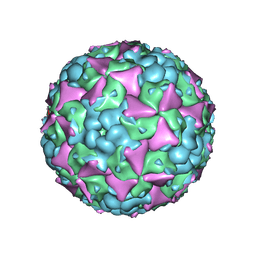
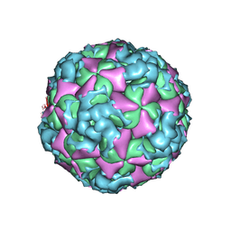
6LHO
 
 | | The cryo-EM structure of coxsackievirus A16 empty particle in complex with Fab 18A7 | | 分子名称: | VP1 protein, VP2 protein, VP3 protein | | 著者 | He, M.Z, Xu, L.F, Zheng, Q.B, Zhu, R, Yin, Z.C, Cheng, T, Li, S.W. | | 登録日 | 2019-12-09 | | 公開日 | 2020-02-05 | | 最終更新日 | 2022-03-23 | | 実験手法 | ELECTRON MICROSCOPY (3.13 Å) | | 主引用文献 | Identification of Antibodies with Non-overlapping Neutralization Sites that Target Coxsackievirus A16.
Cell Host Microbe, 27, 2020
|
|
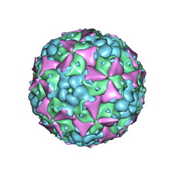
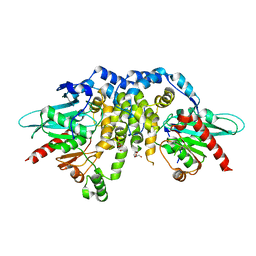
6LHK
 | | The cryo-EM structure of coxsackievirus A16 mature virion in complex with Fab 18A7 | | 分子名称: | SPHINGOSINE, VP1 protein, VP2 protein, ... | | 著者 | He, M.Z, Xu, L.F, Zheng, Q.B, Zhu, R, Yin, Z.C, Cheng, T, Li, S.W. | | 登録日 | 2019-12-09 | | 公開日 | 2020-02-05 | | 最終更新日 | 2022-03-23 | | 実験手法 | ELECTRON MICROSCOPY (2.65 Å) | | 主引用文献 | Identification of Antibodies with Non-overlapping Neutralization Sites that Target Coxsackievirus A16.
Cell Host Microbe, 27, 2020
|
|
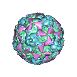
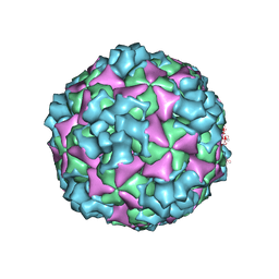
6LHA
 | | The cryo-EM structure of coxsackievirus A16 mature virion | | 分子名称: | SPHINGOSINE, VP1 protein, VP2 protein, ... | | 著者 | He, M.Z, Xu, L.F, Zheng, Q.B, Zhu, R, Yin, Z.C, Cheng, T, Li, S.W. | | 登録日 | 2019-12-07 | | 公開日 | 2020-02-05 | | 最終更新日 | 2022-03-23 | | 実験手法 | ELECTRON MICROSCOPY (3.56 Å) | | 主引用文献 | Identification of Antibodies with Non-overlapping Neutralization Sites that Target Coxsackievirus A16.
Cell Host Microbe, 27, 2020
|
|
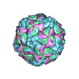
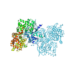
6LHQ
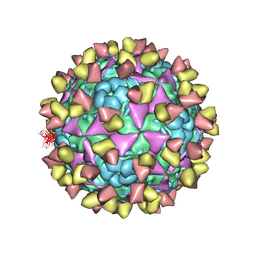 | | The cryo-EM structure of coxsackievirus A16 mature virion in complex with Fab NA9D7 | | 分子名称: | SPHINGOSINE, VP1 protein, VP2 protein, ... | | 著者 | He, M.Z, Xu, L.F, Zheng, Q.B, Zhu, R, Yin, Z.C, Cheng, T, Li, S.W. | | 登録日 | 2019-12-09 | | 公開日 | 2020-02-05 | | 最終更新日 | 2022-03-23 | | 実験手法 | ELECTRON MICROSCOPY (3.06 Å) | | 主引用文献 | Identification of Antibodies with Non-overlapping Neutralization Sites that Target Coxsackievirus A16.
Cell Host Microbe, 27, 2020
|
|
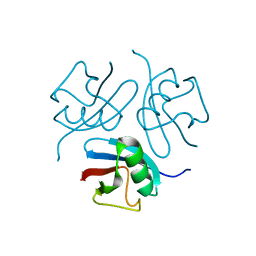
6LHT
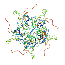 | | Localized reconstruction of coxsackievirus A16 mature virion in complex with Fab 18A7 | | 分子名称: | SPHINGOSINE, VP1 protein, heavy chain variable region of Fab 18A7, ... | | 著者 | He, M.Z, Xu, L.F, Zheng, Q.B, Zhu, R, Yin, Z.C, Cheng, T, Li, S.W. | | 登録日 | 2019-12-10 | | 公開日 | 2020-02-05 | | 最終更新日 | 2020-02-26 | | 実験手法 | ELECTRON MICROSCOPY (3.67 Å) | | 主引用文献 | Identification of Antibodies with Non-overlapping Neutralization Sites that Target Coxsackievirus A16.
Cell Host Microbe, 27, 2020
|
|
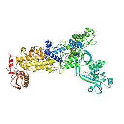
5U0N
 | | Crystal structure of a methyltransferase in complex with the substrate involved in the biosynthesis of gentamicin | | 分子名称: | (1R,2S,3S,4R,6S)-4,6-diamino-3-[(3-amino-3-deoxy-alpha-D-xylopyranosyl)oxy]-2-hydroxycyclohexyl 2-amino-2-deoxy-alpha-D-glucopyranoside, MAGNESIUM ION, Putative gentamicin methyltransferase, ... | | 著者 | Bury, P, Huang, F, Li, S, Sun, Y, Leadlay, P, Dias, M.V.B. | | 登録日 | 2016-11-25 | | 公開日 | 2017-11-01 | | 最終更新日 | 2023-10-04 | | 実験手法 | X-RAY DIFFRACTION (2.115 Å) | | 主引用文献 | Structural Basis of the Selectivity of GenN, an Aminoglycoside N-Methyltransferase Involved in Gentamicin Biosynthesis.
ACS Chem. Biol., 12, 2017
|
|
7CJQ
 | | Structure of DLA-88*001:04 | | 分子名称: | ARG-THR-ILE-SER-TYR-THR-TYR-PRO-PHE, Beta-2-microglobulin, MHC class I DLA-88 | | 著者 | Sun, Y.J, Ma, L.Z, Li, S. | | 登録日 | 2020-07-12 | | 公開日 | 2020-08-26 | | 最終更新日 | 2023-11-29 | | 実験手法 | X-RAY DIFFRACTION (2.7 Å) | | 主引用文献 | Structure of canine MHC class I at 2.7 Angstroms resolution
To Be Published
|
|
3B2V
 | | Crystal structure of the extracellular region of the epidermal growth factor receptor in complex with the Fab fragment of IMC-11F8 | | 分子名称: | 2-acetamido-2-deoxy-beta-D-glucopyranose, 2-acetamido-2-deoxy-beta-D-glucopyranose-(1-4)-2-acetamido-2-deoxy-beta-D-glucopyranose, Epidermal growth factor receptor, ... | | 著者 | Ferguson, K.M, Li, S, Kussie, P. | | 登録日 | 2007-10-19 | | 公開日 | 2008-02-19 | | 最終更新日 | 2023-08-30 | | 実験手法 | X-RAY DIFFRACTION (3.3 Å) | | 主引用文献 | Structural basis for EGF receptor inhibition by the therapeutic antibody IMC-11F8.
Structure, 16, 2008
|
|
5WPR
 | | Crystal structure HpiC1 in C2 space group | | 分子名称: | 12-epi-hapalindole C/U synthase, 2-AMINO-2-HYDROXYMETHYL-PROPANE-1,3-DIOL, CALCIUM ION, ... | | 著者 | Newmister, S.A, Li, S, Garcia-Borras, M, Sanders, J.N, Yang, S, Lowell, A.N, Yu, F, Smith, J.L, Williams, R.M, Houk, K.N, Sherman, D.H. | | 登録日 | 2017-08-07 | | 公開日 | 2018-03-07 | | 最終更新日 | 2023-10-04 | | 実験手法 | X-RAY DIFFRACTION (1.49 Å) | | 主引用文献 | Structural basis of the Cope rearrangement and cyclization in hapalindole biogenesis.
Nat. Chem. Biol., 14, 2018
|
|
4HPH
 | | The crystal structure of isomaltulose synthase mutant E295Q from Erwinia rhapontici NX5 in complex with its natural substrate sucrose | | 分子名称: | CALCIUM ION, GLYCEROL, Sucrose isomerase, ... | | 著者 | Xu, Z, Li, S, Xu, H, Zhou, J. | | 登録日 | 2012-10-23 | | 公開日 | 2013-10-16 | | 最終更新日 | 2023-11-08 | | 実験手法 | X-RAY DIFFRACTION (1.7 Å) | | 主引用文献 | The Structural Basis of Erwinia rhapontici Isomaltulose Synthase
Plos One, 8, 2013
|
|
4HOZ
 | | The crystal structure of isomaltulose synthase mutant D241A from Erwinia rhapontici NX5 in complex with D-glucose | | 分子名称: | CALCIUM ION, GLYCEROL, Sucrose isomerase, ... | | 著者 | Xu, Z, Li, S, Xu, H, Zhou, J. | | 登録日 | 2012-10-23 | | 公開日 | 2013-10-16 | | 最終更新日 | 2023-11-08 | | 実験手法 | X-RAY DIFFRACTION (2 Å) | | 主引用文献 | The Structural Basis of Erwinia rhapontici Isomaltulose Synthase
Plos One, 8, 2013
|
|
1WM2
 | |
2KHT
 | | NMR Structure of human alpha defensin HNP-1 | | 分子名称: | Neutrophil defensin 1 | | 著者 | Zhang, Y, Li, S, Doherty, T.F, Lubkowski, J, Lu, W, Li, J, Barinka, C, Hong, M. | | 登録日 | 2009-04-11 | | 公開日 | 2010-02-09 | | 最終更新日 | 2024-05-01 | | 実験手法 | SOLID-STATE NMR | | 主引用文献 | Resonance assignment and three-dimensional structure determination of a human alpha-defensin, HNP-1, by solid-state NMR.
J.Mol.Biol., 397, 2010
|
|
6ACU
 | | The structure of CVA10 virus mature virion | | 分子名称: | SPHINGOSINE, VP1, VP2, ... | | 著者 | Cui, Y.X, Zheng, Q.B, Zhu, R, Xu, L.F, Li, S.W, Yan, X.D, Zhou, Z.H, Cheng, T. | | 登録日 | 2018-07-27 | | 公開日 | 2018-11-21 | | 最終更新日 | 2022-03-23 | | 実験手法 | ELECTRON MICROSCOPY (3.4 Å) | | 主引用文献 | Discovery and structural characterization of a therapeutic antibody against coxsackievirus A10.
Sci Adv, 4, 2018
|
|
6ACW
 | | The structure of CVA10 virus procapsid particle | | 分子名称: | VP0, VP1, VP3 | | 著者 | Zhu, R, Xu, L.F, Zheng, Q.B, Cui, Y.X, Li, S.W, Yan, X.D, Zhou, Z.H, Cheng, T. | | 登録日 | 2018-07-27 | | 公開日 | 2018-11-21 | | 最終更新日 | 2022-03-23 | | 実験手法 | ELECTRON MICROSCOPY (4 Å) | | 主引用文献 | Discovery and structural characterization of a therapeutic antibody against coxsackievirus A10.
Sci Adv, 4, 2018
|
|
1YOP
 | | The solution structure of Kti11p | | 分子名称: | Kti11p, ZINC ION | | 著者 | Sun, J, Zhang, J, Wu, F, Xu, C, Li, S, Zhao, W, Wu, Z, Wu, J, Zhou, C.-Z, Shi, Y. | | 登録日 | 2005-01-28 | | 公開日 | 2005-04-05 | | 最終更新日 | 2022-03-02 | | 実験手法 | SOLUTION NMR | | 主引用文献 | Solution structure of Kti11p from Saccharomyces cerevisiae reveals a novel zinc-binding module.
Biochemistry, 44, 2005
|
|
6AD1
 | | The structure of CVA10 procapsid from its complex with Fab 2G8 | | 分子名称: | VP0, VP1, VP3 | | 著者 | Zhu, R, Zheng, Q.B, Xu, L.F, Cui, Y.X, Li, S.W, Yan, X.D, Zhou, Z.H, Cheng, T. | | 登録日 | 2018-07-28 | | 公開日 | 2018-11-21 | | 最終更新日 | 2022-03-23 | | 実験手法 | ELECTRON MICROSCOPY (4.2 Å) | | 主引用文献 | Discovery and structural characterization of a therapeutic antibody against coxsackievirus A10.
Sci Adv, 4, 2018
|
|
8WNF
 | | Crystal structure of H. pylori isoleucyl-tRNA synthetase (HpIleRS) in apo form | | 分子名称: | ACETATE ION, GLYCEROL, Isoleucine--tRNA ligase, ... | | 著者 | Guo, Y, Li, S, Zhang, T. | | 登録日 | 2023-10-05 | | 公開日 | 2024-02-14 | | 最終更新日 | 2024-03-27 | | 実験手法 | X-RAY DIFFRACTION (1.9 Å) | | 主引用文献 | Structural basis for substrate and antibiotic recognition by Helicobacter pylori isoleucyl-tRNA synthetase.
Febs Lett., 598, 2024
|
|
6AD0
 | | The structure of CVA10 mature virion in complex with Fab 2G8 | | 分子名称: | SPHINGOSINE, VH of Fab 2G8, VL of Fab 2G8, ... | | 著者 | Zhu, R, Zheng, Q.B, Xu, L.F, Cui, Y.X, Li, S.W, Yan, X.D, Zhou, Z.H, Cheng, T. | | 登録日 | 2018-07-28 | | 公開日 | 2018-11-21 | | 最終更新日 | 2022-03-23 | | 実験手法 | ELECTRON MICROSCOPY (3.9 Å) | | 主引用文献 | Discovery and structural characterization of a therapeutic antibody against coxsackievirus A10.
Sci Adv, 4, 2018
|
|
6ACZ
 | | The structure of CVA10 virus A-particle from its complex with Fab 2G8 | | 分子名称: | VP1, VP2, VP3 | | 著者 | Zhu, R, Zheng, Q.B, Xu, L.F, Cui, Y.X, Li, S.W, Yan, X.D, Zhou, Z.H, Cheng, T. | | 登録日 | 2018-07-28 | | 公開日 | 2018-11-21 | | 最終更新日 | 2022-03-23 | | 実験手法 | ELECTRON MICROSCOPY (4.3 Å) | | 主引用文献 | Discovery and structural characterization of a therapeutic antibody against coxsackievirus A10.
Sci Adv, 4, 2018
|
|
4QLA
 | | Crystal structure of juvenile hormone epoxide hydrolase from the silkworm Bombyx mori | | 分子名称: | Juvenile hormone epoxide hydrolase, PENTAETHYLENE GLYCOL | | 著者 | Zhou, K, Jia, N, Hu, C, Jiang, Y.L, Yang, J.P, Chen, Y, Li, S, Zhou, C.Z. | | 登録日 | 2014-06-11 | | 公開日 | 2014-09-03 | | 最終更新日 | 2023-11-08 | | 実験手法 | X-RAY DIFFRACTION (2.3 Å) | | 主引用文献 | Crystal structure of juvenile hormone epoxide hydrolase from the silkworm Bombyx mori.
Proteins, 82, 2014
|
|
6ACY
 | | The structure of CVA10 virus A-particle | | 分子名称: | VP1, VP2, VP3 | | 著者 | Cui, Y.X, Zheng, Q.B, Zhu, R, Xu, L.F, Li, S.W, Yan, X.D, Zhou, Z.H, Cheng, T. | | 登録日 | 2018-07-28 | | 公開日 | 2018-11-21 | | 最終更新日 | 2022-03-23 | | 実験手法 | ELECTRON MICROSCOPY (3.4 Å) | | 主引用文献 | Discovery and structural characterization of a therapeutic antibody against coxsackievirus A10.
Sci Adv, 4, 2018
|
|
4R0T
 | | Crystal structure of P. aeruginosa TpbA (C132S) in complex with pTyr | | 分子名称: | PHOSPHATE ION, Protein tyrosine phosphatase TpbA, TYROSINE | | 著者 | Xu, K, Li, S, Wang, Y, Bartlam, M. | | 登録日 | 2014-08-01 | | 公開日 | 2015-05-06 | | 最終更新日 | 2024-03-20 | | 実験手法 | X-RAY DIFFRACTION (2.603 Å) | | 主引用文献 | Structural and Biochemical Analysis of Tyrosine Phosphatase Related to Biofilm Formation A (TpbA) from the Opportunistic Pathogen Pseudomonas aeruginosa PAO1
Plos One, 10
|
|
1Z8D
 | | Crystal Structure of Human Muscle Glycogen Phosphorylase a with AMP and Glucose | | 分子名称: | ADENINE, ADENOSINE MONOPHOSPHATE, Glycogen phosphorylase, ... | | 著者 | Lukacs, C.M, Oikonomakos, N.G, Crowther, R.L, Hong, L.N, Kammlott, R.U, Levin, W, Li, S, Liu, C.M, Lucas-McGady, D, Pietranico, S, Reik, L. | | 登録日 | 2005-03-30 | | 公開日 | 2006-03-21 | | 最終更新日 | 2023-11-15 | | 実験手法 | X-RAY DIFFRACTION (2.3 Å) | | 主引用文献 | The crystal structure of human muscle glycogen phosphorylase a with bound glucose and AMP: An intermediate conformation with T-state and R-state features.
Proteins, 63, 2006
|
|
4R0S
 | | Crystal structure of P. aeruginosa TpbA | | 分子名称: | GLYCEROL, PHOSPHATE ION, Protein tyrosine phosphatase TpbA | | 著者 | Xu, K, Li, S, Wang, Y, Bartlam, M. | | 登録日 | 2014-08-01 | | 公開日 | 2015-05-06 | | 最終更新日 | 2024-03-20 | | 実験手法 | X-RAY DIFFRACTION (2.03 Å) | | 主引用文献 | Structural and Biochemical Analysis of Tyrosine Phosphatase Related to Biofilm Formation A (TpbA) from the Opportunistic Pathogen Pseudomonas aeruginosa PAO1
PLoS ONE, 10, 2015
|
|


