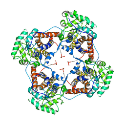
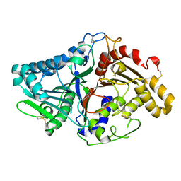
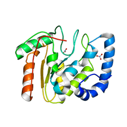
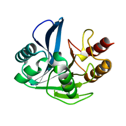
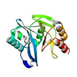
1F0I
 
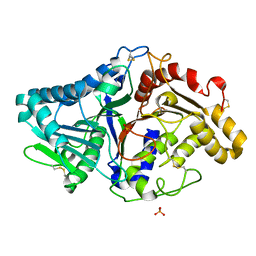 | | THE FIRST CRYSTAL STRUCTURE OF A PHOSPHOLIPASE D | | 分子名称: | PHOSPHATE ION, PHOSPHOLIPASE D | | 著者 | Leiros, I, Secundo, F, Zambonelli, C, Servi, S, Hough, E. | | 登録日 | 2000-05-16 | | 公開日 | 2001-05-16 | | 最終更新日 | 2011-07-13 | | 実験手法 | X-RAY DIFFRACTION (1.4 Å) | | 主引用文献 | The first crystal structure of a phospholipase D.
Structure Fold.Des., 8, 2000
|
|
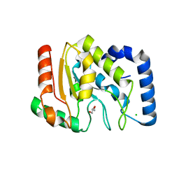
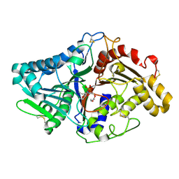
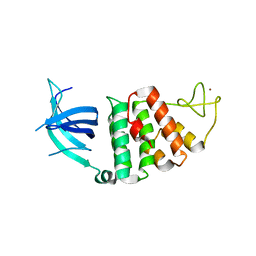
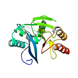
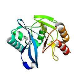
8C10
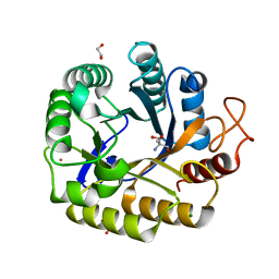 | | Biochemical and structural characterisation of an alkaline family GH5 cellulase from a shipworm symbiont | | 分子名称: | 1,2-ETHANEDIOL, 2-AMINO-2-HYDROXYMETHYL-PROPANE-1,3-DIOL, GH5 Cellulase, ... | | 著者 | Leiros, I, Vaaje-Kolstad, G. | | 登録日 | 2022-12-19 | | 公開日 | 2023-04-19 | | 最終更新日 | 2024-06-19 | | 実験手法 | X-RAY DIFFRACTION (1 Å) | | 主引用文献 | Biochemical and structural characterisation of a family GH5 cellulase from endosymbiont of shipworm P. megotara.
Biotechnol Biofuels Bioprod, 16, 2023
|
|
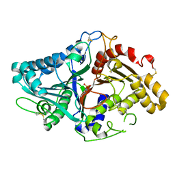
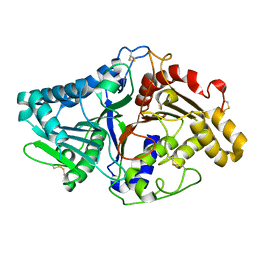
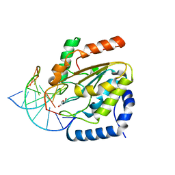
2JHJ
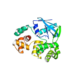 | | 3-methyladenine dna-glycosylase from Archaeoglobus fulgidus | | 分子名称: | 3-METHYLADENINE DNA-GLYCOSYLASE, GLYCEROL, SODIUM ION | | 著者 | Leiros, I, Nabong, M.P, Grosvik, K, Ringvoll, J, Haugland, G.T, Uldal, L, Reite, K, Olsbu, I.K, Knaevelsrud, I, Moe, E, Andersen, O.A, Birkeland, N.K, Ruoff, P, Klungland, A, Bjelland, S. | | 登録日 | 2007-02-22 | | 公開日 | 2007-04-10 | | 最終更新日 | 2023-12-13 | | 実験手法 | X-RAY DIFFRACTION (1.9 Å) | | 主引用文献 | Structural Basis for Enzymatic Excision of N1-Methyladenine and N3-Methylcytosine from DNA
Embo J., 26, 2007
|
|
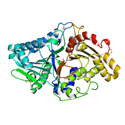
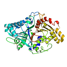
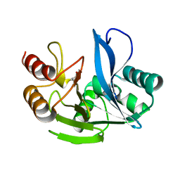
2JHN
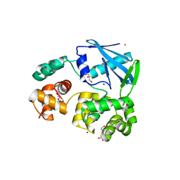 | | 3-methyladenine dna-glycosylase from Archaeoglobus fulgidus | | 分子名称: | 2-(N-MORPHOLINO)-ETHANESULFONIC ACID, 3-METHYLADENINE DNA-GLYCOSYLASE, GLYCEROL, ... | | 著者 | Leiros, I, Nabong, M.P, Grosvik, K, Ringvoll, J, Haugland, G.T, Uldal, L, Reite, K, Olsbu, I.K, Knaevelsrud, I, Moe, E, Andersen, O.A, Birkeland, N.K, Ruoff, P, Klungland, A, Bjelland, S. | | 登録日 | 2007-02-22 | | 公開日 | 2007-04-10 | | 最終更新日 | 2024-05-08 | | 実験手法 | X-RAY DIFFRACTION (1.8 Å) | | 主引用文献 | Structural Basis for Enzymatic Excision of N1-Methyladenine and N3-Methylcytosine from DNA
Embo J., 26, 2007
|
|
2J6X
 | | The crystal structure of lactate oxidase | | 分子名称: | FLAVIN MONONUCLEOTIDE, LACTATE OXIDASE, ZINC ION | | 著者 | Leiros, I, Wang, E, Rasmussen, T, Oksanen, E, Repo, H, Petersen, S.B, Heikinheimo, P, Hough, E. | | 登録日 | 2006-10-05 | | 公開日 | 2006-10-23 | | 最終更新日 | 2023-12-13 | | 実験手法 | X-RAY DIFFRACTION (2.1 Å) | | 主引用文献 | The 2.1 A Structure of Aerococcus Viridans L-Lactate Oxidase (Lox).
Acta Crystallogr.,Sect.F, 62, 2006
|
|
1OKB
 | | crystal structure of Uracil-DNA glycosylase from Atlantic cod (Gadus morhua) | | 分子名称: | CHLORIDE ION, GLYCEROL, URACIL-DNA GLYCOSYLASE | | 著者 | Leiros, I, Moe, E, Lanes, O, Smalas, A.O, Willassen, N.P. | | 登録日 | 2003-07-21 | | 公開日 | 2004-04-05 | | 最終更新日 | 2023-12-13 | | 実験手法 | X-RAY DIFFRACTION (1.9 Å) | | 主引用文献 | The Crystal Structure of Uracil-DNA Glycosylase from Atlantic Cod (Gadus Morhua) Reveals Cold-Adaptation Features
Acta Crystallogr.,Sect.D, 59, 2003
|
|
1V0U
 | |
1V0R
 | |
1V0V
 | |
1V0Y
 | |
1V0S
 | |
1V0W
 | |
1V0T
 | |
2BOO
 | | The crystal structure of Uracil-DNA N-Glycosylase (UNG) from Deinococcus radiodurans. | | 分子名称: | NITRATE ION, URACIL-DNA GLYCOSYLASE | | 著者 | Leiros, I, Moe, E, Smalas, A.O, McSweeney, S. | | 登録日 | 2005-04-13 | | 公開日 | 2005-07-27 | | 最終更新日 | 2023-12-13 | | 実験手法 | X-RAY DIFFRACTION (1.8 Å) | | 主引用文献 | Structure of the Uracil-DNA N-Glycosylase (Ung) from Deinococcus Radiodurans.
Acta Crystallogr.,Sect.D, 61, 2005
|
|
1W3S
 | | The crystal structure of RecO from Deinococcus radiodurans. | | 分子名称: | HYPOTHETICAL PROTEIN DR0819, ZINC ION | | 著者 | Leiros, I, Timmins, J, Hall, D.R, Leonard, G.A, McSweeney, S.M. | | 登録日 | 2004-07-18 | | 公開日 | 2005-02-23 | | 最終更新日 | 2024-05-08 | | 実験手法 | X-RAY DIFFRACTION (2.4 Å) | | 主引用文献 | Crystal Structure and DNA-Binding Analysis of Reco from Deinococcus Radiodurans
Embo J., 24, 2005
|
|
4UQM
 | | Crystal structure determination of uracil-DNA N-glycosylase (UNG) from Deinococcus radiodurans in complex with DNA - new insights into the role of the Leucine-loop for damage recognition and repair | | 分子名称: | 5'-D(*CP*CP*TP*AP*TP*CP*CP*AP*AAB*GP*TP*CP*TP*CP*CP*G)-3', 5'-D(*GP*CP*GP*GP*AP*GP*AP*CP*AP*TP*GP*GP*AP*CP*AP*G)-3', CHLORIDE ION, ... | | 著者 | Pedersen, H.L, Johnson, K.A, McVey, C.E, Leiros, I, Moe, E. | | 登録日 | 2014-06-24 | | 公開日 | 2015-08-12 | | 最終更新日 | 2024-01-10 | | 実験手法 | X-RAY DIFFRACTION (1.35 Å) | | 主引用文献 | Structure determination of uracil-DNA N-glycosylase from Deinococcus radiodurans in complex with DNA.
Acta Crystallogr. D Biol. Crystallogr., 71, 2015
|
|
4D1W
 | | A H224Y mutant for VIM-7 from Pseudomonas aeruginosa | | 分子名称: | METALLO-B-LACTAMASE, ZINC ION | | 著者 | Leiros, H.-K.S, Skagseth, S, Edvardsen, K.S.W, Lorentzen, M.S, Bjerga, G.E.K, Leiros, I, Samuelsen, O. | | 登録日 | 2014-05-05 | | 公開日 | 2014-06-25 | | 最終更新日 | 2023-12-20 | | 実験手法 | X-RAY DIFFRACTION (1.4 Å) | | 主引用文献 | His224 Alters the R2 Drug Binding Site and Phe218 Influences the Catalytic Efficiency in the Metallo-Beta-Lactamase Vim-7.
Antimicrob.Agents Chemother., 58, 2014
|
|
4D1U
 | | A D120A mutant of VIM-7 from Pseudomonas aeruginosa | | 分子名称: | METALLO-B-LACTAMASE, ZINC ION | | 著者 | Leiros, H.-K.S, Skagseth, S, Edvardsen, K.S.W, Lorentzen, M.S, Bjerga, G.E.K, Leiros, I, Samuelsen, O. | | 登録日 | 2014-05-05 | | 公開日 | 2014-06-25 | | 最終更新日 | 2023-12-20 | | 実験手法 | X-RAY DIFFRACTION (1.8 Å) | | 主引用文献 | His224 Alters the R2 Drug Binding Site and Phe218 Influences the Catalytic Efficiency in the Metallo-Beta-Lactamase Vim-7.
Antimicrob.Agents Chemother., 58, 2014
|
|
4D1T
 | | High resolution structure of native tVIM-7 from Pseudomonas aeruginosa | | 分子名称: | METALLO-B-LACTAMASE, ZINC ION | | 著者 | Leiros, H.-K.S, Skagseth, S, Edvardsen, K.S.W, Lorentzen, M.S, Bjerga, G.E.K, Leiros, I, Samuelsen, O. | | 登録日 | 2014-05-05 | | 公開日 | 2014-06-25 | | 最終更新日 | 2023-12-20 | | 実験手法 | X-RAY DIFFRACTION (1.25 Å) | | 主引用文献 | His224 Alters the R2 Drug Binding Site and Phe218 Influences the Catalytic Efficiency in the Metallo-Beta-Lactamase Vim-7.
Antimicrob.Agents Chemother., 58, 2014
|
|
4D1V
 | | A F218Y mutant of VIM-7 from Pseudomonas aeruginosa | | 分子名称: | METALLO-B-LACTAMASE, ZINC ION | | 著者 | Leiros, H.-K.S, Skagseth, S, Edvardsen, K.S.W, Lorentzen, M.S, Bjerga, G.E.K, Leiros, I, Samuelsen, O. | | 登録日 | 2014-05-05 | | 公開日 | 2014-06-25 | | 最終更新日 | 2023-12-20 | | 実験手法 | X-RAY DIFFRACTION (1.7 Å) | | 主引用文献 | His224 Alters the R2 Drug Binding Site and Phe218 Influences the Catalytic Efficiency in the Metallo-Beta-Lactamase Vim-7.
Antimicrob.Agents Chemother., 58, 2014
|
|
1YUO
 | | Optimisation of the surface electrostatics as a strategy for cold adaptation of uracil-DNA N-glycosylase (UNG)from atlantic cod (Gadus morhua) | | 分子名称: | Uracil-DNA glycosylase | | 著者 | Moe, E, Leiros, I, Riise, E.K, Olufsen, M, Lanes, O, Smalas, A.O, Willassen, N.P. | | 登録日 | 2005-02-14 | | 公開日 | 2005-03-01 | | 最終更新日 | 2023-10-25 | | 実験手法 | X-RAY DIFFRACTION (1.95 Å) | | 主引用文献 | Optimisation of the surface electrostatics as a strategy for cold adaptation of uracil-DNA N-glycosylase (UNG) from Atlantic cod (Gadus morhua)
J.Mol.Biol., 343, 2004
|
|
2Y87
 | | Native VIM-7. Structural and computational investigations of VIM-7: Insights into the substrate specificity of VIM metallo-beta- lactamases | | 分子名称: | MAGNESIUM ION, METALLO-B-LACTAMASE, UNKNOWN ATOM OR ION, ... | | 著者 | Saradhi, P, Leiros, H.-K.S, Ahmad, R, Spencer, J, Leiros, I, Walsh, T.R, Sundsfjord, A, Samuelsen, O. | | 登録日 | 2011-02-03 | | 公開日 | 2011-06-15 | | 最終更新日 | 2023-12-20 | | 実験手法 | X-RAY DIFFRACTION (1.86 Å) | | 主引用文献 | Structural and Computational Investigations of Vim- 7: Insights Into the Substrate Specificity of Vim Metallo-Beta-Lactamases
J.Mol.Biol., 411, 2011
|
|
2Y8A
 | | VIM-7 with Oxidised. Structural and computational investigations of VIM-7: Insights into the substrate specificity of VIM metallo-beta- lactamases | | 分子名称: | MAGNESIUM ION, METALLO-B-LACTAMASE, UNKNOWN ATOM OR ION, ... | | 著者 | Saradhi, P, Leiros, H.-K.S, Ahmad, R, Spencer, J, Leiros, I, Walsh, T.R, Sundsfjord, A, Samuelsen, O. | | 登録日 | 2011-02-03 | | 公開日 | 2011-06-15 | | 最終更新日 | 2024-05-08 | | 実験手法 | X-RAY DIFFRACTION (2.33 Å) | | 主引用文献 | Structural and Computational Investigations of Vim- 7: Insights Into the Substrate Specificity of Vim Metallo-Beta-Lactamases
J.Mol.Biol., 411, 2011
|
|
1BZX
 | | THE CRYSTAL STRUCTURE OF ANIONIC SALMON TRYPSIN IN COMPLEX WITH BOVINE PANCREATIC TRYPSIN INHIBITOR | | 分子名称: | CALCIUM ION, PROTEIN (BOVINE PANCREATIC TRYPSIN INHIBITOR), PROTEIN (TRYPSIN) | | 著者 | Helland, R, Leiros, I, Berglund, G.I, Willassen, N.P, Smalas, A.O. | | 登録日 | 1998-11-05 | | 公開日 | 1998-11-11 | | 最終更新日 | 2023-08-09 | | 実験手法 | X-RAY DIFFRACTION (2.1 Å) | | 主引用文献 | The crystal structure of anionic salmon trypsin in complex with bovine pancreatic trypsin inhibitor.
Eur.J.Biochem., 256, 1998
|
|
4AOU
 | | CtIDH bound to NADP. The complex structures of Isocitrate dehydrogenase from Clostridium thermocellum and Desulfotalea psychrophila, support a new active site locking mechanism | | 分子名称: | ISOCITRATE DEHYDROGENASE [NADP], ISOCITRIC ACID, MAGNESIUM ION, ... | | 著者 | Leiros, H.-K.S, Fedoy, A.-E, Leiros, I, Steen, I.H. | | 登録日 | 2012-03-30 | | 公開日 | 2012-07-11 | | 最終更新日 | 2024-05-08 | | 実験手法 | X-RAY DIFFRACTION (2.5 Å) | | 主引用文献 | The complex structures of isocitrate dehydrogenase from Clostridium thermocellum and Desulfotalea psychrophila suggest a new active site locking mechanism.
Febs Open Bio, 2, 2012
|
|


