Loading
PDBj
MenuPDBj@FacebookPDBj@TwitterPDBj@YouTubewwPDB FoundationwwPDB
RCSB PDBPDBeBMRBAdv. SearchSearch help
PDB: 91 results

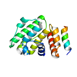
8GUW
DownloadVisualize
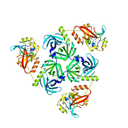
BU of 8guw by Molmil
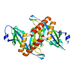
Structure of Aurora Kinase A in complex with activator peptide
Descriptor: ADENOSINE-5'-DIPHOSPHATE, Peptide from Centrosomal protein of 192 kDa,Aurora kinase A
Authors:Lee, I.-G, Park, J.
Deposit date:2022-09-13
Release date:2023-05-31
Last modified:2023-11-29
Method:X-RAY DIFFRACTION (2.7 Å)
Cite:Structural basis for CEP192-mediated regulation of centrosomal AURKA.
Sci Adv, 9, 2023
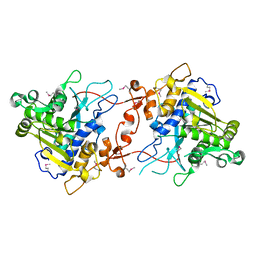
8KIC
DownloadVisualize
BU of 8kic by Molmil
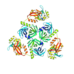
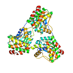
Bacterial serine protease
Descriptor: Lysozyme fragment (unknown sequence), peptidase Do
Authors:Lee, I.-G, Jeon, H.
Deposit date:2023-08-23
Release date:2024-09-04
Method:ELECTRON MICROSCOPY (2.5 Å)
Cite:Structure of bacterial serine protease
To Be Published
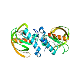
4ZG0
DownloadVisualize
BU of 4zg0 by Molmil
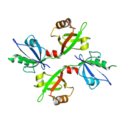
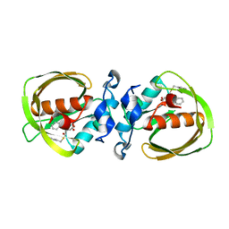
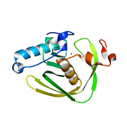
Crystal structure of Mouse Syndesmos protein
Descriptor: Protein syndesmos
Authors:Lee, I, Kim, H, Yoo, J, Cho, H, Lee, W.
Deposit date:2015-04-22
Release date:2016-04-20
Last modified:2023-11-08
Method:X-RAY DIFFRACTION (2.006 Å)
Cite:Crystal structure of syndesmos and its interaction with Syndecan-4 proteoglycan
Biochem.Biophys.Res.Commun., 463, 2015
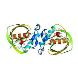
8W69
DownloadVisualize
BU of 8w69 by Molmil
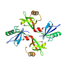
DegQ-b-casein complex
Descriptor: Casein fragment, peptidase Do
Authors:Lee, I.-G, Jeon, H.
Deposit date:2023-08-28
Release date:2024-09-11
Method:ELECTRON MICROSCOPY (3.6 Å)
Cite:Structure of E. coli Deg-b-casein complex
To Be Published
5G1D
DownloadVisualize
BU of 5g1d by Molmil
The complex structure of syntenin-1 PDZ domain with c-terminal extension
Descriptor: SYNDECAN-4, SYNTENIN-1
Authors:Lee, I, Kim, H, Yun, J.H, Lee, W.
Deposit date:2016-03-25
Release date:2016-11-23
Last modified:2024-05-08
Method:X-RAY DIFFRACTION (2.81 Å)
Cite:New Structural Insight of C-Terminal Region of Syntenin-1, Enhancing the Molecular Dimerization and Inhibitory Function Related on Syndecan-4 Signaling.
Sci.Rep., 6, 2016
5G1E
DownloadVisualize
BU of 5g1e by Molmil
The complex structure of syntenin-1 PDZ domain with c-terminal extension
Descriptor: SYNTENIN-1
Authors:Lee, I, Kim, H, Yun, J.H, Lee, W.
Deposit date:2016-03-25
Release date:2016-11-23
Last modified:2024-05-08
Method:X-RAY DIFFRACTION (1.92 Å)
Cite:New Structural Insight of C-Terminal Region of Syntenin-1, Enhancing the Molecular Dimerization and Inhibitory Function Related on Syndecan-4 Signaling.
Sci.Rep., 6, 2016
5JSD
DownloadVisualize
BU of 5jsd by Molmil
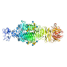
Crystal structure of phiAB6 tailspike in complex with five-repeated oligosaccharides of Acinetobacter baumannii surface polysaccharide
Descriptor: ACETIC ACID, MALONIC ACID, beta-D-galactopyranose-(1-3)-2-amino-2-deoxy-beta-D-galactopyranose-(1-3)-[5,7-bisacetamido-3,5,7,9-tetradeoxy-L-glycero-alpha-L-manno-non-2-ulopyranosonic acid-(2-6)-beta-D-glucopyranose-(1-6)]beta-D-galactopyranose-(1-3)-2-amino-2-deoxy-beta-D-galactopyranose-(1-3)-[beta-D-glucopyranose-(1-6)]beta-D-galactopyranose, ...
Authors:Lee, I.M, Tu, I.F, Huang, K.F, Wu, S.H.
Deposit date:2016-05-08
Release date:2017-03-08
Last modified:2024-03-20
Method:X-RAY DIFFRACTION (1.48 Å)
Cite:Structural basis for fragmenting the exopolysaccharide of Acinetobacter baumannii by bacteriophage Phi AB6 tailspike protein
Sci Rep, 7, 2017
5JS4
DownloadVisualize
BU of 5js4 by Molmil
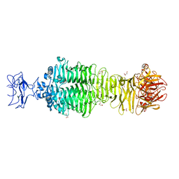
Crystal structure of phiAB6 tailspike
Descriptor: MALONIC ACID, phiAB6 tailspike
Authors:Lee, I.M, Tu, I.F, Huang, K.F, Wu, S.H.
Deposit date:2016-05-07
Release date:2017-03-08
Last modified:2024-03-20
Method:X-RAY DIFFRACTION (1.48 Å)
Cite:Structural basis for fragmenting the exopolysaccharide of Acinetobacter baumannii by bacteriophage Phi AB6 tailspike protein
Sci Rep, 7, 2017
5JSE
DownloadVisualize
BU of 5jse by Molmil
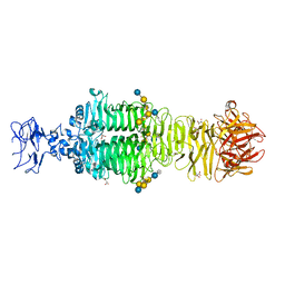
Crystal structure of phiAB6 tailspike in complex with three-repeated oligosaccharides of Acinetobacter baumannii surface polysaccharide
Descriptor: ACETIC ACID, MALONIC ACID, beta-D-galactopyranose-(1-3)-2-amino-2-deoxy-beta-D-galactopyranose-(1-3)-[5,7-bisacetamido-3,5,7,9-tetradeoxy-L-glycero-alpha-L-manno-non-2-ulopyranosonic acid-(2-6)-beta-D-glucopyranose-(1-6)]beta-D-galactopyranose-(1-3)-2-amino-2-deoxy-beta-D-galactopyranose-(1-3)-[beta-D-glucopyranose-(1-6)]beta-D-galactopyranose, ...
Authors:Lee, I.M, Tu, I.F, Huang, K.F, Wu, S.H.
Deposit date:2016-05-08
Release date:2017-03-08
Last modified:2024-03-20
Method:X-RAY DIFFRACTION (1.89 Å)
Cite:Structural basis for fragmenting the exopolysaccharide of Acinetobacter baumannii by bacteriophage Phi AB6 tailspike protein
Sci Rep, 7, 2017
3CMM
DownloadVisualize
BU of 3cmm by Molmil
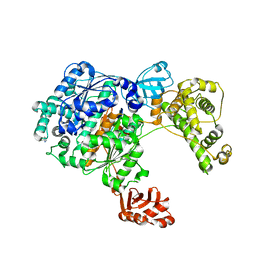
Crystal Structure of the Uba1-Ubiquitin Complex
Descriptor: PROLINE, Ubiquitin, Ubiquitin-activating enzyme E1 1
Authors:Lee, I, Schindelin, H.
Deposit date:2008-03-23
Release date:2008-08-05
Last modified:2024-02-21
Method:X-RAY DIFFRACTION (2.7 Å)
Cite:Structural insights into E1-catalyzed ubiquitin activation and transfer to conjugating enzymes.
Cell(Cambridge,Mass.), 134, 2008
6B9H
DownloadVisualize
BU of 6b9h by Molmil
Complex of Hook Domain with a Dynein Light Intermediate Chain Peptide
Descriptor: Cytoplasmic dynein 1 light intermediate chain 1, Protein Hook homolog 3
Authors:Lee, I.G, Dominguez, R.
Deposit date:2017-10-10
Release date:2018-03-21
Last modified:2023-10-04
Method:X-RAY DIFFRACTION (1.5 Å)
Cite:A conserved interaction of the dynein light intermediate chain with dynein-dynactin effectors necessary for processivity.
Nat Commun, 9, 2018
2YIJ
DownloadVisualize
BU of 2yij by Molmil
Crystal Structure of phospholipase A1
Descriptor: PHOSPHOLIPASE A1-IIGAMMA
Authors:Lee, I.
Deposit date:2011-05-13
Release date:2012-05-30
Last modified:2024-10-16
Method:X-RAY DIFFRACTION (2 Å)
Cite:Crystal Structure of Phospholipase A1
To be Published
6JF8
DownloadVisualize
BU of 6jf8 by Molmil
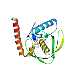
K4U bound crystal structure of class I type b peptide deformylase from Acinetobacter baumannii
Descriptor: L-[(N-HYDROXYAMINO)CARBONYL]PHENYLALANINE, Peptide deformylase, ZINC ION
Authors:Lee, I.H, Ho, T.H, Kang, L.W.
Deposit date:2019-02-08
Release date:2020-02-12
Last modified:2023-11-22
Method:X-RAY DIFFRACTION (1.7 Å)
Cite:K4U bound crystal structure of class I type b peptide deformylase from Acinetobacter baumannii
To be published
6JF4
DownloadVisualize
BU of 6jf4 by Molmil
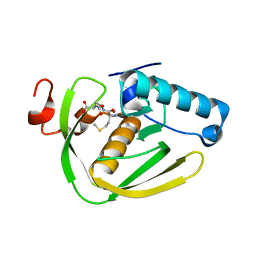
K1U bound crystal structure of class I type b peptide deformylase from Acinetobacter baumannii
Descriptor: (3R)-3-benzyl-4-oxo-4-[(2-oxo-2-phenylethyl)sulfanyl]butanoic acid, Peptide deformylase, ZINC ION
Authors:Lee, I.H, Ho, T.H, Kang, L.W.
Deposit date:2019-02-08
Release date:2020-02-12
Last modified:2023-11-22
Method:X-RAY DIFFRACTION (2 Å)
Cite:K1U bound crystal structure of class I type b peptide deformylase from Acinetobacter baumannii
To be published
6JFF
DownloadVisualize
BU of 6jff by Molmil
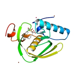
K3U bound crystal structure of class I type b peptide deformylase from Pseudomonas aeruginosa
Descriptor: NICKEL (II) ION, Peptide deformylase, S-(2-oxo-2-phenylethyl) (2R)-2-benzyl-4,4,4-trifluorobutanethioate
Authors:Lee, I.H, Ho, T.H, Kang, L.W.
Deposit date:2019-02-08
Release date:2020-02-12
Last modified:2023-11-22
Method:X-RAY DIFFRACTION (2.1 Å)
Cite:K3U bound crystal structure of class I type b peptide deformylase from Pseudomonas aeruginosa
To be published
6JFA
DownloadVisualize
BU of 6jfa by Molmil
Met-Ala-Ser bound crystal structure of class I type b peptide deformylase from Pseudomonas aeruginosa
Descriptor: MET-ALA-SER, NICKEL (II) ION, Peptide deformylase
Authors:Lee, I.H, Ho, T.H, Kang, L.W.
Deposit date:2019-02-08
Release date:2020-02-12
Last modified:2023-11-22
Method:X-RAY DIFFRACTION (1.93 Å)
Cite:Met-Ala-Ser bound crystal structure of class I type b peptide deformylase from Pseudomonas aeruginosa
To be published
6JF5
DownloadVisualize
BU of 6jf5 by Molmil
K2U bound crystal structure of class I type b peptide deformylase from Acinetobacter baumannii
Descriptor: (3~{R},4~{R})-4-oxidanyl-3-(phenylmethyl)-4-(phenylmethylsulfanyl)butanoic acid, Peptide deformylase, ZINC ION
Authors:Lee, I.H, Ho, T.H, Kang, L.W.
Deposit date:2019-02-08
Release date:2020-02-12
Last modified:2023-11-22
Method:X-RAY DIFFRACTION (2 Å)
Cite:K2U bound crystal structure of class I type b peptide deformylase from Acinetobacter baumannii
To be published
6JFE
DownloadVisualize
BU of 6jfe by Molmil
K2U bound crystal structure of class I type b peptide deformylase from Pseudomonas aeruginosa
Descriptor: NICKEL (II) ION, Peptide deformylase, S-(2-oxo-2-phenylethyl) (2R)-2-benzyl-4,4,4-trifluorobutanethioate
Authors:Lee, I.H, Ho, T.H, Kang, L.W.
Deposit date:2019-02-08
Release date:2020-02-12
Last modified:2023-11-22
Method:X-RAY DIFFRACTION (2.1 Å)
Cite:K2U bound crystal structure of class I type b peptide deformylase from Pseudomonas aeruginosa
To be published
6JF9
DownloadVisualize
BU of 6jf9 by Molmil
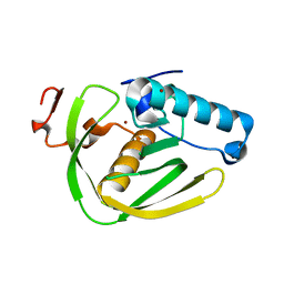
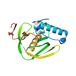
Apo crystal structure of class I type b peptide deformylase from Pseudomonas aeruginosa
Descriptor: GLYCEROL, NICKEL (II) ION, Peptide deformylase
Authors:Lee, I.H, Ho, T.H, Kang, L.W.
Deposit date:2019-02-08
Release date:2020-02-12
Last modified:2023-11-22
Method:X-RAY DIFFRACTION (1.7 Å)
Cite:Apo crystal structure of class I type b peptide deformylase from Pseudomonas aeruginosa
To be published
6JF3
DownloadVisualize
BU of 6jf3 by Molmil
Actinonin bound crystal structure of class I type b peptide deformylase from Acinetobacter baumannii
Descriptor: ACTINONIN, Peptide deformylase, ZINC ION
Authors:Lee, I.H, Ho, T.H, Kang, L.W.
Deposit date:2019-02-08
Release date:2020-02-12
Last modified:2023-11-22
Method:X-RAY DIFFRACTION (2.01 Å)
Cite:Actinonin bound crystal structure of class I type b peptide deformylase from Acinetobacter baumannii
To be published
6JFC
DownloadVisualize
BU of 6jfc by Molmil
Actinonin bound crystal structure of class I type b peptide deformylase from Pseudomonas aeruginosa
Descriptor: ACTINONIN, NICKEL (II) ION, Peptide deformylase
Authors:Lee, I.H, Ho, T.H, Kang, L.W.
Deposit date:2019-02-08
Release date:2020-02-12
Last modified:2023-11-22
Method:X-RAY DIFFRACTION (2.04 Å)
Cite:Actinonin bound crystal structure of class I type b peptide deformylase from Pseudomonas aeruginosa
To be published
6JFN
DownloadVisualize
BU of 6jfn by Molmil
K4U bound crystal structure of class I type b peptide deformylase from Pseudomonas aeruginosa
Descriptor: L-[(N-HYDROXYAMINO)CARBONYL]PHENYLALANINE, NICKEL (II) ION, Peptide deformylase
Authors:Lee, I.H, Ho, T.H, Kang, L.W.
Deposit date:2019-02-11
Release date:2020-03-04
Last modified:2023-11-22
Method:X-RAY DIFFRACTION (2.04 Å)
Cite:K4U bound crystal structure of class I type b peptide deformylase from Pseudomonas aeruginosa
To be published
6JFD
DownloadVisualize
BU of 6jfd by Molmil
K1U bound crystal structure of class I type b peptide deformylase from Pseudomonas aeruginosa
Descriptor: (3R)-3-benzyl-4-oxo-4-[(2-oxo-2-phenylethyl)sulfanyl]butanoic acid, NICKEL (II) ION, Peptide deformylase
Authors:Lee, I.H, Ho, T.H, Kang, L.W.
Deposit date:2019-02-08
Release date:2020-02-12
Last modified:2023-11-22
Method:X-RAY DIFFRACTION (2.4 Å)
Cite:K1U bound crystal structure of class I type b peptide deformylase from Pseudomonas aeruginosa
To be published
6JEV
DownloadVisualize
BU of 6jev by Molmil
K2U bound crystal structure of class I type a peptide deformylase from Acinetobacter baumanii
Descriptor: (3~{R},4~{R})-4-oxidanyl-3-(phenylmethyl)-4-(phenylmethylsulfanyl)butanoic acid, Peptide deformylase, ZINC ION
Authors:Lee, I.H, Ho, T.H, Kang, L.W.
Deposit date:2019-02-07
Release date:2020-02-12
Last modified:2023-11-22
Method:X-RAY DIFFRACTION (1.9 Å)
Cite:K2U bound crystal peptide deformylase from Acinetobacter baumanii
To be published
6IKT
DownloadVisualize
BU of 6ikt by Molmil
K1U complex structure of peptide deformylase from Xanthomonas oryzae pv. oryzae
Descriptor: (3R)-3-benzyl-4-oxo-4-[(2-oxo-2-phenylethyl)sulfanyl]butanoic acid, CADMIUM ION, NICKEL (II) ION, ...
Authors:Lee, I.H, Kang, L.W.
Deposit date:2018-10-16
Release date:2019-10-23
Last modified:2023-11-22
Method:X-RAY DIFFRACTION (1.9 Å)
Cite:K1U complex structure of peptide deformylase from Xanthomonas oryzae pv. oryzae
To be published


1234>

226707

數據於2024-10-30公開中

PDB statisticsPDBj update infoContact PDBjnumon