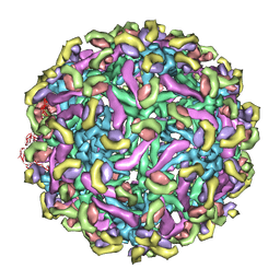
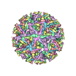
1K4R
 
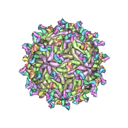
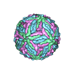 | | Structure of Dengue Virus | | 分子名称: | MAJOR ENVELOPE PROTEIN E | | 著者 | Kuhn, R.J, Zhang, W, Rossmann, M.G, Pletnev, S.V, Corver, J, Lenches, E, Jones, C.T, Mukhopadhyay, S, Chipman, P.R, Strauss, E.G, Baker, T.S, Strauss, J.H. | | 登録日 | 2001-10-08 | | 公開日 | 2002-03-13 | | 最終更新日 | 2018-07-18 | | 実験手法 | ELECTRON MICROSCOPY (24 Å) | | 主引用文献 | Structure of dengue virus: implications for flavivirus organization, maturation, and fusion.
Cell(Cambridge,Mass.), 108, 2002
|
|
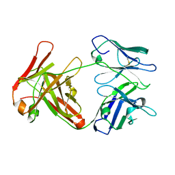
1EGX
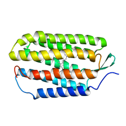
 | | SOLUTION STRUCTURE OF THE ENA-VASP HOMOLOGY 1 (EVH1) DOMAIN OF HUMAN VASODILATOR-STIMULATED PHOSPHOPROTEIN (VASP) | | 分子名称: | VASODILATOR-STIMULATED PHOSPHOPROTEIN | | 著者 | Ball, L, Kuhne, R, Hoffmann, B, Hafner, A, Schmieder, P, Volkmer-Engert, R, Hof, M, Wahl, M, Schneider-Mergener, J, Walter, U, Oschkinat, H, Jarchau, T. | | 登録日 | 2000-02-17 | | 公開日 | 2000-09-20 | | 最終更新日 | 2024-05-01 | | 実験手法 | SOLUTION NMR | | 主引用文献 | Dual epitope recognition by the VASP EVH1 domain modulates polyproline ligand specificity and binding affinity.
EMBO J., 19, 2000
|
|
1R84
 | | NMR structure of the 13-cis-15-syn retinal in dark_adapted bacteriorhodopsin | | 分子名称: | Bacteriorhodopsin, RETINAL | | 著者 | Patzelt, H, Simon, B, Ter Laak, A, Kessler, B, Kuhne, R, Schmieder, P, Oesterhaelt, D, Oschkinat, H. | | 登録日 | 2003-10-23 | | 公開日 | 2003-11-11 | | 最終更新日 | 2022-03-02 | | 実験手法 | SOLUTION NMR | | 主引用文献 | The structures of the active center in dark-adapted bacteriorhodopsin by solution-state NMR spectroscopy
Proc.Natl.Acad.Sci.USA, 99, 2002
|
|
1S3A
 | | NMR Solution Structure of Subunit B8 from Human NADH-Ubiquinone Oxidoreductase Complex I (CI-B8) | | 分子名称: | NADH-ubiquinone oxidoreductase B8 subunit | | 著者 | Brockmann, C, Diehl, A, Rehbein, K, Kuhne, R, Oschkinat, H. | | 登録日 | 2004-01-13 | | 公開日 | 2005-01-25 | | 最終更新日 | 2022-03-02 | | 実験手法 | SOLUTION NMR | | 主引用文献 | The oxidized subunit B8 from human complex I adopts a thioredoxin fold.
Structure, 12, 2004
|
|
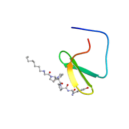
1JMQ
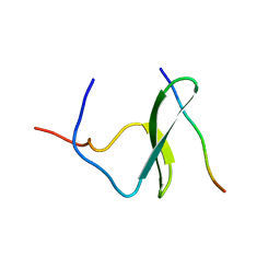 | | YAP65 (L30K mutant) WW domain in Complex with GTPPPPYTVG peptide | | 分子名称: | 65 KDA YES-ASSOCIATED PROTEIN, WW Domain Binding Protein-1 | | 著者 | Pires, J.R, Taha-Nejad, F, Toepert, F, Ast, T, Hoffmuller, U, Schneider-Mergener, J, Kuhne, R, Macias, M.J, Oschkinat, H. | | 登録日 | 2001-07-19 | | 公開日 | 2001-12-21 | | 最終更新日 | 2024-05-22 | | 実験手法 | SOLUTION NMR | | 主引用文献 | Solution structures of the YAP65 WW domain and the variant L30 K in complex with the peptides GTPPPPYTVG, N-(n-octyl)-GPPPY and PLPPY and the application of peptide libraries reveal a minimal binding epitope.
J.Mol.Biol., 314, 2001
|
|
1K9Q
 | | YAP65 WW domain complexed to N-(n-octyl)-GPPPY-NH2 | | 分子名称: | 65 kDa Yes-associated protein, N-OCTANE, WW domain binding protein-1 | | 著者 | Pires, J.R, Taha-Nejad, F, Toepert, F, Ast, T, Hoffmuller, U, Schneider-Mergener, J, Kuhne, R, Macias, M.J, Oschkinat, H. | | 登録日 | 2001-10-30 | | 公開日 | 2001-12-28 | | 最終更新日 | 2021-11-10 | | 実験手法 | SOLUTION NMR | | 主引用文献 | Solution structures of the YAP65 WW domain and the variant L30 K in complex with the peptides GTPPPPYTVG, N-(n-octyl)-GPPPY and PLPPY and the application of peptide libraries reveal a minimal binding epitope.
J.Mol.Biol., 314, 2001
|
|
1K9R
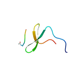 | | YAP65 WW domain complexed to Acetyl-PLPPY | | 分子名称: | 65 kDa Yes-associated protein, WW domain binding protein-1 | | 著者 | Pires, J.R, Taha-Nejad, F, Toepert, F, Ast, T, Hoffmuller, U, Schneider-Mergener, J, Kuhne, R, Macias, M.J, Oschkinat, H. | | 登録日 | 2001-10-30 | | 公開日 | 2001-12-28 | | 最終更新日 | 2021-11-10 | | 実験手法 | SOLUTION NMR | | 主引用文献 | Solution structures of the YAP65 WW domain and the variant L30 K in complex with the peptides GTPPPPYTVG, N-(n-octyl)-GPPPY and PLPPY and the application of peptide libraries reveal a minimal binding epitope.
J.Mol.Biol., 314, 2001
|
|
1OQA
 | | Solution structure of the BRCT-c domain from human BRCA1 | | 分子名称: | Breast cancer type 1 susceptibility protein | | 著者 | Gaiser, O.J, Ball, L.J, Schmieder, P, Leitner, D, Strauss, H, Wahl, M, Kuhne, R, Oschkinat, H, Heinemann, U. | | 登録日 | 2003-03-07 | | 公開日 | 2004-06-15 | | 最終更新日 | 2024-05-22 | | 実験手法 | SOLUTION NMR | | 主引用文献 | Solution structure, backbone dynamics, and association behavior of the C-terminal BRCT domain from the breast cancer-associated protein BRCA1.
Biochemistry, 43, 2004
|
|
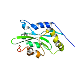
1Q8G
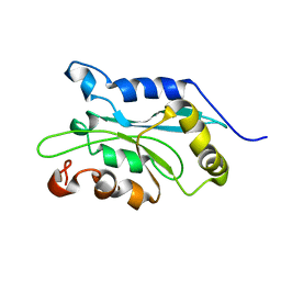 | | NMR structure of human Cofilin | | 分子名称: | Cofilin, non-muscle isoform | | 著者 | Pope, B.J, Zierler-Gould, K.M, Kuhne, R, Weeds, A.G, Ball, L.J. | | 登録日 | 2003-08-21 | | 公開日 | 2004-07-06 | | 最終更新日 | 2024-05-22 | | 実験手法 | SOLUTION NMR | | 主引用文献 | The solution structure of human cofilin: rationalizing actin binding and pH sensitivity
J.Biol.Chem., 279, 2004
|
|
1Q8X
 | | NMR structure of human cofilin | | 分子名称: | Cofilin, non-muscle isoform | | 著者 | Pope, B.J, Zierler-Gould, K.M, Kuhne, R, Weeds, A.G, Ball, L.J. | | 登録日 | 2003-08-22 | | 公開日 | 2004-07-06 | | 最終更新日 | 2024-05-22 | | 実験手法 | SOLUTION NMR | | 主引用文献 | The solution structure of human cofilin: rationalizing actin binding and pH sensitivity
J.Biol.Chem., 279, 2004
|
|
1R2N
 | | NMR structure of the all-trans retinal in dark-adapted Bacteriorhodopsin | | 分子名称: | Bacteriorhodopsin, RETINAL | | 著者 | Patzelt, H, Simon, B, terLaak, A, Kessler, B, Kuhne, R, Schmieder, P, Oesterhaelt, D, Oschkinat, H. | | 登録日 | 2003-09-29 | | 公開日 | 2003-10-28 | | 最終更新日 | 2022-03-02 | | 実験手法 | SOLUTION NMR | | 主引用文献 | The structures of the active center in dark-adapted bacteriorhodopsin by solution-state NMR spectroscopy
Proc.Natl.Acad.Sci.USA, 99, 2002
|
|
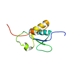
1R4T
 | | Solution structure of exoenzyme S | | 分子名称: | exoenzyme S | | 著者 | Langdon, G.M, Leitner, D, Labudde, D, Kuhne, R, Schmieder, P, Aktories, K, Oschkinat, H.O, Schmidt, G. | | 登録日 | 2003-10-08 | | 公開日 | 2005-04-12 | | 最終更新日 | 2024-05-22 | | 実験手法 | SOLUTION NMR | | 主引用文献 | Solution structure of the N-terminal GTPase activating domain of Pseudomonas aeruginosa exoenzyme S
To be Published
|
|
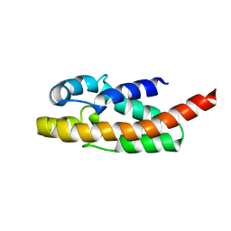
1IEZ
 | | Solution Structure of 3,4-Dihydroxy-2-Butanone 4-Phosphate Synthase of Riboflavin Biosynthesis | | 分子名称: | 3,4-Dihydroxy-2-Butanone 4-Phosphate Synthase | | 著者 | Kelly, M.J.S, Ball, L.J, Kuhne, R, Bacher, A, Oschkinat, H. | | 登録日 | 2001-04-11 | | 公開日 | 2001-11-07 | | 最終更新日 | 2024-05-01 | | 実験手法 | SOLUTION NMR | | 主引用文献 | The NMR structure of the 47-kDa dimeric enzyme 3,4-dihydroxy-2-butanone-4-phosphate synthase and ligand binding studies reveal the location of the active site.
Proc.Natl.Acad.Sci.USA, 98, 2001
|
|
1L2Z
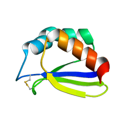
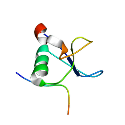 | | CD2BP2-GYF domain in complex with proline-rich CD2 tail segment peptide | | 分子名称: | CD2 ANTIGEN (CYTOPLASMIC TAIL)-BINDING PROTEIN 2, T-CELL SURFACE ANTIGEN CD2 | | 著者 | Freund, C, Kuhne, R, Yang, H, Park, S, Reinherz, E.L, Wagner, G. | | 登録日 | 2002-02-26 | | 公開日 | 2002-11-20 | | 最終更新日 | 2024-05-22 | | 実験手法 | SOLUTION NMR | | 主引用文献 | Dynamic interaction of CD2 with the GYF and the SH3 domain of compartmentalized effector molecules
Embo J., 21, 2002
|
|
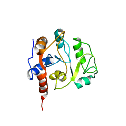
2JNU
 | | Solution structure of the RGS domain of human RGS14 | | 分子名称: | Regulator of G-protein signaling 14 | | 著者 | Dowler, E.F, Diehl, A, Bray, J, Elkins, J, Soundararajan, M, Doyle, D.A, Gileadi, C, Phillips, C, Schoch, G.A, Yang, X, Brockmann, C, Leidert, M, Rehbein, K, Schmieder, P, Kuhne, R, Higman, V.A, Sundstrom, M, Arrowsmith, C, Weigelt, J, Edwards, A, Oschkinat, H, Ball, L.J, Structural Genomics Consortium (SGC) | | 登録日 | 2007-02-02 | | 公開日 | 2007-02-27 | | 最終更新日 | 2024-05-08 | | 実験手法 | SOLUTION NMR | | 主引用文献 | Structural diversity in the RGS domain and its interaction with heterotrimeric G protein alpha-subunits.
Proc.Natl.Acad.Sci.Usa, 105, 2008
|
|
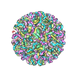
2R6P
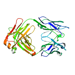
 | | Fit of E protein and Fab 1A1D-2 into 24 angstrom resolution cryoEM map of Fab complexed with dengue 2 virus. | | 分子名称: | Heavy chain of 1A1D-2, Light chain of 1A1D-2, Major envelope protein E | | 著者 | Lok, S.M, Kostyuchenko, V.K, Holdaway, H.A, Chipman, P.R, Kuhn, R.J, Rossmann, M.G. | | 登録日 | 2007-09-06 | | 公開日 | 2007-12-25 | | 最終更新日 | 2024-02-21 | | 実験手法 | ELECTRON MICROSCOPY (24 Å) | | 主引用文献 | Binding of a neutralizing antibody to dengue virus alters the arrangement of surface glycoproteins.
Nat.Struct.Mol.Biol., 15, 2008
|
|
8FE4
 | | Structure of dengue virus (DENV2) in complex with prM13, an anti-PrM monoclonal antibody | | 分子名称: | Envelope protein E, prM protein, prM13 Fab Heavy Chain, ... | | 著者 | Dowd, A.D, Sirohi, D, Speer, S, Mukherjee, S, Govero, J, Aleshnick, M, Larman, B, Sukupolvi-Petty, S, Sevvana, M, Miller, A.S, Klose, T, Zheng, A, Kielian, M, Kuhn, R.J, Diamond, M.S, Pierson, T.C. | | 登録日 | 2022-12-05 | | 公開日 | 2023-02-01 | | 最終更新日 | 2024-06-19 | | 実験手法 | ELECTRON MICROSCOPY (9.8 Å) | | 主引用文献 | prM-reactive antibodies reveal a role for partially mature virions in dengue virus pathogenesis.
Proc.Natl.Acad.Sci.USA, 120, 2023
|
|
8FE3
 | | Structure of dengue virus (DENV2) in complex with prM12, an anti-PrM monoclonal antibody | | 分子名称: | Envelope protein E, prM protein, prM12 Fab Heavy Chain, ... | | 著者 | Dowd, A.D, Sirohi, D, Speer, S, Mukherjee, S, Govero, J, Aleshnick, M, Larman, B, Sukupolvi-Petty, S, Sevvana, M, Miller, A.S, Klose, T, Zheng, A, Kielian, M, Kuhn, R.J, Diamond, M.S, Pierson, T.C. | | 登録日 | 2022-12-05 | | 公開日 | 2023-02-01 | | 最終更新日 | 2024-06-19 | | 実験手法 | ELECTRON MICROSCOPY (10.2 Å) | | 主引用文献 | prM-reactive antibodies reveal a role for partially mature virions in dengue virus pathogenesis.
Proc.Natl.Acad.Sci.USA, 120, 2023
|
|
4UOM
 | | Electron Cryo-microscopy of Venezuelan Equine Encephalitis Virus TC- 83 in complex with neutralizing antibody Fab F5 | | 分子名称: | FAB FRAGMENT HEAVY CHAIN, FAB FRAGMENT LIGHT CHAIN | | 著者 | Porta, J, Jose, J, Roehrig, J.T, Blair, C.D, Kuhn, R.J, Rossmann, M.G. | | 登録日 | 2014-06-05 | | 公開日 | 2014-10-15 | | 最終更新日 | 2017-08-23 | | 実験手法 | ELECTRON MICROSCOPY (17 Å) | | 主引用文献 | Locking and Blocking the Viral Landscape of an Alphavirus with Neutralizing Antibodies.
J.Virol., 88, 2014
|
|
4UOK
 | | Electron Cryo-microscopy of Venezuelan Equine Encephalitis Virus TC-83 in complex with neutralizing antibody Fab 3B4C-4 | | 分子名称: | FAB FRAGMENT HEAVY CHAIN, FAB FRAGMENT LIGHT CHAIN | | 著者 | Porta, J, Jose, J, Roehrig, J.T, Blair, C.D, Kuhn, R.J, Rossmann, M.G. | | 登録日 | 2014-06-04 | | 公開日 | 2014-09-17 | | 最終更新日 | 2019-05-29 | | 実験手法 | ELECTRON MICROSCOPY (18 Å) | | 主引用文献 | Locking and blocking the viral landscape of an alphavirus with neutralizing antibodies.
J.Virol., 88, 2014
|
|
7T17
 | |
6W09
 | |
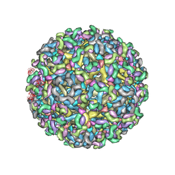
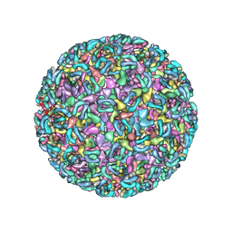
6CO8
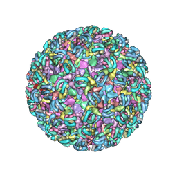
 | | Structure of Zika virus at a resolution of 3.1 Angstrom | | 分子名称: | 2-acetamido-2-deoxy-beta-D-glucopyranose-(1-4)-2-acetamido-2-deoxy-beta-D-glucopyranose, E protein, M protein | | 著者 | Sevvana, M, Long, F, Miller, A.J, Klose, T, Buda, G, Sun, L, Kuhn, R.J, Rossmann, M.R. | | 登録日 | 2018-03-12 | | 公開日 | 2018-07-04 | | 最終更新日 | 2020-07-29 | | 実験手法 | ELECTRON MICROSCOPY (3.1 Å) | | 主引用文献 | Refinement and Analysis of the Mature Zika Virus Cryo-EM Structure at 3.1 angstrom Resolution.
Structure, 26, 2018
|
|
6W2U
 | |
6W1C
 | |


