Loading
PDBj
MenuPDBj@FacebookPDBj@TwitterPDBj@YouTubewwPDB FoundationwwPDB
RCSB PDBPDBeBMRBAdv. SearchSearch help
PDB: 13 results

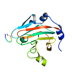
2H7T
DownloadVisualize
BU of 2h7t by Molmil
Solution Structure of the C-terminal Domain of Insulin-like Growth Factor Binding Protein 2 (IGFBP-2)
Descriptor: Insulin-like growth factor-binding protein 2
Authors:Kuang, Z.
Deposit date:2006-06-03
Release date:2007-04-24
Last modified:2024-10-16
Method:SOLUTION NMR
Cite:Structure, dynamics and heparin binding of the C-terminal domain of insulin-like growth factor-binding protein-2 (IGFBP-2)
J.Mol.Biol., 364, 2006
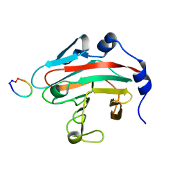
3EK9
DownloadVisualize
BU of 3ek9 by Molmil
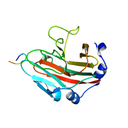
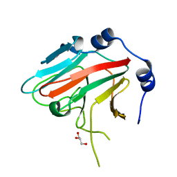
SPRY Domain-containing SOCS Box Protein 2: Crystal Structure and Residues Critical for Protein Binding
Descriptor: GLYCEROL, SPRY domain-containing SOCS box protein 2
Authors:Kuang, Z, Yao, S, Xu, Y, Garrett, T.J.P, Norton, R.S.
Deposit date:2008-09-19
Release date:2009-02-24
Last modified:2023-08-30
Method:X-RAY DIFFRACTION (2.6 Å)
Cite:SPRY domain-containing SOCS box protein 2: crystal structure and residues critical for protein binding.
J.Mol.Biol., 386, 2009
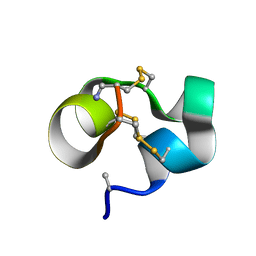
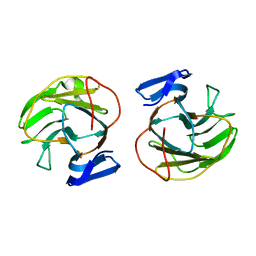
2LOC
DownloadVisualize
BU of 2loc by Molmil
Conotoxin analogue [D-Ala2]BuIIIB
Descriptor: Mu-conotoxin BuIIIB
Authors:Kuang, Z.
Deposit date:2012-01-21
Release date:2013-01-23
Last modified:2024-10-16
Method:SOLUTION NMR
Cite:Mammalian neuronal sodium channel blocker mu-conotoxin BuIIIB has a structured N-terminus that influences potency.
Acs Chem.Biol., 8, 2013
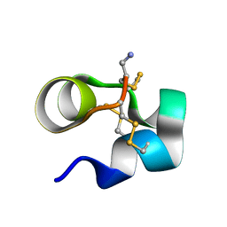
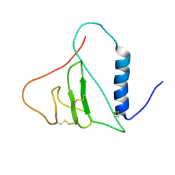
2LO9
DownloadVisualize
BU of 2lo9 by Molmil
NMR solution structure of Mu-contoxin BuIIIB
Descriptor: Mu-conotoxin BuIIIB
Authors:Kuang, Z.
Deposit date:2012-01-20
Release date:2013-01-23
Last modified:2023-06-14
Method:SOLUTION NMR
Cite:Mammalian neuronal sodium channel blocker mu-conotoxin BuIIIB has a structured N-terminus that influences potency.
Acs Chem.Biol., 8, 2013
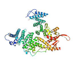
7EWF
DownloadVisualize
BU of 7ewf by Molmil
Selenomethionine-substituted structure of S. cerevisiae Csn12 in complex with Thp3 and Sem1
Descriptor: 26S proteasome complex subunit SEM1, Cop9 signalosome complex subunit 12, Protein THP3
Authors:Kuang, Z, Niu, L.
Deposit date:2021-05-25
Release date:2022-09-07
Last modified:2024-10-16
Method:X-RAY DIFFRACTION (2.85 Å)
Cite:Structural assembly of the nucleic-acid-binding Thp3-Csn12-Sem1 complex functioning in mRNA splicing.
Nucleic Acids Res., 50, 2022
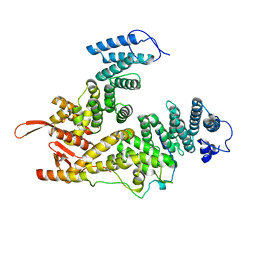
7EWM
DownloadVisualize
BU of 7ewm by Molmil
Native crystal structure of S. cerevisiae Csn12 in complex with Thp3 and Sem1
Descriptor: 26S proteasome complex subunit SEM1, Cop9 signalosome complex subunit 12, Protein THP3
Authors:Kuang, Z, Niu, L.
Deposit date:2021-05-25
Release date:2022-09-07
Last modified:2023-11-29
Method:X-RAY DIFFRACTION (2.9 Å)
Cite:Structural assembly of the nucleic-acid-binding Thp3-Csn12-Sem1 complex functioning in mRNA splicing.
Nucleic Acids Res., 50, 2022
8JAT
DownloadVisualize
BU of 8jat by Molmil
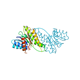
Crystal structure of the 3-ketodihydrosphingosine reductase TSC10 from Cryptococcus neoformans
Descriptor: 3-ketodihydrosphingosine reductase TSC10, NADPH DIHYDRO-NICOTINAMIDE-ADENINE-DINUCLEOTIDE PHOSPHATE
Authors:Zhao, P, Kuang, Z.
Deposit date:2023-05-07
Release date:2023-06-21
Last modified:2024-05-29
Method:X-RAY DIFFRACTION (3.2 Å)
Cite:Crystal structure of the 3-ketodihydrosphingosine reductase TSC10 from Cryptococcus neoformans.
Biochem.Biophys.Res.Commun., 670, 2023
6JKJ
DownloadVisualize
BU of 6jkj by Molmil
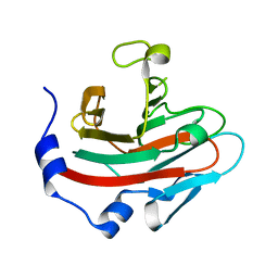
Crystal structure of human SPSB2 in the apo-state
Descriptor: SPRY domain-containing SOCS box protein 2
Authors:Luo, Y, Kuang, Z.
Deposit date:2019-03-01
Release date:2019-07-03
Last modified:2023-11-22
Method:X-RAY DIFFRACTION (1.9 Å)
Cite:Crystal structure of the SPRY domain of human SPSB2 in the apo state.
Acta Crystallogr.,Sect.F, 75, 2019
6JWN
DownloadVisualize
BU of 6jwn by Molmil
Crystal structure of the SPRY domain of SPSB2 in complex with cR9, a cyclic peptide inhibitor of SPSB-iNOS interaction
Descriptor: Nitric oxide synthase, inducible, SPRY domain-containing SOCS box protein 2
Authors:Li, K, Kuang, Z.
Deposit date:2019-04-21
Release date:2020-04-22
Last modified:2024-03-27
Method:X-RAY DIFFRACTION (1.61 Å)
Cite:Crystal structure of SPSB2 in complex with cR9, a cyclic peptide inhibitor of SPSB-iNOS interaction
To Be Published
6KEY
DownloadVisualize
BU of 6key by Molmil
Structural basis for the regulation of inducible nitric oxide synthase (iNOS) by the SPRY domain-containing SOCS box protein 2 (SPSB2)
Descriptor: Nitric oxide synthase, inducible, SPRY domain-containing SOCS box protein 2
Authors:Li, K, Kuang, Z.
Deposit date:2019-07-05
Release date:2020-07-01
Last modified:2024-03-27
Method:X-RAY DIFFRACTION (1.24 Å)
Cite:Structural basis for the regulation of inducible nitric oxide synthase by the SPRY domain-containing SOCS box protein SPSB2, an E3 ubiquitin ligase.
Nitric Oxide, 113-114, 2021
6JWM
DownloadVisualize
BU of 6jwm by Molmil
Crystal structure of the SPRY domain of SPSB2 in complex with cR7, a potent cyclic peptide inhibitor of SPSB2-iNOS interaction
Descriptor: Nitric oxide synthase, inducible, SPRY domain-containing SOCS box protein 2
Authors:Li, K, Kuang, Z.
Deposit date:2019-04-21
Release date:2020-04-22
Last modified:2024-03-27
Method:X-RAY DIFFRACTION (1.23 Å)
Cite:Crystal structure of SPSB2 in complex with cR7, a potent cyclic peptide inhibitor of SPSB2-iNOS interaction
To Be Published
5XN3
DownloadVisualize
BU of 5xn3 by Molmil
Crystal structure of SPSB2 in complex with a rational designed RGD containing cyclic peptide inhibitor of SPSB2-iNOS interaction
Descriptor: SPRY domain-containing SOCS box protein 2, cR8 peptide from NOS2
Authors:You, T, Kuang, Z.
Deposit date:2017-05-17
Release date:2017-06-14
Last modified:2023-11-22
Method:X-RAY DIFFRACTION (1.34 Å)
Cite:Crystal structure of SPSB2 in complex with a rational designed RGD-containing cyclic peptide inhibitor of SPSB2-iNOS interaction.
Biochem. Biophys. Res. Commun., 489, 2017
7CCB
DownloadVisualize
BU of 7ccb by Molmil
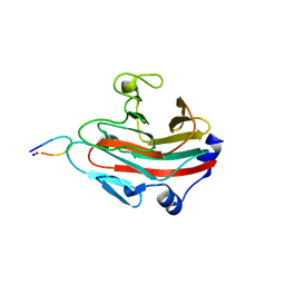
Crystal structure of the SPRY domain-containing protein 7 (SPRY7)
Descriptor: SPRY domain-containing protein 7
Authors:Yang, J, Kuang, Z.
Deposit date:2020-06-16
Release date:2020-10-14
Last modified:2023-11-29
Method:X-RAY DIFFRACTION (1.62 Å)
Cite:Crystal structure of the SPRY domain-containing protein 7 reveals unique structural features.
Biochem.Biophys.Res.Commun., 531, 2020

226707

數據於2024-10-30公開中

PDB statisticsPDBj update infoContact PDBjnumon