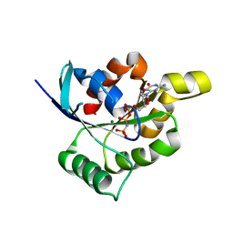
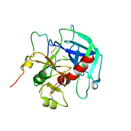
4CDB
 
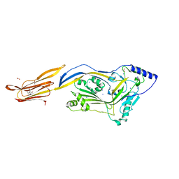 | | Crystal structure of listeriolysin O | | 分子名称: | ACETATE ION, LISTERIOLYSIN O, SODIUM ION, ... | | 著者 | Koester, S, Yildiz, O. | | 登録日 | 2013-10-30 | | 公開日 | 2014-04-23 | | 最終更新日 | 2023-12-20 | | 実験手法 | X-RAY DIFFRACTION (2.15 Å) | | 主引用文献 | Crystal Structure of Listeriolysin O Reveals Molecular Details of Oligomerization and Pore Formation
Nat.Commun., 5, 2014
|
|
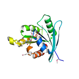
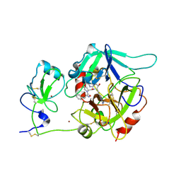
2YGG
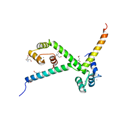 | | Complex of CaMBR and CaM | | 分子名称: | (4R)-2-METHYLPENTANE-2,4-DIOL, (4S)-2-METHYL-2,4-PENTANEDIOL, CALCIUM ION, ... | | 著者 | Koester, S, Yildiz, O. | | 登録日 | 2011-04-15 | | 公開日 | 2011-09-28 | | 最終更新日 | 2024-05-08 | | 実験手法 | X-RAY DIFFRACTION (2.227 Å) | | 主引用文献 | Structure of Human Na+/H+ Exchanger Nhe1 Regulatory Region in Complex with Cam and Ca2+
J.Biol.Chem., 286, 2011
|
|
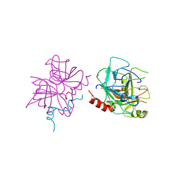
2WJH
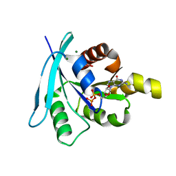 | | Structure and function of the FeoB G-domain from Methanococcus jannaschii | | 分子名称: | CITRATE ANION, FERROUS IRON TRANSPORT PROTEIN B HOMOLOG, GUANOSINE-5'-DIPHOSPHATE, ... | | 著者 | Koester, S, Wehner, M, Herrmann, C, Kuehlbrandt, W, Yildiz, O. | | 登録日 | 2009-05-26 | | 公開日 | 2009-07-28 | | 最終更新日 | 2023-12-13 | | 実験手法 | X-RAY DIFFRACTION (2.1 Å) | | 主引用文献 | Structure and Function of the Feob G-Domain from Methanococcus Jannaschii
J.Mol.Biol., 392, 2009
|
|
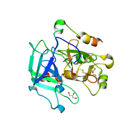
2WJG
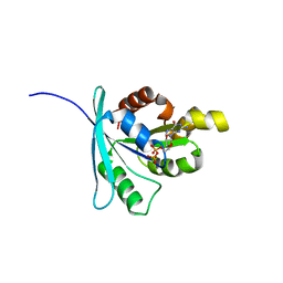 | | Structure and function of the FeoB G-domain from Methanococcus jannaschii | | 分子名称: | FERROUS IRON TRANSPORT PROTEIN B HOMOLOG, GUANOSINE-5'-DIPHOSPHATE, POLYALANINE | | 著者 | Koester, S, Wehner, M, Herrmann, C, Kuehlbrandt, W, Yildiz, O. | | 登録日 | 2009-05-26 | | 公開日 | 2009-07-28 | | 最終更新日 | 2024-05-08 | | 実験手法 | X-RAY DIFFRACTION (2.2 Å) | | 主引用文献 | Structure and Function of the Feob G-Domain from Methanococcus Jannaschii.
J.Mol.Biol., 392, 2009
|
|
2WJI
 | | Structure and function of the FeoB G-domain from Methanococcus jannaschii | | 分子名称: | FERROUS IRON TRANSPORT PROTEIN B HOMOLOG, MAGNESIUM ION, PHOSPHATE ION, ... | | 著者 | Koester, S, Wehner, M, Herrmann, C, Kuehlbrandt, W, Yildiz, O. | | 登録日 | 2009-05-26 | | 公開日 | 2009-07-28 | | 最終更新日 | 2023-12-13 | | 実験手法 | X-RAY DIFFRACTION (1.903 Å) | | 主引用文献 | Structure and Function of the Feob G-Domain from Methanococcus Jannaschii
J.Mol.Biol., 392, 2009
|
|
2WJJ
 | | Structure and function of the FeoB G-domain from Methanococcus jannaschii | | 分子名称: | FERROUS IRON TRANSPORT PROTEIN B HOMOLOG, GUANOSINE-5'-DIPHOSPHATE | | 著者 | Koester, S, Wehner, M, Herrmann, C, Kuehlbrandt, W, Yildiz, O. | | 登録日 | 2009-05-26 | | 公開日 | 2009-07-28 | | 最終更新日 | 2011-07-13 | | 実験手法 | X-RAY DIFFRACTION (2.405 Å) | | 主引用文献 | Structure and Function of the Feob G-Domain from Methanococcus Jannaschii
J.Mol.Biol., 392, 2009
|
|
6P9U
 | | Crystal structure of human thrombin mutant W215A | | 分子名称: | 2-acetamido-2-deoxy-beta-D-glucopyranose, Prothrombin, ZINC ION | | 著者 | Pelc, L.A, Koester, S.K, Chen, Z, Di Cera, E. | | 登録日 | 2019-06-10 | | 公開日 | 2019-09-04 | | 最終更新日 | 2023-10-11 | | 実験手法 | X-RAY DIFFRACTION (3.3 Å) | | 主引用文献 | Residues W215, E217 and E192 control the allosteric E*-E equilibrium of thrombin.
Sci Rep, 9, 2019
|
|
6PXJ
 | | Crystal structure of human thrombin mutant I16T | | 分子名称: | GLYCEROL, MAGNESIUM ION, Thrombin heavy chain, ... | | 著者 | Stojanovski, B, Chen, Z, Koester, S.K, Pelc, L.A, Di Cera, E. | | 登録日 | 2019-07-26 | | 公開日 | 2019-12-18 | | 最終更新日 | 2023-10-11 | | 実験手法 | X-RAY DIFFRACTION (1.7 Å) | | 主引用文献 | Role of the I16-D194 ionic interaction in the trypsin fold.
Sci Rep, 9, 2019
|
|
6PXQ
 | | Crystal structure of human thrombin mutant D194A | | 分子名称: | 2-acetamido-2-deoxy-beta-D-glucopyranose, Thrombin heavy chain, Thrombin light chain | | 著者 | Stojanovski, B, Chen, Z, Koester, S.K, Pelc, L.A, Di Cera, E. | | 登録日 | 2019-07-26 | | 公開日 | 2019-12-18 | | 最終更新日 | 2023-10-11 | | 実験手法 | X-RAY DIFFRACTION (2.8 Å) | | 主引用文献 | Role of the I16-D194 ionic interaction in the trypsin fold.
Sci Rep, 9, 2019
|
|
6PX5
 | | CRYSTAL STRUCTURE OF HUMAN MEIZOTHROMBIN DESF1 MUTANT S195A bound with PPACK | | 分子名称: | 2-acetamido-2-deoxy-beta-D-glucopyranose-(1-4)-2-acetamido-2-deoxy-beta-D-glucopyranose, D-phenylalanyl-N-[(2S,3S)-6-{[amino(iminio)methyl]amino}-1-chloro-2-hydroxyhexan-3-yl]-L-prolinamide, Prothrombin, ... | | 著者 | Pelc, L.A, Koester, S.K, Chen, Z, Gistover, N, Di Cera, E. | | 登録日 | 2019-07-24 | | 公開日 | 2019-09-04 | | 最終更新日 | 2023-10-11 | | 実験手法 | X-RAY DIFFRACTION (2.4 Å) | | 主引用文献 | Residues W215, E217 and E192 control the allosteric E*-E equilibrium of thrombin.
Sci Rep, 9, 2019
|
|


