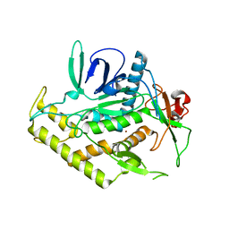
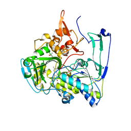
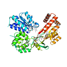
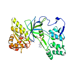
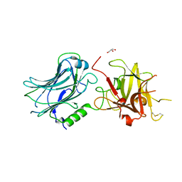
3N7J
 
 | | Crystal structure of botulinum neurotoxin serotype D binding domain | | 分子名称: | Botulinum neurotoxin type D | | 著者 | Fu, Z, Baldwin, M.R, Karalewitz, A, Kroken, A, Barbieri, J.T, Kim, J.-J.P. | | 登録日 | 2010-05-27 | | 公開日 | 2010-09-08 | | 最終更新日 | 2023-09-06 | | 実験手法 | X-RAY DIFFRACTION (2 Å) | | 主引用文献 | Identification of a Unique Ganglioside Binding Loop within Botulinum Neurotoxins C and D-SA .
Biochemistry, 49, 2010
|
|
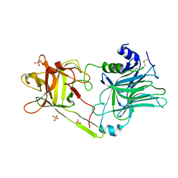
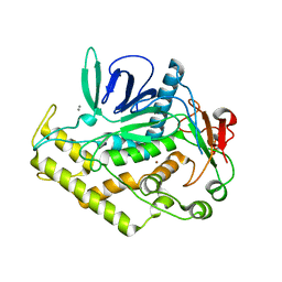
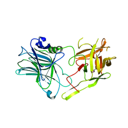
3N7K
 | | Crystal structure of botulinum neurotoxin serotype C1 binding domain | | 分子名称: | Botulinum neurotoxin type C1 | | 著者 | Fu, Z, Kroken, A, Karalewitz, A, Baldwin, M.R, Barbieri, J.T, Kim, J.-J.P. | | 登録日 | 2010-05-27 | | 公開日 | 2010-09-08 | | 最終更新日 | 2023-09-06 | | 実験手法 | X-RAY DIFFRACTION (2.5 Å) | | 主引用文献 | Identification of a Unique Ganglioside Binding Loop within Botulinum Neurotoxins C and D-SA .
Biochemistry, 49, 2010
|
|
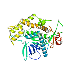
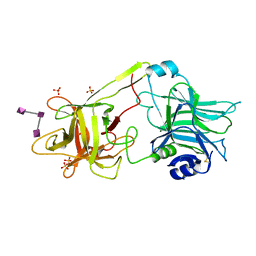
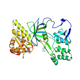
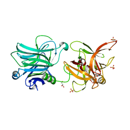
3FUQ
 | | Glycosylated SV2 and Gangliosides as Dual Receptors for Botulinum Neurotoxin Serotype F | | 分子名称: | BoNT/F (Neurotoxin type F) | | 著者 | Fu, Z, Chen, C, Barbieri, J.T, Kim, J.-J.P, Baldwin, M.R. | | 登録日 | 2009-01-14 | | 公開日 | 2009-06-16 | | 最終更新日 | 2023-09-06 | | 実験手法 | X-RAY DIFFRACTION (2.1 Å) | | 主引用文献 | Glycosylated SV2 and gangliosides as dual receptors for botulinum neurotoxin serotype F
Biochemistry, 48, 2009
|
|
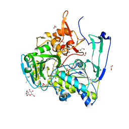
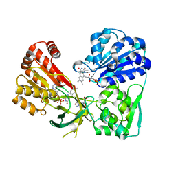
3FUO
 | | The Crystal structure of receptor binding domain of botulinum neurotoxin serotype A | | 分子名称: | Botulinum neurotoxin type A | | 著者 | Fu, Z, Chen, C, Barbieri, J.T, Kim, J.-J.P, Baldwin, M.R. | | 登録日 | 2009-01-14 | | 公開日 | 2009-06-16 | | 最終更新日 | 2023-09-06 | | 実験手法 | X-RAY DIFFRACTION (1.8 Å) | | 主引用文献 | Glycosylated SV2 and gangliosides as dual receptors for botulinum neurotoxin serotype F
Biochemistry, 48, 2009
|
|
2G7P
 | | Structure of the Light Chain of Botulinum Neurotoxin Serotype A Bound to Small Molecule Inhibitors | | 分子名称: | Botulinum neurotoxin type A, ZINC ION | | 著者 | Fu, Z, Baldwin, M.R, Boldt, G.E, Janda, K.D, Barbieri, J.T, Kim, J.-J.P. | | 登録日 | 2006-02-28 | | 公開日 | 2006-08-15 | | 最終更新日 | 2023-08-30 | | 実験手法 | X-RAY DIFFRACTION (2.3 Å) | | 主引用文献 | Light chain of botulinum neurotoxin serotype A: structural resolution of a catalytic intermediate.
Biochemistry, 45, 2006
|
|
3HN1
 | | Crystal structure of HCR/T complexed with GT2 and lactose | | 分子名称: | N-acetyl-alpha-neuraminic acid-(2-8)-N-acetyl-alpha-neuraminic acid, SULFATE ION, Tetanus toxin, ... | | 著者 | Chen, C, Fu, Z, Kim, J.-J.P, Barbieri, J.T, Baldwin, M.R. | | 登録日 | 2009-05-29 | | 公開日 | 2009-07-14 | | 最終更新日 | 2023-09-06 | | 実験手法 | X-RAY DIFFRACTION (2.1 Å) | | 主引用文献 | Gangliosides as high affinity receptors for tetanus neurotoxin.
J.Biol.Chem., 284, 2009
|
|
2G7Q
 | | Structure of the Light Chain of Botulinum Neurotoxin Serotype A Bound to Small Molecule Inhibitors | | 分子名称: | Botulinum neurotoxin type A, N-HYDROXY-L-ARGININAMIDE, ZINC ION | | 著者 | Fu, Z, Baldwin, M.R, Boldt, G.E, Janda, K.D, Barbieri, J.T, Kim, J.-J.P. | | 登録日 | 2006-02-28 | | 公開日 | 2006-08-15 | | 最終更新日 | 2023-08-30 | | 実験手法 | X-RAY DIFFRACTION (2.41 Å) | | 主引用文献 | Light chain of botulinum neurotoxin serotype A: structural resolution of a catalytic intermediate.
Biochemistry, 45, 2006
|
|
2G7K
 | | Structure of the Light Chain of Botulinum Neurotoxin, Serotype A Bound to small Molecule Inhibitors | | 分子名称: | Botulinum neurotoxin type A | | 著者 | Fu, Z, Baldwin, M.R, Boldt, G.E, Crawford, A, Janda, K.D, Barbieri, J.T, Kim, J.-J.P. | | 登録日 | 2006-02-28 | | 公開日 | 2006-08-15 | | 最終更新日 | 2023-08-30 | | 実験手法 | X-RAY DIFFRACTION (2.8 Å) | | 主引用文献 | Light chain of botulinum neurotoxin serotype A: structural resolution of a catalytic intermediate.
Biochemistry, 45, 2006
|
|
2GMJ
 | | Structure of Porcine Electron Transfer Flavoprotein-Ubiquinone Oxidoreductase | | 分子名称: | Electron transfer flavoprotein-ubiquinone oxidoreductase, FLAVIN-ADENINE DINUCLEOTIDE, IRON/SULFUR CLUSTER, ... | | 著者 | Zhang, J, Frerman, F.E, Kim, J.-J.P. | | 登録日 | 2006-04-06 | | 公開日 | 2006-10-17 | | 最終更新日 | 2023-08-30 | | 実験手法 | X-RAY DIFFRACTION (2.6 Å) | | 主引用文献 | Structure of electron transfer flavoprotein-ubiquinone oxidoreductase and electron transfer to the mitochondrial ubiquinone pool.
Proc.Natl.Acad.Sci.Usa, 103, 2006
|
|
3HMY
 | | Crystal structure of HCR/T complexed with GT2 | | 分子名称: | GLYCEROL, N-acetyl-alpha-neuraminic acid-(2-8)-N-acetyl-alpha-neuraminic acid-(2-8)-N-acetyl-alpha-neuraminic acid, SULFATE ION, ... | | 著者 | Chen, C, Fu, Z, Kim, J.-J.P, Barbieri, J.T, Baldwin, M.R. | | 登録日 | 2009-05-29 | | 公開日 | 2009-07-14 | | 最終更新日 | 2023-09-06 | | 実験手法 | X-RAY DIFFRACTION (2 Å) | | 主引用文献 | Gangliosides as high affinity receptors for tetanus neurotoxin.
J.Biol.Chem., 284, 2009
|
|
2GMH
 | | Structure of Porcine Electron Transfer Flavoprotein-Ubiquinone Oxidoreductase in Complexed with Ubiquinone | | 分子名称: | 1,2-ETHANEDIOL, 2,3-DIMETHOXY-5-METHYL-6-(3,11,15,19-TETRAMETHYL-EICOSA-2,6,10,14,18-PENTAENYL)-[1,4]BENZOQUINONE, Electron transfer flavoprotein-ubiquinone oxidoreductase, ... | | 著者 | Zhang, J, Frerman, F.E, Kim, J.-J.P. | | 登録日 | 2006-04-06 | | 公開日 | 2006-10-17 | | 最終更新日 | 2024-02-14 | | 実験手法 | X-RAY DIFFRACTION (2.5 Å) | | 主引用文献 | Structure of electron transfer flavoprotein-ubiquinone oxidoreductase and electron transfer to the mitochondrial ubiquinone pool.
Proc.Natl.Acad.Sci.Usa, 103, 2006
|
|
3OJW
 | | Disulfide crosslinked cytochrome P450 reductase inactive | | 分子名称: | FLAVIN MONONUCLEOTIDE, FLAVIN-ADENINE DINUCLEOTIDE, NADPH-cytochrome P450 reductase | | 著者 | Xia, C, Hamdane, D, Shen, A, Choi, V, Kasper, C, Zhang, H, Im, S.-C, Waskell, L, Kim, J.-J.P. | | 登録日 | 2010-08-23 | | 公開日 | 2011-02-23 | | 最終更新日 | 2023-09-06 | | 実験手法 | X-RAY DIFFRACTION (2.2 Å) | | 主引用文献 | Conformational Changes of NADPH-Cytochrome P450 Oxidoreductase Are Essential for Catalysis and Cofactor Binding.
J.Biol.Chem., 286, 2011
|
|
3QE2
 | | Crystal Structure of Human NADPH-Cytochrome P450 Reductase | | 分子名称: | CALCIUM ION, FLAVIN MONONUCLEOTIDE, FLAVIN-ADENINE DINUCLEOTIDE, ... | | 著者 | Xia, C, Marohnic, C, Panda, S.P, Masters, B.S, Kim, J.-J.P. | | 登録日 | 2011-01-19 | | 公開日 | 2011-08-03 | | 最終更新日 | 2023-09-13 | | 実験手法 | X-RAY DIFFRACTION (1.75 Å) | | 主引用文献 | Structural basis for human NADPH-cytochrome P450 oxidoreductase deficiency.
Proc.Natl.Acad.Sci.USA, 108, 2011
|
|
3QFT
 | | Crystal Structure of NADPH-Cytochrome P450 Reductase (FAD/NADPH domain and R457H Mutant) | | 分子名称: | FLAVIN-ADENINE DINUCLEOTIDE, NADP NICOTINAMIDE-ADENINE-DINUCLEOTIDE PHOSPHATE, NADPH--cytochrome P450 reductase | | 著者 | Xia, C, Marohnic, C, Panda, S.P, Masters, B.S, Kim, J.-J.P. | | 登録日 | 2011-01-22 | | 公開日 | 2011-08-17 | | 最終更新日 | 2023-09-13 | | 実験手法 | X-RAY DIFFRACTION (1.4 Å) | | 主引用文献 | Structural basis for human NADPH-cytochrome P450 oxidoreductase deficiency.
Proc.Natl.Acad.Sci.USA, 108, 2011
|
|
3QFC
 | | Crystal Structure of Human NADPH-Cytochrome P450 (V492E mutant) | | 分子名称: | CALCIUM ION, FLAVIN MONONUCLEOTIDE, FLAVIN-ADENINE DINUCLEOTIDE, ... | | 著者 | Xia, C, Marohnic, C, Panda, S.P, Masters, B.S, Kim, J.-J.P. | | 登録日 | 2011-01-21 | | 公開日 | 2011-08-03 | | 最終更新日 | 2023-09-13 | | 実験手法 | X-RAY DIFFRACTION (1.8 Å) | | 主引用文献 | Structural basis for human NADPH-cytochrome P450 oxidoreductase deficiency.
Proc.Natl.Acad.Sci.USA, 108, 2011
|
|
3QFR
 | | Crystal Structure of Human NADPH-Cytochrome P450 Reductase (R457H Mutant) | | 分子名称: | CALCIUM ION, FLAVIN MONONUCLEOTIDE, FLAVIN-ADENINE DINUCLEOTIDE, ... | | 著者 | Xia, C, Marohnic, C, Panda, S.P, Masters, B.S, Kim, J.-J.P. | | 登録日 | 2011-01-22 | | 公開日 | 2011-08-03 | | 最終更新日 | 2023-09-13 | | 実験手法 | X-RAY DIFFRACTION (2.4 Å) | | 主引用文献 | Structural basis for human NADPH-cytochrome P450 oxidoreductase deficiency.
Proc.Natl.Acad.Sci.USA, 108, 2011
|
|
3QFS
 | | Crystal Structure of NADPH-Cytochrome P450 Reductase (FAD/NADPH domain) | | 分子名称: | FLAVIN-ADENINE DINUCLEOTIDE, NADP NICOTINAMIDE-ADENINE-DINUCLEOTIDE PHOSPHATE, NADPH--cytochrome P450 reductase | | 著者 | Xia, C, Marohnic, C, Panda, S.P, Masters, B.S, Kim, J.-J.P. | | 登録日 | 2011-01-22 | | 公開日 | 2011-08-17 | | 最終更新日 | 2023-09-13 | | 実験手法 | X-RAY DIFFRACTION (1.4 Å) | | 主引用文献 | Structural basis for human NADPH-cytochrome P450 oxidoreductase deficiency.
Proc.Natl.Acad.Sci.USA, 108, 2011
|
|
2G7N
 | | Structure of the Light Chain of Botulinum Neurotoxin Serotype A Bound to small Molecule Inhibitors | | 分子名称: | Botulinum neurotoxin type A, SILVER ION, ZINC ION | | 著者 | Fu, Z, Baldwin, M.R, Boldt, G.E, Janda, K.D, Barbieri, J.T, Kim, J.-J.P. | | 登録日 | 2006-02-28 | | 公開日 | 2007-03-13 | | 最終更新日 | 2023-08-30 | | 実験手法 | X-RAY DIFFRACTION (1.9 Å) | | 主引用文献 | Structure of the Light Chain of Botulinum Neurotoxin Serotype A bound to Small Molecule Inhibitors
To be Published
|
|
3N7L
 | | Crystal structure of botulinum neurotoxin serotype D/C VPI 5993 binding domain | | 分子名称: | GLYCEROL, Neurotoxin, SULFATE ION | | 著者 | Fu, Z, Karalewitz, A, Kroken, A, Baldwin, M.R, Barbieri, J.T, Kim, J.-J.P. | | 登録日 | 2010-05-27 | | 公開日 | 2010-09-08 | | 最終更新日 | 2023-09-06 | | 実験手法 | X-RAY DIFFRACTION (2 Å) | | 主引用文献 | Identification of a Unique Ganglioside Binding Loop within Botulinum Neurotoxins C and D-SA .
Biochemistry, 49, 2010
|
|
3OJX
 | | Disulfide crosslinked cytochrome P450 reductase inactive | | 分子名称: | FLAVIN MONONUCLEOTIDE, FLAVIN-ADENINE DINUCLEOTIDE, NADP NICOTINAMIDE-ADENINE-DINUCLEOTIDE PHOSPHATE, ... | | 著者 | Xia, C, Hamdane, D, Shen, A, Choi, V, Kasper, C, Zhang, H, Im, S.-C, Waskell, L, Kim, J.-J.P. | | 登録日 | 2010-08-23 | | 公開日 | 2011-02-23 | | 最終更新日 | 2023-09-06 | | 実験手法 | X-RAY DIFFRACTION (2.5 Å) | | 主引用文献 | Conformational Changes of NADPH-Cytochrome P450 Oxidoreductase Are Essential for Catalysis and Cofactor Binding.
J.Biol.Chem., 286, 2011
|
|
3RMX
 | | Crystal structure of HCR/D F1240A mutant | | 分子名称: | Botulinum neurotoxin type D | | 著者 | Fu, Z, Karalewitz, A, Kroken, A, Kim, J.-J.P, Barbieri, J.T. | | 登録日 | 2011-04-21 | | 公開日 | 2011-06-01 | | 最終更新日 | 2023-09-13 | | 実験手法 | X-RAY DIFFRACTION (2.75 Å) | | 主引用文献 | Novel Ganglioside-mediated Entry of Botulinum Neurotoxin Serotype D into Neurons.
J.Biol.Chem., 286, 2011
|
|
3RMY
 | | Crystal structure of HCR/D W1238A mutant | | 分子名称: | Botulinum neurotoxin type D, GLYCEROL | | 著者 | Fu, Z, Karalewitz, A, Kroken, A, Kim, J.-J.P, Barbieri, J.T. | | 登録日 | 2011-04-21 | | 公開日 | 2011-06-01 | | 最終更新日 | 2023-09-13 | | 実験手法 | X-RAY DIFFRACTION (2.3 Å) | | 主引用文献 | Novel Ganglioside-mediated Entry of Botulinum Neurotoxin Serotype D into Neurons.
J.Biol.Chem., 286, 2011
|
|
3RSJ
 | | Structure of HCRF in complex with Ganglioside GD1a | | 分子名称: | BoNT/F, N-acetyl-alpha-neuraminic acid-(2-3)-beta-D-galactopyranose-(1-3)-2-acetamido-2-deoxy-beta-D-galactopyranose, N-acetyl-alpha-neuraminic acid-(2-3)-beta-D-galactopyranose-(1-3)-2-acetamido-2-deoxy-beta-D-galactopyranose-(1-4)-[N-acetyl-alpha-neuraminic acid-(2-3)]beta-D-galactopyranose, ... | | 著者 | Fu, Z, Benson, M.A, Barbieri, J.T, Kim, J.-J.P, Baldwin, M.R. | | 登録日 | 2011-05-02 | | 公開日 | 2011-08-17 | | 最終更新日 | 2023-09-13 | | 実験手法 | X-RAY DIFFRACTION (2 Å) | | 主引用文献 | Unique ganglioside recognition strategies for clostridial neurotoxins.
J.Biol.Chem., 286, 2011
|
|
1Q25
 | | Crystal structure of N-terminal 3 domains of CI-MPR | | 分子名称: | 2-acetamido-2-deoxy-beta-D-glucopyranose-(1-4)-2-acetamido-2-deoxy-beta-D-glucopyranose, GLYCEROL, alpha-D-mannopyranose-(1-3)-[alpha-D-mannopyranose-(1-6)]beta-D-mannopyranose-(1-4)-2-acetamido-2-deoxy-beta-D-glucopyranose-(1-4)-2-acetamido-2-deoxy-beta-D-glucopyranose, ... | | 著者 | Olson, L.J, Dahms, N.M, Kim, J.-J.P. | | 登録日 | 2003-07-23 | | 公開日 | 2004-06-29 | | 最終更新日 | 2020-07-29 | | 実験手法 | X-RAY DIFFRACTION (1.8 Å) | | 主引用文献 | Structure of uPAR, plasminogen, and sugar-binding sites of the 300 kDa mannose 6-phosphate receptor
Embo J., 23, 2004
|
|
3FH4
 | | Crystal Structure of Recombinant Vibrio proteolyticus aminopeptidase | | 分子名称: | 2-AMINO-2-HYDROXYMETHYL-PROPANE-1,3-DIOL, Bacterial leucyl aminopeptidase, SODIUM ION, ... | | 著者 | Yong, W, Kim, J.-J.P, Hartley, M, Bennett, B. | | 登録日 | 2008-12-08 | | 公開日 | 2009-11-17 | | 最終更新日 | 2023-09-06 | | 実験手法 | X-RAY DIFFRACTION (1.95 Å) | | 主引用文献 | Heterologous expression and purification of Vibrio proteolyticus (Aeromonas proteolytica) aminopeptidase: a rapid protocol
Protein Expr.Purif., 66, 2009
|
|


