6JL3
 
 | |
4PG3
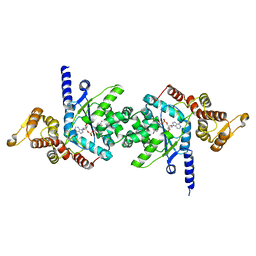
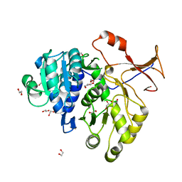
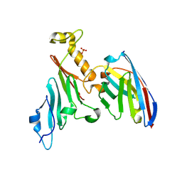
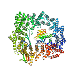
 | | Crystal structure of KRS complexed with inhibitor | | 分子名称: | LYSINE, Lysine--tRNA ligase, cladosporin | | 著者 | Sharma, A, Yogavel, M, Khan, S, Sharma, A, Belrhali, H. | | 登録日 | 2014-05-01 | | 公開日 | 2014-07-16 | | 最終更新日 | 2023-09-27 | | 実験手法 | X-RAY DIFFRACTION (2.696 Å) | | 主引用文献 | Structural basis of malaria parasite lysyl-tRNA synthetase inhibition by cladosporin.
J. Struct. Funct. Genomics, 15, 2014
|
|
4OO9
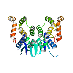
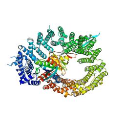
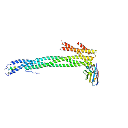
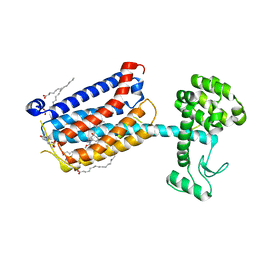 | | Structure of the human class C GPCR metabotropic glutamate receptor 5 transmembrane domain in complex with the negative allosteric modulator mavoglurant | | 分子名称: | 2-(N-MORPHOLINO)-ETHANESULFONIC ACID, Mavoglurant, Metabotropic glutamate receptor 5, ... | | 著者 | Dore, A.S, Okrasa, K, Patel, J.C, Serrano-Vega, M, Bennett, K, Cooke, R.M, Errey, J.C, Jazayeri, A, Khan, S, Tehan, B, Weir, M, Wiggin, G.R, Marshall, F.H. | | 登録日 | 2014-01-31 | | 公開日 | 2014-07-02 | | 最終更新日 | 2023-09-20 | | 実験手法 | X-RAY DIFFRACTION (2.6 Å) | | 主引用文献 | Structure of class C GPCR metabotropic glutamate receptor 5 transmembrane domain.
Nature, 511, 2014
|
|
3VGJ
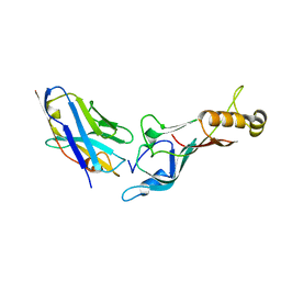
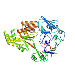
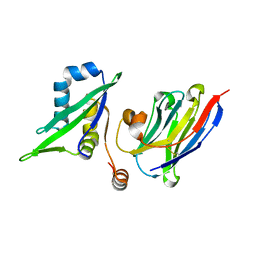
 | | Crystal of Plasmodium falciparum tyrosyl-tRNA synthetase (PfTyrRS)in complex with adenylate analog | | 分子名称: | ADENOSINE MONOPHOSPHATE, TYROSINE, Tyrosyl-tRNA synthetase, ... | | 著者 | Banday, M.M, Yogavel, M, Bhatt, T.K, Khan, S, Sharma, A, Sharma, A. | | 登録日 | 2011-08-14 | | 公開日 | 2012-07-25 | | 最終更新日 | 2024-03-20 | | 実験手法 | X-RAY DIFFRACTION (2.212 Å) | | 主引用文献 | Malaria parasite tyrosyl-tRNA synthetase secretion triggers pro-inflammatory responses.
Nat Commun, 2, 2011
|
|
5ZKH
 | |
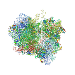
4V63
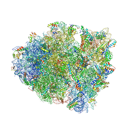 | | Structural basis for translation termination on the 70S ribosome. | | 分子名称: | 16S RRNA, 23S RRNA, 30S ribosomal protein S10, ... | | 著者 | Laurberg, M, Asahara, H, Korostelev, A, Zhu, J, Trakhanov, S, Noller, H.F. | | 登録日 | 2008-05-16 | | 公開日 | 2014-07-09 | | 最終更新日 | 2023-09-20 | | 実験手法 | X-RAY DIFFRACTION (3.207 Å) | | 主引用文献 | Structural basis for translation termination on the 70S ribosome
Nature, 454, 2008
|
|
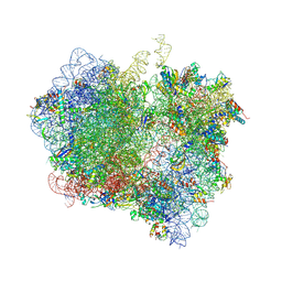
4V4J
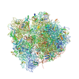 | | Interactions and Dynamics of the Shine-Dalgarno Helix in the 70S Ribosome. | | 分子名称: | 16S RNA, 23S LARGE SUBUNIT RIBOSOMAL RNA, 30S ribosomal protein S10, ... | | 著者 | Korostelev, A, Trakhanov, S, Asahara, H, Laurberg, M, Noller, H.F. | | 登録日 | 2007-07-18 | | 公開日 | 2014-07-09 | | 最終更新日 | 2023-09-20 | | 実験手法 | X-RAY DIFFRACTION (3.83 Å) | | 主引用文献 | Interactions and dynamics of the Shine Dalgarno helix in the 70S ribosome.
Proc.Natl.Acad.Sci.Usa, 104, 2007
|
|
4V4I
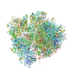 | | Crystal Structure of a 70S Ribosome-tRNA Complex Reveals Functional Interactions and Rearrangements. | | 分子名称: | 16S SMALL SUBUNIT RIBOSOMAL RNA, 23S LARGE SUBUNIT RIBOSOMAL RNA, 30S ribosomal protein S10, ... | | 著者 | Korostelev, A, Trakhanov, S, Laurberg, M, Noller, H.F. | | 登録日 | 2007-02-15 | | 公開日 | 2014-07-09 | | 最終更新日 | 2024-04-03 | | 実験手法 | X-RAY DIFFRACTION (3.71 Å) | | 主引用文献 | Crystal Structure of a 70S Ribosome-tRNA Complex Reveals Functional Interactions and Rearrangements
Cell(Cambridge,Mass.), 126, 2006
|
|
4V85
 | | Crystal Structure of Release Factor RF3 Trapped in the GTP State on a Rotated Conformation of the Ribosome. | | 分子名称: | 16S rRNA, 23S rRNA, 30S ribosomal protein S10, ... | | 著者 | Zhou, J, Lancaster, L, Trakhanov, S, Noller, H.F. | | 登録日 | 2011-06-13 | | 公開日 | 2014-07-09 | | 最終更新日 | 2019-07-17 | | 実験手法 | X-RAY DIFFRACTION (3.2 Å) | | 主引用文献 | Crystal structure of release factor RF3 trapped in the GTP state on a rotated conformation of the ribosome.
Rna, 18, 2012
|
|
4V89
 | | Crystal Structure of Release Factor RF3 Trapped in the GTP State on a Rotated Conformation of the Ribosome (without viomycin) | | 分子名称: | 16S rRNA, 23S rRNA, 30S ribosomal protein S10, ... | | 著者 | Zhou, J, Lancaster, L, Trakhanov, S, Noller, H.F. | | 登録日 | 2011-11-17 | | 公開日 | 2014-07-09 | | 最終更新日 | 2019-07-17 | | 実験手法 | X-RAY DIFFRACTION (3.7 Å) | | 主引用文献 | Crystal structure of release factor RF3 trapped in the GTP state on a rotated conformation of the ribosome.
Rna, 18, 2012
|
|
12E8
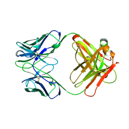 | | 2E8 FAB FRAGMENT | | 分子名称: | IGG1-KAPPA 2E8 FAB (HEAVY CHAIN), IGG1-KAPPA 2E8 FAB (LIGHT CHAIN) | | 著者 | Rupp, B, Trakhanov, S. | | 登録日 | 1998-03-14 | | 公開日 | 1998-08-05 | | 最終更新日 | 2023-08-02 | | 実験手法 | X-RAY DIFFRACTION (1.9 Å) | | 主引用文献 | Structure of a monoclonal 2E8 Fab antibody fragment specific for the low-density lipoprotein-receptor binding region of apolipoprotein E refined at 1.9 A.
Acta Crystallogr.,Sect.D, null, 1999
|
|
1HSL
 | |
3QYA
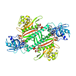
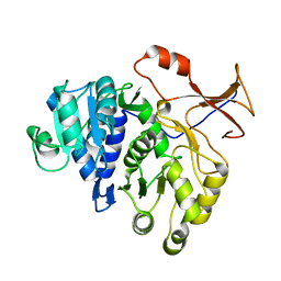
 | | Crystal structure of a red-emitter mutant of Lampyris turkestanicus luciferase | | 分子名称: | 1,2-ETHANEDIOL, DI(HYDROXYETHYL)ETHER, GLYCEROL, ... | | 著者 | Kheirabadi, M, Gohlke, U, Hossein Khani, S, Heinemann, U, Naderi-Manesh, H. | | 登録日 | 2011-03-03 | | 公開日 | 2012-03-07 | | 最終更新日 | 2023-09-13 | | 実験手法 | X-RAY DIFFRACTION (2.13 Å) | | 主引用文献 | Crystal structure of native and a mutant of Lampyris turkestanicus luciferase implicate in bioluminescence color shift.
Biochim.Biophys.Acta, 1834, 2013
|
|
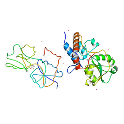
6Q84
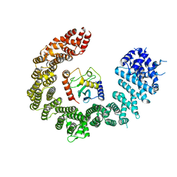
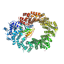
 | | Crystal structure of RanGTP-Pdr6-eIF5A export complex | | 分子名称: | Eukaryotic translation initiation factor 5A-1, GTP-binding nuclear protein Ran, GUANOSINE-5'-TRIPHOSPHATE, ... | | 著者 | Aksu, M, Trakhanov, S, Vera-Rodriguez, A, Gorlich, D. | | 登録日 | 2018-12-14 | | 公開日 | 2019-05-01 | | 最終更新日 | 2024-05-15 | | 実験手法 | X-RAY DIFFRACTION (3.7 Å) | | 主引用文献 | Structural basis for the nuclear import and export functions of the biportin Pdr6/Kap122.
J.Cell Biol., 218, 2019
|
|
5E0Q
 | |
6Q83
 | | Crystal structure of the biportin Pdr6 in complex with UBC9 | | 分子名称: | Importin beta-like protein KAP122, UBC9 | | 著者 | Aksu, M, Trakhanov, S, Vera-Rodriguez, A, Gorlich, D. | | 登録日 | 2018-12-14 | | 公開日 | 2019-05-01 | | 最終更新日 | 2024-01-24 | | 実験手法 | X-RAY DIFFRACTION (4.53 Å) | | 主引用文献 | Structural basis for the nuclear import and export functions of the biportin Pdr6/Kap122.
J.Cell Biol., 218, 2019
|
|
6Q82
 | | Crystal structure of the biportin Pdr6 in complex with RanGTP | | 分子名称: | GTP-binding nuclear protein Ran, GUANOSINE-5'-TRIPHOSPHATE, Importin beta-like protein KAP122, ... | | 著者 | Aksu, M, Vera-Rodriguez, A, Trakhanov, S, Gorlich, D. | | 登録日 | 2018-12-14 | | 公開日 | 2019-05-01 | | 最終更新日 | 2019-06-12 | | 実験手法 | X-RAY DIFFRACTION (2.994 Å) | | 主引用文献 | Structural basis for the nuclear import and export functions of the biportin Pdr6/Kap122.
J.Cell Biol., 218, 2019
|
|
7NOW
 | |
7NQA
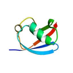
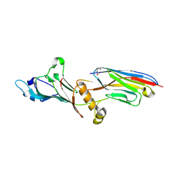 | | Crystal structure of Nucleoporin-98 nanobody MS98-6 complex solved at 2.2A resolution | | 分子名称: | 2-AMINO-2-HYDROXYMETHYL-PROPANE-1,3-DIOL, Anti-Nup98 Nanobody MS98-6, Nuclear pore complex protein Nup98-Nup96, ... | | 著者 | Sola-Colom, M, Trakhanov, S, Goerlich, D. | | 登録日 | 2021-03-01 | | 公開日 | 2021-07-21 | | 最終更新日 | 2024-05-22 | | 実験手法 | X-RAY DIFFRACTION (2.2 Å) | | 主引用文献 | A checkpoint function for Nup98 in nuclear pore formation suggested by novel inhibitory nanobodies.
Embo J., 2024
|
|
1DPE
 | |
5DLQ
 | | Crystal structure of RanGTP-Exportin 4-eIF5A complex | | 分子名称: | Eukaryotic translation initiation factor 5A-1, Exportin-4, GTP-binding nuclear protein Ran, ... | | 著者 | Aksu, M, Trakhanov, S, Gorlich, D. | | 登録日 | 2015-09-07 | | 公開日 | 2016-06-22 | | 最終更新日 | 2016-06-29 | | 実験手法 | X-RAY DIFFRACTION (3.2 Å) | | 主引用文献 | Structure of the exportin Xpo4 in complex with RanGTP and the hypusine-containing translation factor eIF5A.
Nat Commun, 7, 2016
|
|
5C3L
 | | Structure of the metazoan Nup62.Nup58.Nup54 nucleoporin complex. | | 分子名称: | Nanobody Nb15, Nucleoporin Nup58, Nucleoporin Nup62, ... | | 著者 | Chug, H, Trakhanov, S, Hulsmann, B.B, Pleiner, T, Gorlich, D. | | 登録日 | 2015-06-17 | | 公開日 | 2015-08-26 | | 最終更新日 | 2024-01-10 | | 実験手法 | X-RAY DIFFRACTION (2.9 Å) | | 主引用文献 | Crystal structure of the metazoan Nup62Nup58Nup54 nucleoporin complex.
Science, 350, 2015
|
|
5C2U
 | | Ferredoxin-like domain of nucleoporin Nup54 bound to a nanobody | | 分子名称: | Nanobody, Nup54 | | 著者 | Chug, H, Trakhanov, S, Hulsmann, B.B, Pleiner, T, Gorlich, D. | | 登録日 | 2015-06-16 | | 公開日 | 2015-08-26 | | 最終更新日 | 2024-01-10 | | 実験手法 | X-RAY DIFFRACTION (1.55 Å) | | 主引用文献 | Crystal structure of the metazoan Nup62Nup58Nup54 nucleoporin complex.
Science, 350, 2015
|
|
4M46
 | |


