8P2K
 
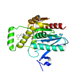
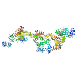
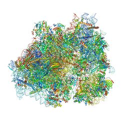 | | Ternary complex of translating ribosome, NAC and METAP1 | | 分子名称: | 18s rRNA, 28S rRNA, 40S ribosomal protein S11, ... | | 著者 | Jia, M, Jaskolowski, M, Scaiola, A, Jomaa, A, Ban, N. | | 登録日 | 2023-05-16 | | 公開日 | 2023-07-19 | | 最終更新日 | 2024-04-24 | | 実験手法 | ELECTRON MICROSCOPY (2.9 Å) | | 主引用文献 | NAC controls cotranslational N-terminal methionine excision in eukaryotes.
Science, 380, 2023
|
|
6FQ1
 | |
6FQR
 | |
4G5S
 | | Structure of LGN GL3/Galphai3 complex | | 分子名称: | CITRIC ACID, G-protein-signaling modulator 2, GUANOSINE-5'-DIPHOSPHATE, ... | | 著者 | Jia, M, Li, J, Zhu, J, Wen, W, Zhang, M, Wang, W. | | 登録日 | 2012-07-18 | | 公開日 | 2012-09-05 | | 最終更新日 | 2024-03-20 | | 実験手法 | X-RAY DIFFRACTION (3.62 Å) | | 主引用文献 | Crystal Structures of the scaffolding protein LGN reveal the general mechanism by which GoLoco binding motifs inhibit the release of GDP from Galphai subunits in G-coupled heterotrimeric proteins
To be Published
|
|
4G5R
 | | Structure of LGN GL4/Galphai3 complex | | 分子名称: | CITRIC ACID, G-protein-signaling modulator 2, GUANOSINE-5'-DIPHOSPHATE, ... | | 著者 | Jia, M, Li, J, Zhu, J, Wen, W, Zhang, M, Wang, W. | | 登録日 | 2012-07-18 | | 公開日 | 2012-09-05 | | 最終更新日 | 2024-03-20 | | 実験手法 | X-RAY DIFFRACTION (3.481 Å) | | 主引用文献 | Crystal Structures of the scaffolding protein LGN reveal the general mechanism by which GoLoco binding motifs inhibit the release of GDP from Galphai subunits in G-coupled heterotrimeric proteins
To be Published
|
|
4G5O
 | | Structure of LGN GL4/Galphai3(Q147L) complex | | 分子名称: | CITRIC ACID, G-protein-signaling modulator 2, GUANOSINE-5'-DIPHOSPHATE, ... | | 著者 | Jia, M, Li, J, Zhu, J, Wen, W, Zhang, M, Wang, W. | | 登録日 | 2012-07-18 | | 公開日 | 2012-09-05 | | 最終更新日 | 2024-03-20 | | 実験手法 | X-RAY DIFFRACTION (2.9 Å) | | 主引用文献 | Crystal Structures of the scaffolding protein LGN reveal the general mechanism by which GoLoco binding motifs inhibit the release of GDP from Galphai subunits in G-coupled heterotrimeric proteins
To be Published
|
|
4G5Q
 | | Structure of LGN GL4/Galphai1 complex | | 分子名称: | CITRIC ACID, G-protein-signaling modulator 2, GUANOSINE-5'-DIPHOSPHATE, ... | | 著者 | Jia, M, Li, J, Zhu, J, Wen, W, Zhang, M, Wang, W. | | 登録日 | 2012-07-18 | | 公開日 | 2012-09-05 | | 最終更新日 | 2024-03-20 | | 実験手法 | X-RAY DIFFRACTION (2.9 Å) | | 主引用文献 | Crystal Structures of the scaffolding protein LGN reveal the general mechanism by which GoLoco binding motifs inhibit the release of GDP from Galphai subunits in G-coupled heterotrimeric proteins
To be Published
|
|
6GX6
 | | Crystal structure of IMP3 RRM12 in complex with RNA (ACAC) | | 分子名称: | 1,2-ETHANEDIOL, Insulin-like growth factor 2 mRNA-binding protein 3, PHOSPHATE ION, ... | | 著者 | Jia, M, Gut, H, Chao, A.J. | | 登録日 | 2018-06-26 | | 公開日 | 2018-09-05 | | 最終更新日 | 2024-01-17 | | 実験手法 | X-RAY DIFFRACTION (2 Å) | | 主引用文献 | Structural basis of IMP3 RRM12 recognition of RNA.
RNA, 24, 2018
|
|
8AQF
 | | CRYSTAL STRUCTURE OF HUMAN MONOGLYCERIDE LIPASE WITH COMPOUND LEI-515 | | 分子名称: | 1-[(~{R})-[2-chloranyl-4-[(2~{S},3~{S})-4-(3-chlorophenyl)-2,3-dimethyl-piperazin-1-yl]carbonyl-phenyl]sulfinyl]-3,3-bis(fluoranyl)pentan-2-one, Monoglyceride lipase | | 著者 | Jiang, M, Huizenga, M, Wirt, J, Paloczi, J, Amedi, A, van der Berg, R, Benz, J, Collin, L, Deng, H, Driever, W, Florea, B, Grether, U, Janssen, A, Heitman, L, Lam, T.W, Mohr, F, Pavlovic, A, Ruf, I, Rutjes, H, Stevens, F, van der Vliet, D, van der Wel, T, Wittwer, M, Boeckel, C, Pacher, P, Hohmann, A, van der Stelt, M. | | 登録日 | 2022-08-12 | | 公開日 | 2023-08-23 | | 実験手法 | X-RAY DIFFRACTION (1.55 Å) | | 主引用文献 | Discovery of a peripheral restricted, reversible monoacylglycerol lipase inhibitor that reduces liver injury and chemotherapy-induced neuropathy
To Be Published
|
|
6J4K
 | |
6J4R
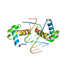 | | Structural basis for the target DNA recognition and binding by the MYB domain of phosphate starvation response regulator 1 | | 分子名称: | DNA (5'-D(*CP*AP*GP*TP*AP*TP*AP*TP*AP*CP*C)-3'), DNA (5'-D(*CP*CP*AP*TP*AP*TP*AP*TP*GP*AP*C)-3'), DNA (5'-D(*GP*GP*TP*AP*TP*AP*TP*AP*CP*TP*G)-3'), ... | | 著者 | Jiang, M.Q, Sun, L.F, Isupov, M.N. | | 登録日 | 2019-01-10 | | 公開日 | 2019-04-24 | | 最終更新日 | 2024-03-27 | | 実験手法 | X-RAY DIFFRACTION (2.8 Å) | | 主引用文献 | Structural basis for the Target DNA recognition and binding by the MYB domain of phosphate starvation response 1.
Febs J., 286, 2019
|
|
6J5B
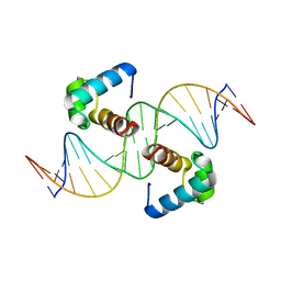 | | Structural basis for the target DNA recognition and binding by the MYB domain of phosphate starvation response regulator 1 | | 分子名称: | DNA (5'-D(*GP*GP*TP*AP*CP*AP*GP*TP*AP*TP*AP*TP*AP*CP*CP*AP*TP*AP*AP*A)-3'), DNA (5'-D(*TP*TP*TP*AP*TP*GP*GP*TP*AP*TP*AP*TP*AP*CP*TP*GP*TP*AP*CP*C)-3'), Protein PHOSPHATE STARVATION RESPONSE 1 | | 著者 | Jiang, M.Q, Sun, L.F, Isupov, M.N, Wu, Y.K. | | 登録日 | 2019-01-10 | | 公開日 | 2019-04-24 | | 最終更新日 | 2024-03-27 | | 実験手法 | X-RAY DIFFRACTION (2.7 Å) | | 主引用文献 | Structural basis for the Target DNA recognition and binding by the MYB domain of phosphate starvation response 1.
Febs J., 286, 2019
|
|
7N97
 | | State 2 of TcdB and FZD2 at pH5 | | 分子名称: | Frizzled-2, Toxin B | | 著者 | Jiang, M, Zhang, J. | | 登録日 | 2021-06-17 | | 公開日 | 2022-03-02 | | 実験手法 | ELECTRON MICROSCOPY (5.1 Å) | | 主引用文献 | Structural Basis for Receptor Recognition of the Clostridium difficile Toxin B and its Dissociation upon Acidification
To Be Published
|
|
7N8X
 | | Partial C. difficile TcdB and CSPG4 fragment | | 分子名称: | Chondroitin sulfate proteoglycan 4, Toxin B | | 著者 | Jiang, M, Zhang, J. | | 登録日 | 2021-06-16 | | 公開日 | 2022-03-02 | | 実験手法 | ELECTRON MICROSCOPY (3.4 Å) | | 主引用文献 | Structural Basis for Receptor Recognition of Clostridium difficile Toxin B and its Dissociation upon Acidification
To Be Published
|
|
7N9Q
 | | State 3 of TcdB and FZD2 at pH5 | | 分子名称: | Toxin B | | 著者 | Jiang, M, Zhang, J. | | 登録日 | 2021-06-18 | | 公開日 | 2022-03-02 | | 実験手法 | ELECTRON MICROSCOPY (4.6 Å) | | 主引用文献 | Structural Basis for Receptor Recognition of Clostridium difficile Toxin B and its Dissociation upon Acidification
To Be Published
|
|
7N9S
 | | TcdB and frizzled-2 CRD complex | | 分子名称: | Frizzled-2, Toxin B | | 著者 | Jiang, M, Zhang, J. | | 登録日 | 2021-06-18 | | 公開日 | 2022-03-02 | | 実験手法 | ELECTRON MICROSCOPY (5.1 Å) | | 主引用文献 | Structural Basis for Receptor Recognition of Clostridium difficile Toxin B and its Dissociation upon Acidification
To Be Published
|
|
7N9R
 | | state 4 of TcdB and FZD2 at pH5 | | 分子名称: | Toxin B | | 著者 | Jiang, M, Zhang, J. | | 登録日 | 2021-06-18 | | 公開日 | 2022-03-02 | | 実験手法 | ELECTRON MICROSCOPY (5.9 Å) | | 主引用文献 | Structural Basis for Receptor Recognition of Clostridium difficile Toxin B and its Dissociation upon Acidification
To Be Published
|
|
7N9Y
 | | Full-length TcdB and CSPG4 (401-560) complex | | 分子名称: | Chondroitin sulfate proteoglycan 4, Toxin B | | 著者 | Jiang, M, Zhang, J. | | 登録日 | 2021-06-18 | | 公開日 | 2022-03-02 | | 実験手法 | ELECTRON MICROSCOPY (4.8 Å) | | 主引用文献 | Structural Basis for Receptor Recognition of Clostridium difficile Toxin B and its Dissociation upon Acidification
To Be Published
|
|
7N95
 | | state 1 of TcdB and FZD2 at pH5 | | 分子名称: | Frizzled-2, Toxin B | | 著者 | Jiang, M, Zhang, J. | | 登録日 | 2021-06-16 | | 公開日 | 2022-03-02 | | 実験手法 | ELECTRON MICROSCOPY (4.1 Å) | | 主引用文献 | Structural Basis for Receptor Recognition of Clostridium difficile Toxin B and its Dissociation upon Acidification
To Be Published
|
|
8FGW
 | | Human IFT-A complex structures provide molecular insights into ciliary transport | | 分子名称: | Intraflagellar transport protein 122 homolog, Intraflagellar transport protein 140 homolog, Intraflagellar transport protein 43 homolog, ... | | 著者 | Jiang, M, Palicharla, V.R, Miller, D, Hwang, S.H, Zhu, H, Hixson, P, Mukhopadhyay, S, Sun, J. | | 登録日 | 2022-12-12 | | 公開日 | 2023-02-22 | | 最終更新日 | 2023-04-12 | | 実験手法 | ELECTRON MICROSCOPY (3.7 Å) | | 主引用文献 | Human IFT-A complex structures provide molecular insights into ciliary transport.
Cell Res., 33, 2023
|
|
8FH3
 | | Human IFT-A complex structures provide molecular insights into ciliary transport | | 分子名称: | Intraflagellar transport protein 122 homolog, Intraflagellar transport protein 140 homolog, Tubby-related protein 3, ... | | 著者 | Jiang, M, Palicharla, V.R, Miller, D, Hwang, S.H, Zhu, H, Hixson, P, Mukhopadhyay, S, Sun, J. | | 登録日 | 2022-12-13 | | 公開日 | 2023-02-22 | | 最終更新日 | 2023-04-12 | | 実験手法 | ELECTRON MICROSCOPY (4.3 Å) | | 主引用文献 | Human IFT-A complex structures provide molecular insights into ciliary transport.
Cell Res., 33, 2023
|
|
7UJJ
 | | Stx2a and DARPin complex | | 分子名称: | 1,2-ETHANEDIOL, 3-PYRIDINIUM-1-YLPROPANE-1-SULFONATE, DARPin, ... | | 著者 | Jiang, M, Zhang, J. | | 登録日 | 2022-03-30 | | 公開日 | 2023-04-12 | | 実験手法 | ELECTRON MICROSCOPY (6.5 Å) | | 主引用文献 | A Multi-Specific DARPin Potently Neutralizes Shiga Toxin 2 via Simultaneous Modulation of Both Toxin Subunits.
Bioengineering (Basel), 9, 2022
|
|
1EHC
 | | STRUCTURE OF SIGNAL TRANSDUCTION PROTEIN CHEY | | 分子名称: | CHEY, SULFATE ION | | 著者 | Jiang, M, Bourret, R, Simon, M, Volz, K. | | 登録日 | 1996-03-05 | | 公開日 | 1997-05-15 | | 最終更新日 | 2024-02-07 | | 実験手法 | X-RAY DIFFRACTION (2.26 Å) | | 主引用文献 | Uncoupled phosphorylation and activation in bacterial chemotaxis. The 2.3 A structure of an aspartate to lysine mutant at position 13 of CheY.
J.Biol.Chem., 272, 1997
|
|
2E3U
 | |
5TDH
 | | The crystal structure of the dominant negative mutant G protein alpha(i)-1-beta-1-gamma-2 G203A/A326S | | 分子名称: | GUANOSINE-5'-DIPHOSPHATE, Guanine nucleotide-binding protein G(I)/G(S)/G(O) subunit gamma-2, Guanine nucleotide-binding protein G(I)/G(S)/G(T) subunit beta-1, ... | | 著者 | Liu, P, Jia, M.-Z, Zhou, X.E, de Waal, P.W, Dickson, B.M, Liu, B, Hou, L, Yin, Y.-T, Kang, Y.-Y, Shi, Y, Melcher, K, Xu, H.E, Jiang, Y. | | 登録日 | 2016-09-19 | | 公開日 | 2016-11-09 | | 最終更新日 | 2024-03-20 | | 実験手法 | X-RAY DIFFRACTION (3 Å) | | 主引用文献 | The structural basis of the dominant negative phenotype of the G alpha i1 beta 1 gamma 2 G203A/A326S heterotrimer
Acta Pharmacol.Sin., 37, 2016
|
|


