Loading
PDBj
MenuPDBj@FacebookPDBj@TwitterPDBj@YouTubewwPDB FoundationwwPDB
RCSB PDBPDBeBMRBAdv. SearchSearch help
PDB: 38 results

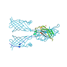
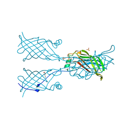
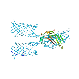
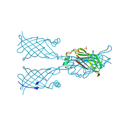
5W17
DownloadVisualize
BU of 5w17 by Molmil
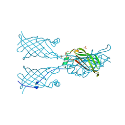
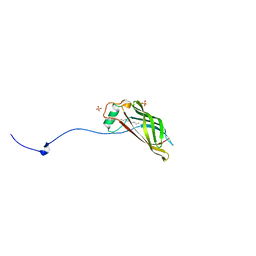
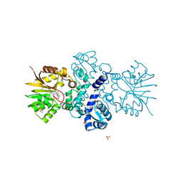
Crystal structure of Campylobacter jejuni YCEI protein that crystallizes with large solvent channels for nanotechnology applications
Descriptor: EICOSANE, Putative periplasmic protein, SULFATE ION
Authors:Huber, T.R, Snow, C.D.
Deposit date:2017-06-01
Release date:2018-01-03
Last modified:2023-10-04
Method:X-RAY DIFFRACTION (2.58 Å)
Cite:Installing Guest Molecules at Specific Sites within Scaffold Protein Crystals.
Bioconjug. Chem., 29, 2018
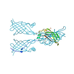
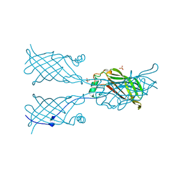
5W2V
DownloadVisualize
BU of 5w2v by Molmil
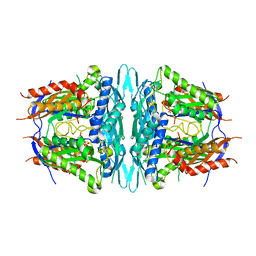
Crystal structure of mutant CJ YCEI protein (CJ-G34C) with selenocysteine guest structure
Descriptor: Putative periplasmic protein, SELENOCYSTEINE, SULFATE ION, ...
Authors:Huber, T.R, Snow, C.D.
Deposit date:2017-06-07
Release date:2018-01-03
Last modified:2023-10-04
Method:X-RAY DIFFRACTION (2.9 Å)
Cite:Installing Guest Molecules at Specific Sites within Scaffold Protein Crystals.
Bioconjug. Chem., 29, 2018
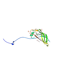
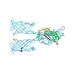
5W2D
DownloadVisualize
BU of 5w2d by Molmil
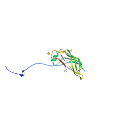
Crystal structure of mutant CJ YCEI protein (CJ-G34C) for nanotechnology applications
Descriptor: Polyisoprenoid-binding protein, SULFATE ION, UNKNOWN LIGAND
Authors:Huber, T.R, Snow, C.D.
Deposit date:2017-06-06
Release date:2018-01-03
Last modified:2023-10-04
Method:X-RAY DIFFRACTION (2.7 Å)
Cite:Installing Guest Molecules at Specific Sites within Scaffold Protein Crystals.
Bioconjug. Chem., 29, 2018
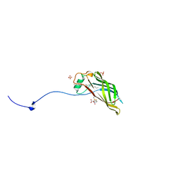
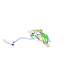
5W30
DownloadVisualize
BU of 5w30 by Molmil
Crystal structure of mutant CJ YCEI protein (CJ-N48C) with monobromobimane guest structure
Descriptor: 3-(bromomethyl)-2,5,6-trimethyl-1H,7H-pyrazolo[1,2-a]pyrazole-1,7-dione, Putative periplasmic protein, SULFATE ION, ...
Authors:Huber, T.R, Snow, C.D.
Deposit date:2017-06-07
Release date:2018-01-03
Last modified:2023-10-04
Method:X-RAY DIFFRACTION (2.75 Å)
Cite:Installing Guest Molecules at Specific Sites within Scaffold Protein Crystals.
Bioconjug. Chem., 29, 2018
5W39
DownloadVisualize
BU of 5w39 by Molmil
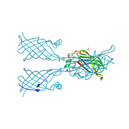
Crystal structure of mutant CJ YCEI protein (CJ-N182C) with monobromobimane guest structure
Descriptor: 3-(bromomethyl)-2,5,6-trimethyl-1H,7H-pyrazolo[1,2-a]pyrazole-1,7-dione, EICOSANE, Polyisoprenoid-binding protein, ...
Authors:Huber, T.R, Snow, C.D.
Deposit date:2017-06-07
Release date:2018-01-03
Last modified:2023-10-04
Method:X-RAY DIFFRACTION (2.48 Å)
Cite:Installing Guest Molecules at Specific Sites within Scaffold Protein Crystals.
Bioconjug. Chem., 29, 2018
5W3A
DownloadVisualize
BU of 5w3a by Molmil
Crystal structure of mutant CJ YCEI protein (CJ-N182C) with 5-mercapto-2-nitrobenzoic acid guest structure
Descriptor: 5-MERCAPTO-2-NITRO-BENZOIC ACID, EICOSANE, Polyisoprenoid-binding protein, ...
Authors:Huber, T.R, Snow, C.D.
Deposit date:2017-06-07
Release date:2018-01-03
Last modified:2023-10-04
Method:X-RAY DIFFRACTION (2.76 Å)
Cite:Installing Guest Molecules at Specific Sites within Scaffold Protein Crystals.
Bioconjug. Chem., 29, 2018
5W32
DownloadVisualize
BU of 5w32 by Molmil
Crystal structure of mutant CJ YCEI protein (CJ-N48C) with selenocysteine guest structure
Descriptor: Putative periplasmic protein, SELENOCYSTEINE, SULFATE ION, ...
Authors:Huber, T.R, Snow, C.D.
Deposit date:2017-06-07
Release date:2018-01-03
Last modified:2023-10-04
Method:X-RAY DIFFRACTION (2.6 Å)
Cite:Installing Guest Molecules at Specific Sites within Scaffold Protein Crystals.
Bioconjug. Chem., 29, 2018
5W3C
DownloadVisualize
BU of 5w3c by Molmil
Crystal structure of mutant CJ YCEI protein (CJ-N182C) with selenocysteine guest structure
Descriptor: EICOSANE, Polyisoprenoid-binding protein, SELENOCYSTEINE, ...
Authors:Huber, T.R, Snow, C.D.
Deposit date:2017-06-07
Release date:2018-01-03
Last modified:2023-10-04
Method:X-RAY DIFFRACTION (2.87 Å)
Cite:Installing Guest Molecules at Specific Sites within Scaffold Protein Crystals.
Bioconjug. Chem., 29, 2018
5W2Z
DownloadVisualize
BU of 5w2z by Molmil
Crystal structure of mutant CJ YCEI protein (CJ-N48C) with 5-mercapto-2-nitrobenzoic acid guest structure
Descriptor: 5-MERCAPTO-2-NITRO-BENZOIC ACID, Putative periplasmic protein, SULFATE ION, ...
Authors:Huber, T.R, Snow, C.D.
Deposit date:2017-06-07
Release date:2018-01-03
Last modified:2023-10-04
Method:X-RAY DIFFRACTION (2.8 Å)
Cite:Installing Guest Molecules at Specific Sites within Scaffold Protein Crystals.
Bioconjug. Chem., 29, 2018
5W2K
DownloadVisualize
BU of 5w2k by Molmil
Crystal structure of mutant CJ YCEI protein (CJ-G34C) with hydroxymercuribenzoic acid guest structure
Descriptor: MERCURIBENZOIC ACID, Polyisoprenoid-binding protein, SULFATE ION, ...
Authors:Huber, T.R, Snow, C.D.
Deposit date:2017-06-06
Release date:2018-01-03
Last modified:2023-10-04
Method:X-RAY DIFFRACTION (2.78 Å)
Cite:Installing Guest Molecules at Specific Sites within Scaffold Protein Crystals.
Bioconjug. Chem., 29, 2018
5W2R
DownloadVisualize
BU of 5w2r by Molmil
Crystal structure of mutant CJ YCEI protein (CJ-G34C) with 5-mercapto-2-nitrobenzoic acid guest structure
Descriptor: 5-MERCAPTO-2-NITRO-BENZOIC ACID, Polyisoprenoid-binding protein, SULFATE ION, ...
Authors:Huber, T.R, Snow, C.D.
Deposit date:2017-06-06
Release date:2018-01-03
Last modified:2023-10-04
Method:X-RAY DIFFRACTION (2.9 Å)
Cite:Installing Guest Molecules at Specific Sites within Scaffold Protein Crystals.
Bioconjug. Chem., 29, 2018
5W2X
DownloadVisualize
BU of 5w2x by Molmil
Crystal structure of mutant CJ YCEI protein (CJ-N48C) for nanotechnology applications
Descriptor: Polyisoprenoid-binding protein, SULFATE ION, UNKNOWN LIGAND
Authors:Huber, T.R, Snow, C.D.
Deposit date:2017-06-07
Release date:2018-01-03
Last modified:2023-10-04
Method:X-RAY DIFFRACTION (2.73 Å)
Cite:Installing Guest Molecules at Specific Sites within Scaffold Protein Crystals.
Bioconjug. Chem., 29, 2018
5W31
DownloadVisualize
BU of 5w31 by Molmil
Crystal structure of mutant CJ YCEI protein (CJ-N48C) with mercuribenzoic acid guest structure
Descriptor: MERCURIBENZOIC ACID, Putative periplasmic protein, SULFATE ION, ...
Authors:Huber, T.R, Snow, C.D.
Deposit date:2017-06-07
Release date:2018-01-03
Last modified:2023-10-04
Method:X-RAY DIFFRACTION (2.56 Å)
Cite:Installing Guest Molecules at Specific Sites within Scaffold Protein Crystals.
Bioconjug. Chem., 29, 2018
5W3B
DownloadVisualize
BU of 5w3b by Molmil
Crystal structure of mutant CJ YCEI protein (CJ-N182C) with mercuribenzoic acid guest structure
Descriptor: EICOSANE, MERCURIBENZOIC ACID, Polyisoprenoid-binding protein, ...
Authors:Huber, T.R, Snow, C.D.
Deposit date:2017-06-07
Release date:2018-01-03
Last modified:2023-10-04
Method:X-RAY DIFFRACTION (2.7 Å)
Cite:Installing Guest Molecules at Specific Sites within Scaffold Protein Crystals.
Bioconjug. Chem., 29, 2018
5W37
DownloadVisualize
BU of 5w37 by Molmil
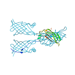
Crystal structure of mutant CJ YCEI protein (CJ-N182C) for nanotechnology applications
Descriptor: EICOSANE, Polyisoprenoid-binding protein, SULFATE ION
Authors:Huber, T.R, Snow, C.D.
Deposit date:2017-06-07
Release date:2018-01-03
Last modified:2023-10-04
Method:X-RAY DIFFRACTION (2.52 Å)
Cite:Installing Guest Molecules at Specific Sites within Scaffold Protein Crystals.
Bioconjug. Chem., 29, 2018
6O6I
DownloadVisualize
BU of 6o6i by Molmil
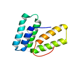
Endoplasmic reticulum protein 29 (ERp29) C-terminal domain: Structure Determination from Backbone Amide Pseudocontact Shifts Generated by Double-histidine Cobalt Tags
Descriptor: Endoplasmic reticulum resident protein 29
Authors:Bahramzadeh, A, Huber, T, Otting, G.
Deposit date:2019-03-06
Release date:2019-07-24
Last modified:2020-01-01
Method:SOLUTION NMR
Cite:Three-Dimensional Protein Structure Determination Using Pseudocontact Shifts of Backbone Amide Protons Generated by Double-Histidine Co2+-Binding Motifs at Multiple Sites.
Biochemistry, 58, 2019
6P9V
DownloadVisualize
BU of 6p9v by Molmil
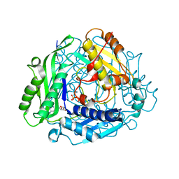
Crystal Structure of hMAT Mutant K289L
Descriptor: ADENOSINE, MAGNESIUM ION, POTASSIUM ION, ...
Authors:Miller, M.D, Xu, W, Huber, T.D, Clinger, J.A, Liu, Y, Thorson, J.S, Phillips Jr, G.N.
Deposit date:2019-06-10
Release date:2020-04-22
Last modified:2023-10-11
Method:X-RAY DIFFRACTION (2.051 Å)
Cite:Methionine Adenosyltransferase Engineering to Enable Bioorthogonal Platforms for AdoMet-Utilizing Enzymes.
Acs Chem.Biol., 15, 2020
7R6O
DownloadVisualize
BU of 7r6o by Molmil
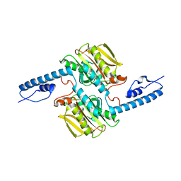
Pyrrolysyl-tRNA synthetase from methanogenic archaeon ISO4-G1 (G1PylRS)
Descriptor: 1,2-ETHANEDIOL, Pyrrolysyl-tRNA synthetase PylS, SULFATE ION
Authors:Frkic, R.L, Huber, T, Jackson, C.J.
Deposit date:2021-06-23
Release date:2022-03-02
Last modified:2023-10-18
Method:X-RAY DIFFRACTION (2.2 Å)
Cite:Genetic Encoding of Cyanopyridylalanine for In-Cell Protein Macrocyclization by the Nitrile-Aminothiol Click Reaction.
Angew.Chem.Int.Ed.Engl., 61, 2022
5JR3
DownloadVisualize
BU of 5jr3 by Molmil
Crystal structure of carminomycin-4-O-methyltransferase DnrK in complex with SAH and 4-methylumbelliferone
Descriptor: 7-hydroxy-4-methyl-2H-chromen-2-one, Carminomycin 4-O-methyltransferase DnrK, S-ADENOSYL-L-HOMOCYSTEINE, ...
Authors:Wang, F, Johnson, B.R, Huber, T.D, Singh, S, Thorson, J.S, Phillips Jr, G.N, Enzyme Discovery for Natural Product Biosynthesis (NatPro)
Deposit date:2016-05-05
Release date:2016-06-01
Last modified:2023-10-25
Method:X-RAY DIFFRACTION (1.84 Å)
Cite:Crystal structure of carminomycin-4-O-methyltransferase DnrK in complex with SAH and 4-methylumbelliferone (to be published)
To Be Published
4WS9
DownloadVisualize
BU of 4ws9 by Molmil
Crystal structure of sMAT N159G from Sulfolobus solfataricus
Descriptor: PHOSPHATE ION, S-adenosylmethionine synthase
Authors:Wang, F, Brady, E.L, Singh, S, Clinger, J.A, Huber, T.D, Thorson, J.S, Phillips Jr, G.N.
Deposit date:2014-10-26
Release date:2014-11-05
Last modified:2023-09-27
Method:X-RAY DIFFRACTION (2.803 Å)
Cite:Crystal structure of sMAT N159G from Sulfolobus solfataricus.
To Be Published
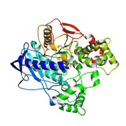
4W1R
DownloadVisualize
BU of 4w1r by Molmil
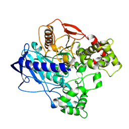
KINETIC CRYSTALLOGRAPHY OF ALPHA_E7-CARBOXYLESTERSE FROM LUCILLA CUPRINA - ABSORBED X-RAY DOSE 9.24 MGy TEMP 150K
Descriptor: DIETHYL HYDROGEN PHOSPHATE, E3
Authors:Jackson, C.J, Carr, P.D, Weik, M, Huber, T, Meirelles, T, Correy, G.
Deposit date:2014-08-14
Release date:2015-08-19
Last modified:2023-11-08
Method:X-RAY DIFFRACTION (2.23 Å)
Cite:Mapping the Accessible Conformational Landscape of an Insect Carboxylesterase Using Conformational Ensemble Analysis and Kinetic Crystallography
Structure, 24, 2016
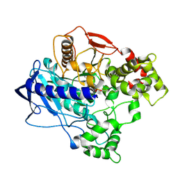
4W1P
DownloadVisualize
BU of 4w1p by Molmil
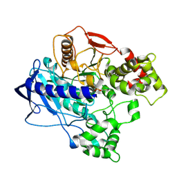
KINETIC CRYSTALLOGRAPHY OF ALPHA_E7-CARBOXYLESTERSE FROM LUCILLA CUPRINA - ABSORBED X-RAY DOSE 5.54 MGy TEMP 150K
Descriptor: DIETHYL HYDROGEN PHOSPHATE, E3
Authors:Jackson, C.J, Carr, P.D, Weik, M, Huber, T, Meirelles, T, Correy, G.
Deposit date:2014-08-14
Release date:2015-08-19
Last modified:2023-11-08
Method:X-RAY DIFFRACTION (2.1 Å)
Cite:Mapping the Accessible Conformational Landscape of an Insect Carboxylesterase Using Conformational Ensemble Analysis and Kinetic Crystallography
Structure, 24, 2016
4W1Q
DownloadVisualize
BU of 4w1q by Molmil
KINETIC CRYSTALLOGRAPHY OF ALPHA_E7-CARBOXYLESTERSE FROM LUCILLA CUPRINA - ABSORBED X-RAY DOSE 7.39 MGy TEMP 150K
Descriptor: DIETHYL HYDROGEN PHOSPHATE, E3
Authors:Jackson, C.J, Carr, P.D, Weik, M, Huber, T, Meirelles, T, Correy, G.
Deposit date:2014-08-14
Release date:2015-08-19
Last modified:2023-11-08
Method:X-RAY DIFFRACTION (2.12 Å)
Cite:Mapping the Accessible Conformational Landscape of an Insect Carboxylesterase Using Conformational Ensemble Analysis and Kinetic Crystallography
Structure, 24, 2016
4W1S
DownloadVisualize
BU of 4w1s by Molmil
KINETIC CRYSTALLOGRAPHY OF ALPHA_E7-CARBOXYLESTERSE FROM LUCILLA CUPRINA - ABSORBED X-RAY DOSE 11.09 MGy TEMP 150K
Descriptor: DIETHYL HYDROGEN PHOSPHATE, E3
Authors:jackson, C.j, carr, p.d, weik, m, huber, t, meirelles, t, correy, g.
Deposit date:2014-08-14
Release date:2015-08-19
Last modified:2023-11-08
Method:X-RAY DIFFRACTION (2.3 Å)
Cite:Mapping the Accessible Conformational Landscape of an Insect Carboxylesterase Using Conformational Ensemble Analysis and Kinetic Crystallography
Structure, 24, 2016
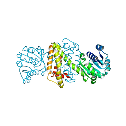
4OQZ
DownloadVisualize
BU of 4oqz by Molmil
Streptomyces aurantiacus imine reductase
Descriptor: Putative oxidoreductase YfjR
Authors:Schneider, L.K, Huber, T, Gerhardt, S, Muller, M, Einsle, O.
Deposit date:2014-02-10
Release date:2014-10-22
Last modified:2024-02-28
Method:X-RAY DIFFRACTION (1.96 Å)
Cite:Direct Reductive Amination of Ketones: Structure and Activity of S-Selective Imine Reductases from Streptomyces.
CHEMCATCHEM, 2014


12>

218853

PDB entries from 2024-04-24

PDB statisticsPDBj update infoContact PDBjnumon