8T51
 
 | |
6UH5
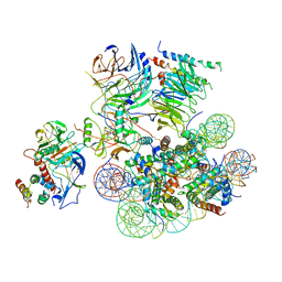 | | Structural basis of COMPASS eCM recognition of the H2Bub nucleosome | | 分子名称: | Bre2, DNA (146-MER), H3 N-terminus, ... | | 著者 | Hsu, P.L, Shi, H, Zheng, N. | | 登録日 | 2019-09-26 | | 公開日 | 2019-11-20 | | 最終更新日 | 2019-12-18 | | 実験手法 | ELECTRON MICROSCOPY (3.5 Å) | | 主引用文献 | Structural Basis of H2B Ubiquitination-Dependent H3K4 Methylation by COMPASS.
Mol.Cell, 76, 2019
|
|
6UGM
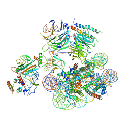 | | Structural basis of COMPASS eCM recognition of an unmodified nucleosome | | 分子名称: | Bre2, DNA (146-MER), H3 N-terminus, ... | | 著者 | Hsu, P.L, Shi, H, Zheng, N. | | 登録日 | 2019-09-26 | | 公開日 | 2019-11-20 | | 最終更新日 | 2024-03-20 | | 実験手法 | ELECTRON MICROSCOPY (3.7 Å) | | 主引用文献 | Structural Basis of H2B Ubiquitination-Dependent H3K4 Methylation by COMPASS.
Mol.Cell, 76, 2019
|
|
6CHG
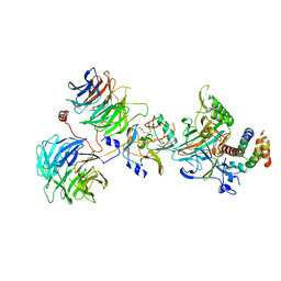 | | Crystal structure of the yeast COMPASS catalytic module | | 分子名称: | H3, Histone-lysine N-methyltransferase, H3 lysine-4 specific, ... | | 著者 | Hsu, P.L, Li, H, Zheng, N. | | 登録日 | 2018-02-22 | | 公開日 | 2018-08-22 | | 最終更新日 | 2023-10-04 | | 実験手法 | X-RAY DIFFRACTION (2.985 Å) | | 主引用文献 | Crystal Structure of the COMPASS H3K4 Methyltransferase Catalytic Module.
Cell, 174, 2018
|
|
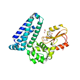
4M3O
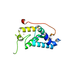 | | Crystal structure of K.lactis Rtr1 NTD | | 分子名称: | KLLA0F12672p, ZINC ION | | 著者 | Hsu, P.L, Yang, W, Zheng, N, Varani, G. | | 登録日 | 2013-08-06 | | 公開日 | 2014-07-16 | | 最終更新日 | 2024-02-28 | | 実験手法 | X-RAY DIFFRACTION (2.1 Å) | | 主引用文献 | Rtr1 Is a Dual Specificity Phosphatase That Dephosphorylates Tyr1 and Ser5 on the RNA Polymerase II CTD.
J.Mol.Biol., 426, 2014
|
|
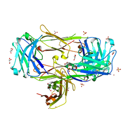
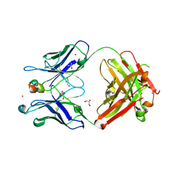
3AUV
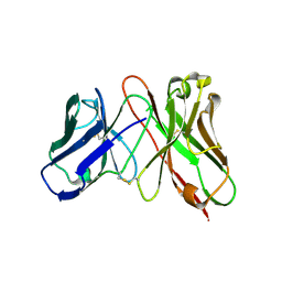 | | Predicting Amino Acid Preferences in the Complementarity Determining Regions of an Antibody-Antigen Recognition Interface | | 分子名称: | sc-dsFv derived from the G6-Fab | | 著者 | Yu, C.M, Peng, H.P, Chen, I.C, Lee, Y.C, Chen, J.B, Tsai, K.C, Chen, C.T, Chang, J.Y, Yang, E.W, Hsu, P.C, Jian, J.W, Hsu, H.J, Chang, H.J, Hsu, W.L, Huang, K.F, Ma, A.C, Yang, A.S. | | 登録日 | 2011-02-16 | | 公開日 | 2012-02-22 | | 最終更新日 | 2012-04-25 | | 実験手法 | X-RAY DIFFRACTION (2.4 Å) | | 主引用文献 | Rationalization and design of the complementarity determining region sequences in an antibody-antigen recognition interface
Plos One, 7, 2012
|
|
6EEY
 | |
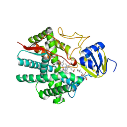
8VW5
 | | Crystal structure of Cbl-b TKB bound to compound 2 | | 分子名称: | CALCIUM ION, E3 ubiquitin-protein ligase CBL-B, MAGNESIUM ION, ... | | 著者 | Yu, C, Murray, J, Hsu, P.L. | | 登録日 | 2024-01-31 | | 公開日 | 2024-07-03 | | 実験手法 | X-RAY DIFFRACTION (1.76 Å) | | 主引用文献 | Optimization of a Novel DEL Hit That Binds in the Cbl-b SH2 Domain and Blocks Substrate Binding.
Acs Med.Chem.Lett., 15, 2024
|
|
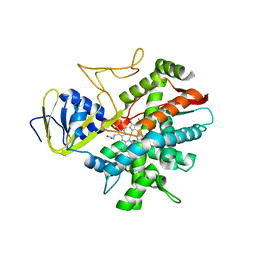
8VW4
 | | Crystal structure of Cbl-b TKB bound to compound 26 | | 分子名称: | (7-methoxy-2-{2-[(1S,3S,4S)-3-(3-methoxy-2-methyl-5-nitrophenyl)-1-methyl-5-oxo-1,5-dihydroimidazo[1,5-a]pyridin-2(3H)-yl]-2-oxoethoxy}quinolin-8-yl)acetic acid, DI(HYDROXYETHYL)ETHER, E3 ubiquitin-protein ligase CBL-B, ... | | 著者 | Yu, C, Murray, J, Hsu, P.L. | | 登録日 | 2024-01-31 | | 公開日 | 2024-07-03 | | 実験手法 | X-RAY DIFFRACTION (2.4 Å) | | 主引用文献 | Optimization of a Novel DEL Hit That Binds in the Cbl-b SH2 Domain and Blocks Substrate Binding.
Acs Med.Chem.Lett., 15, 2024
|
|
8T59
 | |
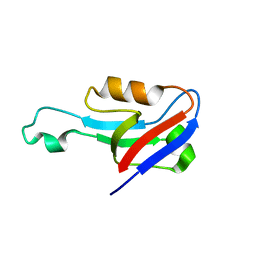
6B3X
 | | Crystal structure of CstF-50 in complex with CstF-77 | | 分子名称: | Cleavage stimulation factor subunit 1, Cleavage stimulation factor subunit 3 | | 著者 | Yang, W, Hsu, P, Yang, F, Song, J.E, Varani, G. | | 登録日 | 2017-09-25 | | 公開日 | 2017-11-29 | | 最終更新日 | 2024-04-03 | | 実験手法 | X-RAY DIFFRACTION (2.3 Å) | | 主引用文献 | Reconstitution of the CstF complex unveils a regulatory role for CstF-50 in recognition of 3'-end processing signals.
Nucleic Acids Res., 46, 2018
|
|
3B99
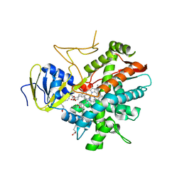 | | Crystal structure of zebrafish prostacyclin synthase (cytochrome P450 8A1) in complex with substrate analog U51605 | | 分子名称: | (5Z)-7-{(1R,4S,5R,6R)-6-[(1E)-oct-1-en-1-yl]-2,3-diazabicyclo[2.2.1]hept-2-en-5-yl}hept-5-enoic acid, PROTOPORPHYRIN IX CONTAINING FE, Prostaglandin I2 synthase | | 著者 | Li, Y.-C, Chiang, C.-W, Yeh, H.-C, Hsu, P.-Y, Whitby, F.G, Wang, L.-H, Chan, N.-L. | | 登録日 | 2007-11-03 | | 公開日 | 2007-11-20 | | 最終更新日 | 2023-11-01 | | 実験手法 | X-RAY DIFFRACTION (2.5 Å) | | 主引用文献 | Structures of Prostacyclin Synthase and Its Complexes with Substrate Analog and Inhibitor Reveal a Ligand-specific Heme Conformation Change
J.Biol.Chem., 283, 2008
|
|
3B6H
 | | Crystal structure of human prostacyclin synthase in complex with inhibitor minoxidil | | 分子名称: | 6-PIPERIDIN-1-YLPYRIMIDINE-2,4-DIAMINE 3-OXIDE, PROTOPORPHYRIN IX CONTAINING FE, Prostacyclin synthase, ... | | 著者 | Li, Y.-C, Chiang, C.-W, Yeh, H.-C, Hsu, P.-Y, Whitby, F.G, Wang, L.-H, Chan, N.-L. | | 登録日 | 2007-10-29 | | 公開日 | 2007-11-20 | | 最終更新日 | 2023-11-01 | | 実験手法 | X-RAY DIFFRACTION (1.62 Å) | | 主引用文献 | Structures of Prostacyclin Synthase and Its Complexes with Substrate Analog and Inhibitor Reveal a Ligand-specific Heme Conformation Change
J.Biol.Chem., 283, 2008
|
|
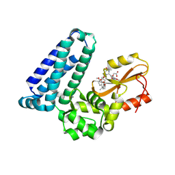
3B98
 | | Crystal structure of zebrafish prostacyclin synthase (cytochrome P450 8A1) | | 分子名称: | PROTOPORPHYRIN IX CONTAINING FE, Prostaglandin I2 synthase | | 著者 | Li, Y.-C, Chiang, C.-W, Yeh, H.-C, Hsu, P.-Y, Whitby, F.G, Wang, L.-H, Chan, N.-L. | | 登録日 | 2007-11-03 | | 公開日 | 2007-11-20 | | 最終更新日 | 2023-11-01 | | 実験手法 | X-RAY DIFFRACTION (2.08 Å) | | 主引用文献 | Structures of Prostacyclin Synthase and Its Complexes with Substrate Analog and Inhibitor Reveal a Ligand-specific Heme Conformation Change
J.Biol.Chem., 283, 2008
|
|


