6QV5
 
 | |
5LPZ
 | |
5LPW
 | |
2PE8
 | |
3BXJ
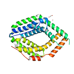
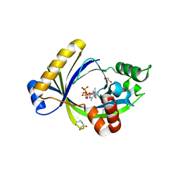
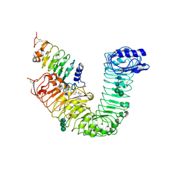
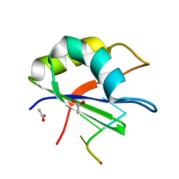
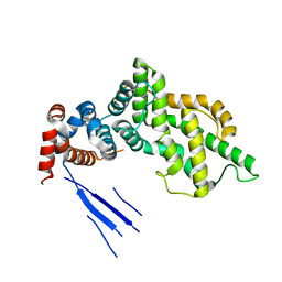 | | Crystal Structure of the C2-GAP Fragment of synGAP | | 分子名称: | Ras GTPase-activating protein SynGAP | | 著者 | Pena, V, Hothorn, M, Eberth, A, Kaschau, N, Parret, A, Gremer, L, Bonneau, F, Ahmadian, M.R, Scheffzek, K. | | 登録日 | 2008-01-14 | | 公開日 | 2008-03-25 | | 最終更新日 | 2024-02-21 | | 実験手法 | X-RAY DIFFRACTION (3 Å) | | 主引用文献 | The C2 domain of SynGAP is essential for stimulation of the Rap GTPase reaction.
Embo Rep., 9, 2008
|
|
3CB5
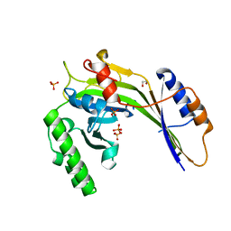
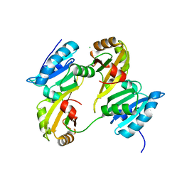
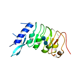
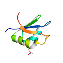
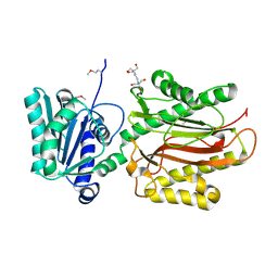 | | Crystal Structure of the S. pombe Peptidase Homology Domain of FACT complex subunit Spt16 (form A) | | 分子名称: | 2-[3-(2-HYDROXY-1,1-DIHYDROXYMETHYL-ETHYLAMINO)-PROPYLAMINO]-2-HYDROXYMETHYL-PROPANE-1,3-DIOL, FACT complex subunit spt16 | | 著者 | Stuwe, T, Hothorn, M, Lejeune, E, Bortfeld-Miller, M, Scheffzek, K, Ladurner, A.G. | | 登録日 | 2008-02-21 | | 公開日 | 2008-06-17 | | 最終更新日 | 2011-07-13 | | 実験手法 | X-RAY DIFFRACTION (2.05 Å) | | 主引用文献 | The FACT Spt16 "peptidase" domain is a histone H3-H4 binding module
Proc.Natl.Acad.Sci.USA, 105, 2008
|
|
5A61
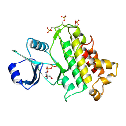
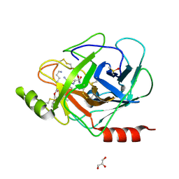
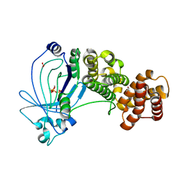 | | Crystal structure of full-length E. coli ygiF in complex with tripolyphosphate and two manganese ions. | | 分子名称: | 1,2-ETHANEDIOL, INORGANIC TRIPHOSPHATASE, MANGANESE (II) ION, ... | | 著者 | Martinez, J, Truffault, V, Hothorn, M. | | 登録日 | 2015-06-23 | | 公開日 | 2015-08-05 | | 最終更新日 | 2024-05-08 | | 実験手法 | X-RAY DIFFRACTION (1.5 Å) | | 主引用文献 | Structural Determinants for Substrate Binding and Catalysis in Triphosphate Tunnel Metalloenzymes.
J.Biol.Chem., 290, 2015
|
|
5A68
 | |
4NHG
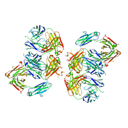
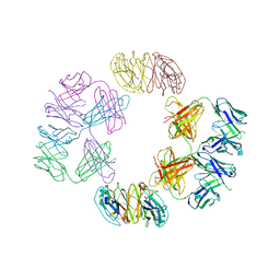 | | Crystal Structure of 2G12 IgG Dimer | | 分子名称: | 2G12 IgG dimer heavy chain, 2G12 IgG dimer light chain, Hepatitis B virus receptor binding protein | | 著者 | Wu, Y, West Jr, A.P, Kim, H.J, Thornton, M.E, Ward, A.B, Bjorkman, P.J. | | 登録日 | 2013-11-05 | | 公開日 | 2014-02-26 | | 最終更新日 | 2019-07-17 | | 実験手法 | X-RAY DIFFRACTION (8.001 Å) | | 主引用文献 | Structural basis for enhanced HIV-1 neutralization by a dimeric immunoglobulin G form of the glycan-recognizing antibody 2G12.
Cell Rep, 5, 2013
|
|
3G3T
 | | Crystal structure of a eukaryotic polyphosphate polymerase in complex with orthophosphate | | 分子名称: | 1,2-ETHANEDIOL, PHOSPHATE ION, Vacuolar transporter chaperone 4 | | 著者 | Lenherr, E.D, Hothorn, M, Scheffzek, K. | | 登録日 | 2009-02-02 | | 公開日 | 2009-05-05 | | 最終更新日 | 2023-09-06 | | 実験手法 | X-RAY DIFFRACTION (1.85 Å) | | 主引用文献 | Catalytic core of a membrane-associated eukaryotic polyphosphate polymerase.
Science, 324, 2009
|
|
3E0P
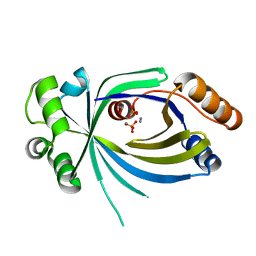
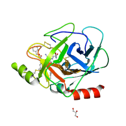 | | The X-ray structure of Human Prostasin in complex with a covalent benzoxazole inhibitor | | 分子名称: | GLYCEROL, Prostasin, benzyl [(1R)-1-({(2S,4R)-2-({(1S)-5-amino-1-[(S)-1,3-benzoxazol-2-yl(hydroxy)methyl]pentyl}carbamoyl)-4-[(4-methylbenzyl)oxy]pyrrolidin-1-yl}carbonyl)-3-phenylpropyl]carbamate | | 著者 | Spraggon, G, Hornsby, M, Shipway, A, Harris, J.L, Lesley, S.A. | | 登録日 | 2008-07-31 | | 公開日 | 2008-09-09 | | 最終更新日 | 2021-10-20 | | 実験手法 | X-RAY DIFFRACTION (1.7 Å) | | 主引用文献 | Discovery of inhibitors of the channel-activating protease prostasin (CAP1/PRSS8) utilizing structure-based design.
Bioorg.Med.Chem.Lett., 18, 2008
|
|
5A60
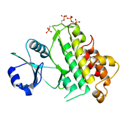
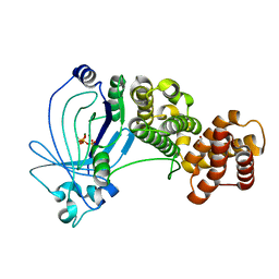 | | Crystal structure of full-length E. coli ygiF in complex with tripolyphosphate and two magnesium ions | | 分子名称: | 1,2-ETHANEDIOL, INORGANIC TRIPHOSPHATASE, MAGNESIUM ION, ... | | 著者 | Martinez, J, Truffault, V, Hothorn, M. | | 登録日 | 2015-06-23 | | 公開日 | 2015-08-05 | | 最終更新日 | 2024-05-08 | | 実験手法 | X-RAY DIFFRACTION (1.82 Å) | | 主引用文献 | Structural Determinants for Substrate Binding and Catalysis in Triphosphate Tunnel Metalloenzymes.
J.Biol.Chem., 290, 2015
|
|
5A64
 | | Crystal structure of mouse thiamine triphosphatase in complex with thiamine triphosphate. | | 分子名称: | 1,2-ETHANEDIOL, THIAMINE TRIPHOSPHATASE, TRIETHYLENE GLYCOL, ... | | 著者 | Martinez, J, Truffault, V, Hothorn, M. | | 登録日 | 2015-06-24 | | 公開日 | 2015-08-05 | | 最終更新日 | 2024-01-10 | | 実験手法 | X-RAY DIFFRACTION (2.1 Å) | | 主引用文献 | Structural Determinants for Substrate Binding and Catalysis in Triphosphate Tunnel Metalloenzymes.
J.Biol.Chem., 290, 2015
|
|
3DXB
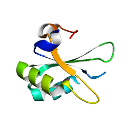
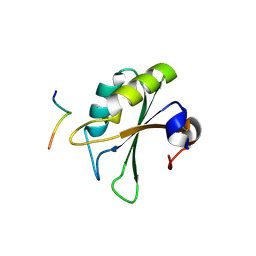
 | | Structure of the UHM domain of Puf60 fused to thioredoxin | | 分子名称: | 1,2-ETHANEDIOL, CHLORIDE ION, thioredoxin N-terminally fused to Puf60(UHM) | | 著者 | Corsini, L, Hothorn, M, Scheffzek, K, Stier, G, Sattler, M. | | 登録日 | 2008-07-24 | | 公開日 | 2008-10-28 | | 最終更新日 | 2023-08-30 | | 実験手法 | X-RAY DIFFRACTION (2.2 Å) | | 主引用文献 | Dimerization and Protein Binding Specificity of the U2AF Homology Motif of the Splicing Factor Puf60.
J.Biol.Chem., 284, 2009
|
|
3E16
 | | X-ray structure of human prostasin in complex with Benzoxazole warhead peptidomimic, lysine in P3 | | 分子名称: | DIMETHYL SULFOXIDE, GLYCEROL, Prostasin, ... | | 著者 | Spraggon, G, Hornsby, M, Shipway, A, Harris, J.L, Lesley, S.A. | | 登録日 | 2008-08-01 | | 公開日 | 2008-09-09 | | 最終更新日 | 2021-10-20 | | 実験手法 | X-RAY DIFFRACTION (1.6 Å) | | 主引用文献 | Discovery of inhibitors of the channel-activating protease prostasin (CAP1/PRSS8) utilizing structure-based design.
Bioorg.Med.Chem.Lett., 18, 2008
|
|
2PEH
 | | Crystal structure of the UHM domain of human SPF45 in complex with SF3b155-ULM5 | | 分子名称: | Splicing factor 3B subunit 1, Splicing factor 45 | | 著者 | Corsini, L, Basquin, J, Hothorn, M, Sattler, M. | | 登録日 | 2007-04-03 | | 公開日 | 2007-06-26 | | 最終更新日 | 2023-08-30 | | 実験手法 | X-RAY DIFFRACTION (2.11 Å) | | 主引用文献 | U2AF-homology motif interactions are required for alternative splicing regulation by SPF45.
Nat.Struct.Mol.Biol., 14, 2007
|
|
4LSX
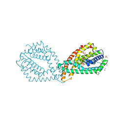
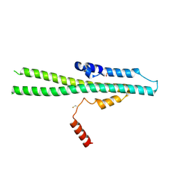
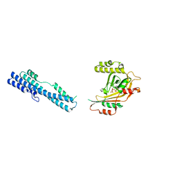
 | | Plant steroid receptor ectodomain bound to brassinolide and SERK1 co-receptor ectodomain | | 分子名称: | 2-acetamido-2-deoxy-beta-D-glucopyranose, 2-acetamido-2-deoxy-beta-D-glucopyranose-(1-4)-2-acetamido-2-deoxy-beta-D-glucopyranose, Brassinolide, ... | | 著者 | Santiago, J, Henzler, C, Hothorn, M. | | 登録日 | 2013-07-23 | | 公開日 | 2013-09-04 | | 最終更新日 | 2023-09-20 | | 実験手法 | X-RAY DIFFRACTION (3.302 Å) | | 主引用文献 | Molecular mechanism for plant steroid receptor activation by somatic embryogenesis co-receptor kinases.
Science, 341, 2013
|
|
4NHH
 | | Structure of 2G12 IgG Dimer | | 分子名称: | 2G12 IgG dimer heavy chain, 2G12 IgG dimer light chain, Hepatitis B virus receptor binding protein | | 著者 | Wu, Y, West Jr, A.P, Kim, H.J, Thornton, M.E, Ward, A.B, Bjorkman, P.J. | | 登録日 | 2013-11-05 | | 公開日 | 2014-02-26 | | 実験手法 | X-RAY DIFFRACTION (6.5 Å) | | 主引用文献 | Structural basis for enhanced HIV-1 neutralization by a dimeric immunoglobulin G form of the glycan-recognizing antibody 2G12.
Cell Rep, 5, 2013
|
|
6QVA
 | |
6R1H
 | |
2XNR
 | |
2XNQ
 | |
5IJH
 | |
5IIQ
 | |
5IJJ
 | |


