Loading
PDBj
MenuPDBj@FacebookPDBj@TwitterPDBj@YouTubewwPDB FoundationwwPDB
RCSB PDBPDBeBMRBAdv. SearchSearch help
PDB: 276 results

3LCK
DownloadVisualize
BU of 3lck by Molmil
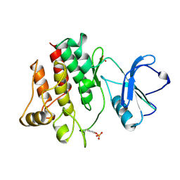
THE KINASE DOMAIN OF HUMAN LYMPHOCYTE KINASE (LCK), ACTIVATED FORM (AUTO-PHOSPHORYLATED ON TYR394)
Descriptor: PROTO-ONCOGENE TYROSINE-PROTEIN KINASE, SULFATE ION
Authors:Yamaguchi, H, Hendrickson, W.A.
Deposit date:1997-04-08
Release date:1997-12-03
Last modified:2024-10-30
Method:X-RAY DIFFRACTION (1.7 Å)
Cite:Structural basis for activation of human lymphocyte kinase Lck upon tyrosine phosphorylation.
Nature, 384, 1996
3LIA
DownloadVisualize
BU of 3lia by Molmil
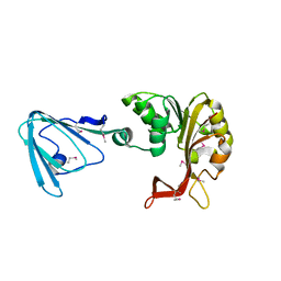
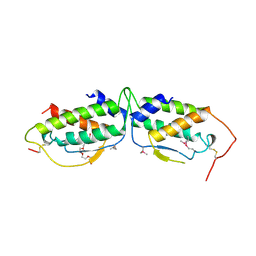
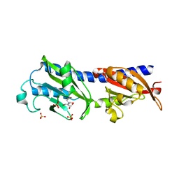
Crystal Structure of the extracellular domain of the putative histidine kinase mmHK1S-Z2
Descriptor: 2-[BIS-(2-HYDROXY-ETHYL)-AMINO]-2-HYDROXYMETHYL-PROPANE-1,3-DIOL, Hypothetical sensory transduction histidine kinase, SULFATE ION
Authors:Zhang, Z, Hendrickson, W.A.
Deposit date:2010-01-24
Release date:2010-05-05
Last modified:2024-02-21
Method:X-RAY DIFFRACTION (1.99 Å)
Cite:Structural characterization of the predominant family of histidine kinase sensor domains.
J.Mol.Biol., 400, 2010
3M72
DownloadVisualize
BU of 3m72 by Molmil
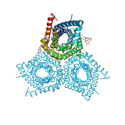
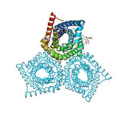
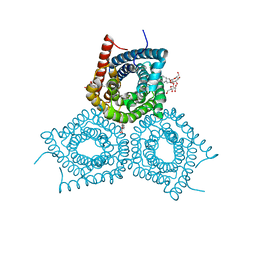
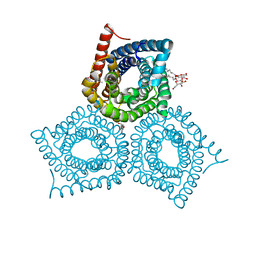
Crystal Structure of Plant SLAC1 homolog TehA
Descriptor: Tellurite resistance protein tehA homolog, octyl beta-D-glucopyranoside
Authors:Chen, Y.-H, Hendrickson, W.A, New York Consortium on Membrane Protein Structure (NYCOMPS)
Deposit date:2010-03-16
Release date:2010-05-12
Last modified:2024-02-21
Method:X-RAY DIFFRACTION (1.7 Å)
Cite:Crystal Structure of Plant SLAC1 homolog TehA
To be Published
3M74
DownloadVisualize
BU of 3m74 by Molmil
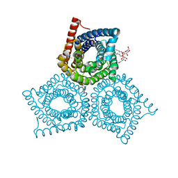
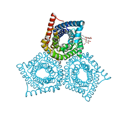
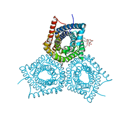
Crystal Structure of Plant SLAC1 homolog TehA
Descriptor: Tellurite resistance protein tehA homolog, octyl beta-D-glucopyranoside
Authors:Chen, Y.-H, Hendrickson, W.A, New York Consortium on Membrane Protein Structure (NYCOMPS)
Deposit date:2010-03-16
Release date:2010-05-12
Last modified:2024-02-21
Method:X-RAY DIFFRACTION (1.65 Å)
Cite:Homologue structure of the SLAC1 anion channel for closing stomata in leaves.
Nature, 467, 2010
3M7E
DownloadVisualize
BU of 3m7e by Molmil
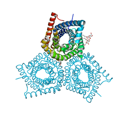
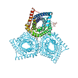
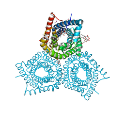
Crystal Structure of Plant SLAC1 homolog TehA
Descriptor: Tellurite resistance protein tehA homolog, octyl beta-D-glucopyranoside
Authors:Chen, Y.-H, Hendrickson, W.A, New York Consortium on Membrane Protein Structure (NYCOMPS)
Deposit date:2010-03-16
Release date:2010-05-12
Last modified:2024-02-21
Method:X-RAY DIFFRACTION (1.8 Å)
Cite:Crystal Structure of Plant SLAC1 homolog TehA
To be Published
3M71
DownloadVisualize
BU of 3m71 by Molmil
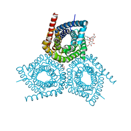
Crystal Structure of Plant SLAC1 homolog TehA
Descriptor: Tellurite resistance protein tehA homolog, octyl beta-D-glucopyranoside
Authors:Chen, Y.-H, Hu, L, Punta, M, Bruni, R, Hillerich, B, Kloss, B, Rost, B, Love, J, Siegelbaum, S.A, Hendrickson, W.A, New York Consortium on Membrane Protein Structure (NYCOMPS)
Deposit date:2010-03-16
Release date:2010-05-12
Last modified:2024-02-21
Method:X-RAY DIFFRACTION (1.2 Å)
Cite:Homologue structure of the SLAC1 anion channel for closing stomata in leaves.
Nature, 467, 2010
3M7C
DownloadVisualize
BU of 3m7c by Molmil
Crystal Structure of Plant SLAC1 homolog TehA
Descriptor: Tellurite resistance protein tehA homolog, octyl beta-D-glucopyranoside
Authors:Chen, Y.-H, Hendrickson, W.A, New York Consortium on Membrane Protein Structure (NYCOMPS)
Deposit date:2010-03-16
Release date:2010-05-12
Last modified:2024-02-21
Method:X-RAY DIFFRACTION (1.7 Å)
Cite:Crystal Structure of Plant SLAC1 homolog TehA
To be Published
3M76
DownloadVisualize
BU of 3m76 by Molmil
Crystal Structure of Plant SLAC1 homolog TehA
Descriptor: Tellurite resistance protein tehA homolog, octyl beta-D-glucopyranoside
Authors:Chen, Y.-H, Hendrickson, W.A, New York Consortium on Membrane Protein Structure (NYCOMPS)
Deposit date:2010-03-16
Release date:2010-05-12
Last modified:2024-02-21
Method:X-RAY DIFFRACTION (1.5 Å)
Cite:Homologue structure of the SLAC1 anion channel for closing stomata in leaves.
Nature, 467, 2010
3M77
DownloadVisualize
BU of 3m77 by Molmil
Crystal Structure of Plant SLAC1 homolog TehA
Descriptor: Tellurite resistance protein tehA homolog, octyl beta-D-glucopyranoside
Authors:Chen, Y.-H, Hendrickson, W.A, New York Consortium on Membrane Protein Structure (NYCOMPS)
Deposit date:2010-03-16
Release date:2010-05-12
Last modified:2024-02-21
Method:X-RAY DIFFRACTION (1.5 Å)
Cite:Crystal Structure of Plant SLAC1 homolog TehA
To be Published
3M70
DownloadVisualize
BU of 3m70 by Molmil
Crystal Structure of TehB from Haemophilus influenzae
Descriptor: Tellurite resistance protein tehB homolog
Authors:Chen, Y.-H, Hendrickson, W.A, New York Consortium on Membrane Protein Structure (NYCOMPS)
Deposit date:2010-03-16
Release date:2010-05-12
Last modified:2017-11-08
Method:X-RAY DIFFRACTION (1.95 Å)
Cite:Crystal Structure of TehB from Haemophilus influenzae
To be Published
3M75
DownloadVisualize
BU of 3m75 by Molmil
Crystal Structure of Plant SLAC1 homolog TehA
Descriptor: Tellurite resistance protein tehA homolog, octyl beta-D-glucopyranoside
Authors:Chen, Y.-H, Hendrickson, W.A, New York Consortium on Membrane Protein Structure (NYCOMPS)
Deposit date:2010-03-16
Release date:2010-05-12
Last modified:2024-02-21
Method:X-RAY DIFFRACTION (1.6 Å)
Cite:Homologue structure of the SLAC1 anion channel for closing stomata in leaves.
Nature, 467, 2010
3M73
DownloadVisualize
BU of 3m73 by Molmil
Crystal Structure of Plant SLAC1 homolog TehA
Descriptor: Tellurite resistance protein tehA homolog, octyl beta-D-glucopyranoside
Authors:Chen, Y.-H, Hendrickson, W.A, New York Consortium on Membrane Protein Structure (NYCOMPS)
Deposit date:2010-03-16
Release date:2010-05-12
Last modified:2024-02-21
Method:X-RAY DIFFRACTION (1.15 Å)
Cite:Homologue structure of the SLAC1 anion channel for closing stomata in leaves.
Nature, 467, 2010
3M7B
DownloadVisualize
BU of 3m7b by Molmil
Crystal Structure of Plant SLAC1 homolog TehA
Descriptor: Tellurite resistance protein tehA homolog, octyl beta-D-glucopyranoside
Authors:Chen, Y.-H, Hendrickson, W.A, New York Consortium on Membrane Protein Structure (NYCOMPS)
Deposit date:2010-03-16
Release date:2010-05-12
Last modified:2024-02-21
Method:X-RAY DIFFRACTION (1.5 Å)
Cite:Crystal Structure of Plant SLAC1 homolog TehA
To be Published
3M7L
DownloadVisualize
BU of 3m7l by Molmil
Crystal Structure of Plant SLAC1 homolog TehA
Descriptor: Tellurite resistance protein tehA homolog, octyl beta-D-glucopyranoside
Authors:Chen, Y.-H, Hendrickson, W.A, New York Consortium on Membrane Protein Structure (NYCOMPS)
Deposit date:2010-03-16
Release date:2010-05-12
Last modified:2020-07-29
Method:X-RAY DIFFRACTION (1.5 Å)
Cite:Homologue structure of the SLAC1 anion channel for closing stomata in leaves.
Nature, 467, 2010
3O1H
DownloadVisualize
BU of 3o1h by Molmil
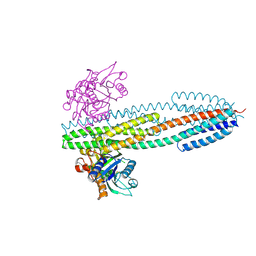
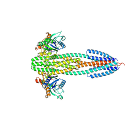
Crystal Structure of the TorS sensor domain - TorT complex in the presence of TMAO
Descriptor: Periplasmic protein TorT, Sensor protein TorS, trimethylamine oxide
Authors:Moore, J.O, Hendrickson, W.A.
Deposit date:2010-07-21
Release date:2011-12-21
Last modified:2024-10-09
Method:X-RAY DIFFRACTION (3.1 Å)
Cite:An asymmetry-to-symmetry switch in signal transmission by the histidine kinase receptor for TMAO.
Structure, 20, 2012
3M78
DownloadVisualize
BU of 3m78 by Molmil
Crystal Structure of Plant SLAC1 homolog TehA
Descriptor: Tellurite resistance protein tehA homolog, octyl beta-D-glucopyranoside
Authors:Chen, Y.-H, Hendrickson, W.A, New York Consortium on Membrane Protein Structure (NYCOMPS)
Deposit date:2010-03-16
Release date:2010-05-12
Last modified:2024-02-21
Method:X-RAY DIFFRACTION (2.6 Å)
Cite:Crystal Structure of Plant SLAC1 homolog TehA
To be Published
3O1J
DownloadVisualize
BU of 3o1j by Molmil
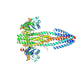
Crystal Structure of the TorS sensor domain - TorT complex in the absence of isopropanol
Descriptor: ISOPROPYL ALCOHOL, Periplasmic protein TorT, Sensor protein TorS
Authors:Moore, J.O, Hendrickson, W.A.
Deposit date:2010-07-21
Release date:2011-12-21
Last modified:2024-10-30
Method:X-RAY DIFFRACTION (2.95 Å)
Cite:An asymmetry-to-symmetry switch in signal transmission by the histidine kinase receptor for TMAO.
Structure, 20, 2012
3O1I
DownloadVisualize
BU of 3o1i by Molmil
Crystal Structure of the TorS sensor domain - TorT complex in the absence of ligand
Descriptor: 2-{2-[2-(2-{2-[2-(2-ETHOXY-ETHOXY)-ETHOXY]-ETHOXY}-ETHOXY)-ETHOXY]-ETHOXY}-ETHANOL, Periplasmic protein TorT, Sensor protein TorS
Authors:Moore, J.O, Hendrickson, W.A.
Deposit date:2010-07-21
Release date:2011-12-21
Last modified:2024-04-03
Method:X-RAY DIFFRACTION (2.8 Å)
Cite:An asymmetry-to-symmetry switch in signal transmission by the histidine kinase receptor for TMAO.
Structure, 20, 2012
1SCF
DownloadVisualize
BU of 1scf by Molmil
HUMAN RECOMBINANT STEM CELL FACTOR
Descriptor: CALCIUM ION, PENTAETHYLENE GLYCOL, STEM CELL FACTOR
Authors:Jiang, X, Gurel, O, Langley, K.E, Hendrickson, W.A.
Deposit date:1998-06-04
Release date:2000-07-07
Last modified:2024-10-30
Method:X-RAY DIFFRACTION (2.2 Å)
Cite:Structure of the active core of human stem cell factor and analysis of binding to its receptor kit.
EMBO J., 19, 2000
1T20
DownloadVisualize
BU of 1t20 by Molmil
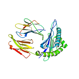
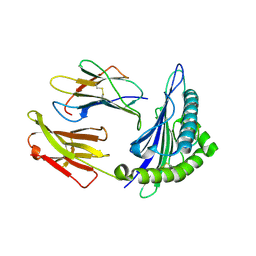
Structural basis for degenerate recognition of HIV peptide variants by cytotoxic lymphocyte, variant SL9-6I
Descriptor: Beta-2-microglobulin, GAG PEPTIDE, HLA class I histocompatibility antigen, ...
Authors:Martinez-Hackert, E, Anikeeva, N, Kalams, S.A, Walker, B.D, Hendrickson, W.A, Sykulev, Y.
Deposit date:2004-04-19
Release date:2005-09-06
Last modified:2024-10-30
Method:X-RAY DIFFRACTION (2.2 Å)
Cite:Structural Basis for Degenerate Recognition of Natural HIV Peptide Variants by Cytotoxic Lymphocytes.
J.Biol.Chem., 281, 2006
1T22
DownloadVisualize
BU of 1t22 by Molmil
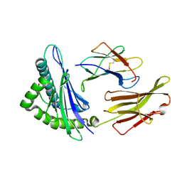
Structural basis for degenerate recognition of HIV peptide variants by cytotoxic lymphocyte, variant SL9, orthorhombic crystal
Descriptor: Beta-2-microglobulin, GAG PEPTIDE, HLA class I histocompatibility antigen, ...
Authors:Martinez-Hackert, E, Anikeeva, N, Kalams, S.A, Walker, B.D, Hendrickson, W.A, Sykulev, Y.
Deposit date:2004-04-19
Release date:2005-09-06
Last modified:2024-10-30
Method:X-RAY DIFFRACTION (2.2 Å)
Cite:Structural Basis for Degenerate Recognition of Natural HIV Peptide Variants by Cytotoxic Lymphocytes.
J.Biol.Chem., 281, 2006
1TEN
DownloadVisualize
BU of 1ten by Molmil
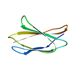
STRUCTURE OF A FIBRONECTIN TYPE III DOMAIN FROM TENASCIN PHASED BY MAD ANALYSIS OF THE SELENOMETHIONYL PROTEIN
Descriptor: TENASCIN
Authors:Leahy, D.J, Hendrickson, W.A, Aukhil, I, Erickson, H.P.
Deposit date:1992-08-28
Release date:1993-10-31
Last modified:2024-02-14
Method:X-RAY DIFFRACTION (1.8 Å)
Cite:Structure of a fibronectin type III domain from tenascin phased by MAD analysis of the selenomethionyl protein.
Science, 258, 1992
1TTU
DownloadVisualize
BU of 1ttu by Molmil
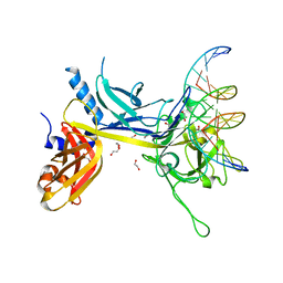
Crystal Structure of CSL bound to DNA
Descriptor: 1,2-ETHANEDIOL, 5'-D(*AP*AP*TP*CP*TP*TP*TP*CP*CP*CP*AP*CP*AP*GP*T)-3', 5'-D(*TP*TP*AP*CP*TP*GP*TP*GP*GP*GP*AP*AP*AP*GP*A)-3', ...
Authors:Kovall, R.A, Hendrickson, W.A.
Deposit date:2004-06-23
Release date:2004-08-31
Last modified:2024-02-14
Method:X-RAY DIFFRACTION (2.85 Å)
Cite:Crystal structure of the nuclear effector of Notch signaling, CSL, bound to DNA
Embo J., 23, 2004
1RZ8
DownloadVisualize
BU of 1rz8 by Molmil
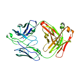
CRYSTAL STRUCTURE OF HUMAN ANTI-HIV-1 GP120-REACTIVE ANTIBODY 17B
Descriptor: Fab 17b heavy chain, Fab 17b light chain
Authors:Huang, C.C, Venturi, M, Majeed, S, Moore, M.J, Phogat, S, Zhang, M.-Y, Dimitrov, D.S, Hendrickson, W.A, Robinson, J, Sodroski, J, Wyatt, R, Choe, H, Farzan, M, Kwong, P.D.
Deposit date:2003-12-24
Release date:2004-02-03
Last modified:2024-10-16
Method:X-RAY DIFFRACTION (2.3 Å)
Cite:Structural basis of tyrosine sulfation and VH-gene usage in antibodies that recognize the HIV type 1 coreceptor-binding site on gp120
Proc.Natl.Acad.Sci.USA, 101, 2004
1T1Z
DownloadVisualize
BU of 1t1z by Molmil
Structural basis for degenerate recognition of HIV peptide variants by cytotoxic lymphocyte, variant SL9-6A
Descriptor: Beta-2-microglobulin, GAG PEPTIDE, HLA class I histocompatibility antigen, ...
Authors:Martinez-Hackert, E, Anikeeva, N, Kalams, S.A, Walker, B.D, Hendrickson, W.A, Sykulev, Y.
Deposit date:2004-04-19
Release date:2005-09-06
Last modified:2024-10-30
Method:X-RAY DIFFRACTION (1.9 Å)
Cite:Structural Basis for Degenerate Recognition of Natural HIV Peptide Variants by Cytotoxic Lymphocytes.
J.Biol.Chem., 281, 2006

226707

PDB entries from 2024-10-30

PDB statisticsPDBj update infoContact PDBjnumon