7SOU
 
 | |
7SOW
 | |
5K35
 | | Structure of the Legionella effector, AnkB, in complex with human Skp1 | | 分子名称: | Ankyrin-repeat protein B, S-phase kinase-associated protein 1 | | 著者 | Wong, K, Kozlov, G, Gehring, K, Montreal-Kingston Bacterial Structural Genomics Initiative (BSGI) | | 登録日 | 2016-05-19 | | 公開日 | 2017-01-25 | | 最終更新日 | 2024-03-06 | | 実験手法 | X-RAY DIFFRACTION (2.85 Å) | | 主引用文献 | Structural Mimicry by a Bacterial F Box Effector Hijacks the Host Ubiquitin-Proteasome System.
Structure, 25, 2017
|
|
3KTR
 | |
3KTP
 | |
3KUT
 | |
3KUJ
 | |
3KUS
 | |
3KUI
 | |
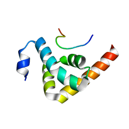
2IMT
 | | The X-ray Structure of a Bak Homodimer Reveals an Inhibitory Zinc Binding Site | | 分子名称: | Apoptosis regulator BAK, ZINC ION | | 著者 | Moldoveanu, T, Liu, Q, Tocilj, A, Watson, M, Shore, G.C, Gehring, K.B. | | 登録日 | 2006-10-04 | | 公開日 | 2007-01-23 | | 最終更新日 | 2023-08-30 | | 実験手法 | X-RAY DIFFRACTION (1.49 Å) | | 主引用文献 | The X-ray structure of a BAK homodimer reveals an inhibitory zinc binding site.
Mol.Cell, 24, 2006
|
|
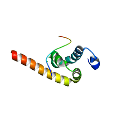
2IMS
 | | The X-ray Structure of a Bak Homodimer Reveals an Inhibitory Zinc Binding Site | | 分子名称: | Apoptosis regulator BAK, ZINC ION | | 著者 | Moldoveanu, T, Liu, Q, Tocilj, A, Watson, M, Shore, G.C, Gehring, K.B. | | 登録日 | 2006-10-04 | | 公開日 | 2006-12-26 | | 最終更新日 | 2017-10-18 | | 実験手法 | X-RAY DIFFRACTION (1.48 Å) | | 主引用文献 | The X-Ray Structure of a BAK Homodimer Reveals an Inhibitory Zinc Binding Site
Mol.Cell, 24, 2006
|
|
2OOB
 | |
4JRD
 | | Crystal structure of the parallel double-stranded helix of poly(A) RNA | | 分子名称: | AMMONIUM ION, RNA (5'-R(*AP*AP*AP*AP*AP*AP*AP*AP*AP*AP*A)-3') | | 著者 | Safaee, N, Noronha, A.M, Kozlov, G, Rodionov, D, Wilds, C.J, Sheldrick, G.M, Gehring, K. | | 登録日 | 2013-03-21 | | 公開日 | 2013-06-05 | | 最終更新日 | 2024-02-28 | | 実験手法 | X-RAY DIFFRACTION (1 Å) | | 主引用文献 | Structure of the parallel duplex of poly(A) RNA: evaluation of a 50 year-old prediction.
Angew.Chem.Int.Ed.Engl., 52, 2013
|
|
5BU2
 | | Structure of the C-terminal domain of lpg1496 from Legionella pneumophila in complex with nucleotide | | 分子名称: | ADENOSINE MONOPHOSPHATE, ADENOSINE-5'-DIPHOSPHATE, alpha-D-ribofuranose, ... | | 著者 | Wong, K, Kozlov, G, Gehring, K, Montreal-Kingston Bacterial Structural Genomics Initiative (BSGI) | | 登録日 | 2015-06-03 | | 公開日 | 2015-08-26 | | 最終更新日 | 2023-09-27 | | 実験手法 | X-RAY DIFFRACTION (2.11 Å) | | 主引用文献 | Structure of the Legionella Effector, lpg1496, Suggests a Role in Nucleotide Metabolism.
J.Biol.Chem., 290, 2015
|
|
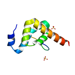
2X5N
 | | Crystal Structure of the SpRpn10 VWA domain | | 分子名称: | 26S PROTEASOME REGULATORY SUBUNIT RPN10, SULFATE ION | | 著者 | Riedinger, C, Boehringer, J, Trempe, J.-F, Lowe, E.D, Brown, N.R, Gehring, K, Noble, M.E.M, Gordon, C, Endicott, J.A. | | 登録日 | 2010-02-10 | | 公開日 | 2010-08-25 | | 最終更新日 | 2024-05-08 | | 実験手法 | X-RAY DIFFRACTION (1.3 Å) | | 主引用文献 | The Structure of Rpn10 and its Interactions with Polyubiquitin Chains and the Proteasome Subunit Rpn12.
J.Biol.Chem., 285, 2010
|
|
4EEW
 | |
7MSU
 | | Crystal structure of an archaeal CNNM, MtCorB, CBS-pair domain in complex with Mg2+-ATP | | 分子名称: | 1,2-ETHANEDIOL, ADENOSINE-5'-TRIPHOSPHATE, Hemolysin, ... | | 著者 | Chen, Y.S, Gehring, K. | | 登録日 | 2021-05-12 | | 公開日 | 2021-06-16 | | 最終更新日 | 2023-10-18 | | 実験手法 | X-RAY DIFFRACTION (2.07 Å) | | 主引用文献 | Crystal structure of an archaeal CorB magnesium transporter.
Nat Commun, 12, 2021
|
|
7M1U
 | | Crystal structure of an archaeal CNNM, MtCorB, R235L mutant with C-terminal deletion | | 分子名称: | Hemolysin, contains CBS domains | | 著者 | Chen, Y.S, Kozlov, G, Gehring, K. | | 登録日 | 2021-03-15 | | 公開日 | 2021-06-16 | | 最終更新日 | 2023-10-18 | | 実験手法 | X-RAY DIFFRACTION (3.8 Å) | | 主引用文献 | Crystal structure of an archaeal CorB magnesium transporter.
Nat Commun, 12, 2021
|
|
2MJC
 | | Zn-binding domain of eukaryotic translation initiation factor 3, subunit G | | 分子名称: | Eukaryotic translation initiation factor 3 subunit G, ZINC ION | | 著者 | Al-Abdul-Wahid, M, Menade, M, Xie, J, Kozlov, G, Gehring, K. | | 登録日 | 2014-01-03 | | 公開日 | 2015-01-07 | | 最終更新日 | 2024-05-15 | | 実験手法 | SOLUTION NMR | | 主引用文献 | Solution NMR structure of the Zn-binding domain of eukaryotic translation initiation factor 3, subunit G
To be Published
|
|
3IQL
 | | Crystal structure of the rat endophilin-A1 SH3 domain | | 分子名称: | CHLORIDE ION, Endophilin-A1 | | 著者 | Trempe, J.F, Kozlov, G, Camacho, E.M, Gehring, K. | | 登録日 | 2009-08-20 | | 公開日 | 2009-11-10 | | 最終更新日 | 2023-09-06 | | 実験手法 | X-RAY DIFFRACTION (1.4 Å) | | 主引用文献 | SH3 domains from a subset of BAR proteins define a Ubl-binding domain and implicate parkin in synaptic ubiquitination.
Mol.Cell, 36, 2009
|
|
1ME0
 | | Chimeric hairpin with 2',5'-linked RNA loop and DNA stem | | 分子名称: | DNA/RNA (5'-D(*GP*GP*AP*C)-R(P*(U25)P*(U25)P*(C25)P*(5GP))-D(P*GP*TP*CP*C)-3') | | 著者 | Denisov, A.Y, Hannoush, R.N, Gehring, K, Damha, M.J. | | 登録日 | 2002-08-07 | | 公開日 | 2003-08-19 | | 最終更新日 | 2024-05-01 | | 実験手法 | SOLUTION NMR | | 主引用文献 | A Novel RNA Motif Based on the Structure of Unusually Stable 2',5'-Linked r(UUCG) Loops
J.AM.CHEM.SOC., 125, 2003
|
|
5BTX
 | | Structure of the N-terminal domain of lpg1496 from Legionella pneumophila in complex with nucleotide | | 分子名称: | ADENOSINE-3',5'-CYCLIC-MONOPHOSPHATE, lpg1496 | | 著者 | Wong, K, Kozlov, G, Gehring, K, Montreal-Kingston Bacterial Structural Genomics Initiative (BSGI) | | 登録日 | 2015-06-03 | | 公開日 | 2015-08-26 | | 最終更新日 | 2023-09-27 | | 実験手法 | X-RAY DIFFRACTION (2.1 Å) | | 主引用文献 | Structure of the Legionella Effector, lpg1496, Suggests a Role in Nucleotide Metabolism.
J.Biol.Chem., 290, 2015
|
|
5BU0
 | | Structure of the C-terminal domain of lpg1496 from Legionella pneumophila | | 分子名称: | lpg1496 | | 著者 | Wong, K, Kozlov, G, Gehring, K, Montreal-Kingston Bacterial Structural Genomics Initiative (BSGI) | | 登録日 | 2015-06-03 | | 公開日 | 2015-08-26 | | 最終更新日 | 2024-03-06 | | 実験手法 | X-RAY DIFFRACTION (2.35 Å) | | 主引用文献 | Structure of the Legionella Effector, lpg1496, Suggests a Role in Nucleotide Metabolism.
J.Biol.Chem., 290, 2015
|
|
4G3O
 | |
2OOA
 | |


