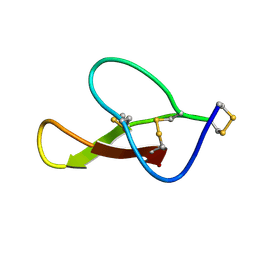
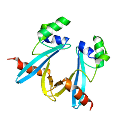
8H2J
 
 | | Structure of Acb2 complexed with 3',3'-cGAMP | | 分子名称: | 2-amino-9-[(2R,3R,3aS,5R,7aR,9R,10R,10aS,12R,14aR)-9-(6-amino-9H-purin-9-yl)-3,5,10,12-tetrahydroxy-5,12-dioxidooctahydro-2H,7H-difuro[3,2-d:3',2'-j][1,3,7,9,2,8]tetraoxadiphosphacyclododecin-2-yl]-1,9-dihydro-6H-purin-6-one, p26 | | 著者 | Feng, Y, Cao, X.L. | | 登録日 | 2022-10-06 | | 公開日 | 2023-02-22 | | 最終更新日 | 2024-04-03 | | 実験手法 | X-RAY DIFFRACTION (2.4 Å) | | 主引用文献 | Bacteriophages inhibit and evade cGAS-like immune function in bacteria.
Cell, 186, 2023
|
|
5H9U
 | |
3CVA
 | | Human Bcl-xL containing a Trp to Ala mutation at position 137 | | 分子名称: | Apoptosis regulator Bcl-X | | 著者 | Feng, Y, Zhang, L, Hu, T, Shen, X, Chen, K, Jiang, H, Liu, D. | | 登録日 | 2008-04-18 | | 公開日 | 2009-03-24 | | 最終更新日 | 2023-11-01 | | 実験手法 | X-RAY DIFFRACTION (2.7 Å) | | 主引用文献 | A conserved hydrophobic core at Bcl-x(L) mediates its structural stability and binding affinity with BH3-domain peptide of pro-apoptotic protein
Arch.Biochem.Biophys., 484, 2009
|
|
5ZIG
 | |
5ZHB
 | |
3KXT
 | | Crystal structure of Sulfolobus Cren7-dsDNA complex | | 分子名称: | 5'-D(*GP*CP*GP*AP*TP*CP*GP*C)-3', Chromatin protein Cren7 | | 著者 | Feng, Y, Wang, J. | | 登録日 | 2009-12-03 | | 公開日 | 2010-06-09 | | 最終更新日 | 2023-11-01 | | 実験手法 | X-RAY DIFFRACTION (1.602 Å) | | 主引用文献 | Crystal structure of the crenarchaeal conserved chromatin protein Cren7 and double-stranded DNA complex
Protein Sci., 19, 2010
|
|
8K0H
 | |
8K0J
 | |
2HR9
 | |
8JP3
 | | FCP trimer in diatom Thalassiosira pseudonana | | 分子名称: | (3S,3'S,5R,5'R,6S,6'R,8'R)-3,5'-dihydroxy-8-oxo-6',7'-didehydro-5,5',6,6',7,8-hexahydro-5,6-epoxy-beta,beta-caroten-3'- yl acetate, 1,2-DISTEAROYL-MONOGALACTOSYL-DIGLYCERIDE, CHLOROPHYLL A, ... | | 著者 | Feng, Y, Li, Z, Zhou, C.C, Liu, C, Shen, J.R, Wang, W. | | 登録日 | 2023-06-10 | | 公開日 | 2024-06-12 | | 最終更新日 | 2024-07-31 | | 実験手法 | ELECTRON MICROSCOPY (2.73 Å) | | 主引用文献 | Structural and spectroscopic insights into fucoxanthin chlorophyll a/c-binding proteins of diatoms in diverse oligomeric states.
Plant Commun., 2024
|
|
8K0K
 | |
8HJD
 | |
8HJC
 | |
6IGM
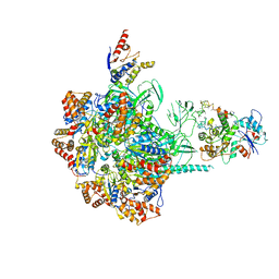 | | Cryo-EM Structure of Human SRCAP Complex | | 分子名称: | Actin-related protein 6, Helicase SRCAP, RuvB-like 1, ... | | 著者 | Feng, Y, Tian, Y, Wu, Z, Xu, Y. | | 登録日 | 2018-09-25 | | 公開日 | 2019-07-24 | | 最終更新日 | 2024-03-27 | | 実験手法 | ELECTRON MICROSCOPY (4 Å) | | 主引用文献 | Cryo-EM structure of human SRCAP complex.
Cell Res., 28, 2018
|
|
6INW
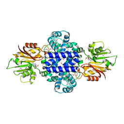 | | A Pericyclic Reaction enzyme | | 分子名称: | O-methyltransferase lepI, S-ADENOSYLMETHIONINE | | 著者 | Feng, Y, Chang, M, Wang, H, Liu, Z, Zhou, Y. | | 登録日 | 2018-10-28 | | 公開日 | 2019-07-03 | | 実験手法 | X-RAY DIFFRACTION (1.798 Å) | | 主引用文献 | Crystal structure of the multifunctional SAM-dependent enzyme LepI provides insights into its catalytic mechanism.
Biochem.Biophys.Res.Commun., 515, 2019
|
|
6KGC
 | | Crystal structure of CaDoc0917(R49D)-CaCohA2 complex at pH 5.4 | | 分子名称: | And cellulose-binding endoglucanase family 9 CelL ortholog dockerin domain, CALCIUM ION, Probably cellulosomal scaffolding protein, ... | | 著者 | Feng, Y, Yao, X. | | 登録日 | 2019-07-11 | | 公開日 | 2020-07-08 | | 最終更新日 | 2023-11-22 | | 実験手法 | X-RAY DIFFRACTION (1.6 Å) | | 主引用文献 | Discovery and mechanism of a pH-dependent dual-binding-site switch in the interaction of a pair of protein modules.
Sci Adv, 6, 2020
|
|
7CMW
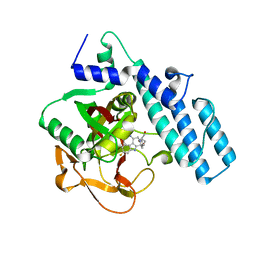 | | Complex structure of PARP1 catalytic domain with pamiparib | | 分子名称: | (2R)-14-fluoro-2-methyl-6,9,10,19-tetrazapentacyclo[14.2.1.02,6.08,18.012,17]nonadeca-1(18),8,12(17),13,15-pentaen-11-one, GLYCEROL, Poly [ADP-ribose] polymerase 1 | | 著者 | Feng, Y.C, Peng, H, Hong, Y, Liu, Y. | | 登録日 | 2020-07-29 | | 公開日 | 2020-12-16 | | 最終更新日 | 2023-11-29 | | 実験手法 | X-RAY DIFFRACTION (2.7 Å) | | 主引用文献 | Discovery of Pamiparib (BGB-290), a Potent and Selective Poly (ADP-ribose) Polymerase (PARP) Inhibitor in Clinical Development.
J.Med.Chem., 63, 2020
|
|
6KGD
 | | Crystal structure of CaDoc0917(R49D)-CaCohA2 complex at pH 8.0 | | 分子名称: | And cellulose-binding endoglucanase family 9 CelL ortholog dockerin domain, CALCIUM ION, Probably cellulosomal scaffolding protein, ... | | 著者 | Feng, Y, Yao, X. | | 登録日 | 2019-07-11 | | 公開日 | 2020-07-08 | | 最終更新日 | 2023-11-22 | | 実験手法 | X-RAY DIFFRACTION (1.65 Å) | | 主引用文献 | Discovery and mechanism of a pH-dependent dual-binding-site switch in the interaction of a pair of protein modules.
Sci Adv, 6, 2020
|
|
6KG9
 | |
6KGF
 | | Crystal structure of CaDoc0917(R16D)-CaCohA2 complex at pH 8.2 | | 分子名称: | And cellulose-binding endoglucanase family 9 CelL ortholog dockerin domain, CALCIUM ION, Probably cellulosomal scaffolding protein, ... | | 著者 | Feng, Y, Yao, X. | | 登録日 | 2019-07-11 | | 公開日 | 2020-07-08 | | 最終更新日 | 2023-11-22 | | 実験手法 | X-RAY DIFFRACTION (2.3 Å) | | 主引用文献 | Discovery and mechanism of a pH-dependent dual-binding-site switch in the interaction of a pair of protein modules.
Sci Adv, 6, 2020
|
|
6KGE
 | | Crystal structure of CaDoc0917(R16D)-CaCohA2 complex at pH 5.5 | | 分子名称: | And cellulose-binding endoglucanase family 9 CelL ortholog dockerin domain, CALCIUM ION, Probably cellulosomal scaffolding protein, ... | | 著者 | Feng, Y, Yao, X. | | 登録日 | 2019-07-11 | | 公開日 | 2020-07-08 | | 最終更新日 | 2023-11-22 | | 実験手法 | X-RAY DIFFRACTION (2 Å) | | 主引用文献 | Discovery and mechanism of a pH-dependent dual-binding-site switch in the interaction of a pair of protein modules.
Sci Adv, 6, 2020
|
|
6KG8
 | |
7WIN
 | |
8IWH
 | | Structure and characteristics of a photosystem II supercomplex containing monomeric LHCX and dimeric FCPII antennae from the diatom Thalassiosira pseudonana | | 分子名称: | (1~{R})-3,5,5-trimethyl-4-[(1~{E},3~{E},5~{E},7~{E},9~{E},11~{E},13~{E},15~{E})-3,7,12,16-tetramethyl-18-[(4~{R})-2,6,6-trimethyl-4-oxidanyl-cyclohexen-1-yl]octadeca-1,3,5,7,9,11,13,15-octaen-17-ynyl]cyclohex-3-en-1-ol, (3S,3'R,5R,6S,7cis)-7',8'-didehydro-5,6-dihydro-5,6-epoxy-beta,beta-carotene-3,3'-diol, (3S,3'S,5R,5'R,6S,6'R,8'R)-3,5'-dihydroxy-8-oxo-6',7'-didehydro-5,5',6,6',7,8-hexahydro-5,6-epoxy-beta,beta-caroten-3'- yl acetate, ... | | 著者 | Feng, Y, Li, Z.H, Wang, W.D, Shen, J.R. | | 登録日 | 2023-03-30 | | 公開日 | 2023-10-25 | | 最終更新日 | 2023-11-08 | | 実験手法 | ELECTRON MICROSCOPY (2.68 Å) | | 主引用文献 | Structure of a diatom photosystem II supercomplex containing a member of Lhcx family and dimeric FCPII.
Sci Adv, 9, 2023
|
|
2JSN
 | |


