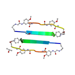
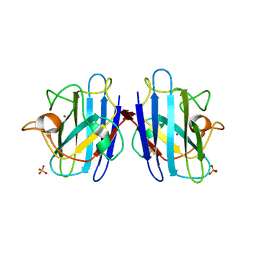
1F0V
 
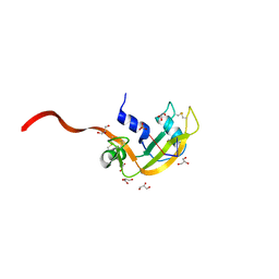 | | Crystal structure of an Rnase A dimer displaying a new type of 3D domain swapping | | 分子名称: | 5'-D(*CP*G)-3', GLYCEROL, PHOSPHATE ION, ... | | 著者 | Liu, Y.S, Gotte, G, Libonati, M, Eisenberg, D.S. | | 登録日 | 2000-05-17 | | 公開日 | 2001-02-21 | | 最終更新日 | 2011-07-13 | | 実験手法 | X-RAY DIFFRACTION (1.7 Å) | | 主引用文献 | A domain-swapped RNase A dimer with implications for amyloid formation
Nat.Struct.Biol., 8, 2001
|
|
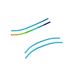
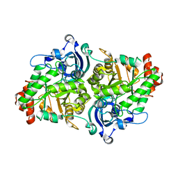
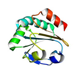
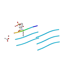
2A88
 | | Crystal structure of A Pantothenate synthetase, apo enzyme in C2 space group | | 分子名称: | GLYCEROL, Pantoate--beta-alanine ligase, SULFATE ION | | 著者 | Wang, S, Eisenberg, D, TB Structural Genomics Consortium (TBSGC) | | 登録日 | 2005-07-07 | | 公開日 | 2006-02-21 | | 最終更新日 | 2023-08-23 | | 実験手法 | X-RAY DIFFRACTION (1.7 Å) | | 主引用文献 | Crystal Structure of the Pantothenate Synthetase from Mycobacterium tuberculosis, Snapshots of the Enzyme in Action.
Biochemistry, 45, 2006
|
|
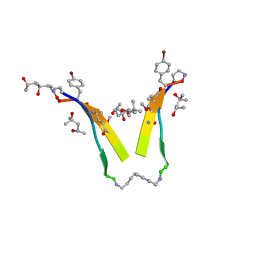
1AL1
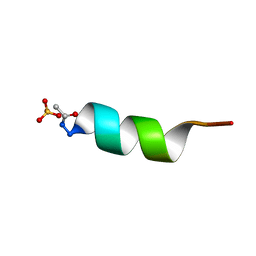 | | CRYSTAL STRUCTURE OF ALPHA1: IMPLICATIONS FOR PROTEIN DESIGN | | 分子名称: | ALPHA HELIX PEPTIDE: ELLKKLLEELKG, SULFATE ION | | 著者 | Hill, C.P, Anderson, D.H, Wesson, L, Degrado, W.F, Eisenberg, D. | | 登録日 | 1990-07-02 | | 公開日 | 1991-10-15 | | 最終更新日 | 2024-06-05 | | 実験手法 | X-RAY DIFFRACTION (2.7 Å) | | 主引用文献 | Crystal structure of alpha 1: implications for protein design.
Science, 249, 1990
|
|
1DFN
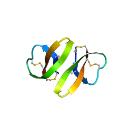 | | CRYSTAL STRUCTURE OF DEFENSIN HNP-3, AN AMPHIPHILIC DIMER: MECHANISMS OF MEMBRANE PERMEABILIZATION | | 分子名称: | DEFENSIN HNP-3 | | 著者 | Hill, C.P, Yee, J, Selsted, M.E, Eisenberg, D. | | 登録日 | 1991-01-18 | | 公開日 | 1992-07-15 | | 最終更新日 | 2017-11-29 | | 実験手法 | X-RAY DIFFRACTION (1.9 Å) | | 主引用文献 | Crystal structure of defensin HNP-3, an amphiphilic dimer: mechanisms of membrane permeabilization.
Science, 251, 1991
|
|
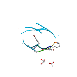
3PPD
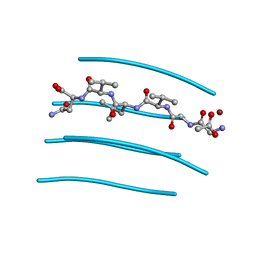 | | GGVLVN segment from Human Prostatic Acid Phosphatase Residues 260-265, involved in Semen-Derived Enhancer of Viral Infection | | 分子名称: | ACETIC ACID, GGVLVN peptide, amyloid forming segment, ... | | 著者 | Zhao, A, Sawaya, M.R, Eisenberg, D. | | 登録日 | 2010-11-24 | | 公開日 | 2011-06-29 | | 最終更新日 | 2024-02-21 | | 実験手法 | X-RAY DIFFRACTION (1.5 Å) | | 主引用文献 | Structure-based design of non-natural amino-acid inhibitors of amyloid fibril formation.
Nature, 475, 2011
|
|
3NI3
 | | 54-Membered ring macrocyclic beta-sheet peptide | | 分子名称: | 54-membered ring macrocyclic beta-sheet peptide, ISOPROPYL ALCOHOL | | 著者 | Sawaya, M.R, Eisenberg, D, Nowick, J.S, Korman, T.P, Khakshoor, O. | | 登録日 | 2010-06-15 | | 公開日 | 2010-09-15 | | 最終更新日 | 2023-11-15 | | 実験手法 | X-RAY DIFFRACTION (1.34 Å) | | 主引用文献 | X-ray crystallographic structure of an artificial beta-sheet dimer.
J.Am.Chem.Soc., 132, 2010
|
|
2OMM
 | |
1S4Q
 | | Crystal Structure of Guanylate Kinase from Mycobacterium tuberculosis (Rv1389) | | 分子名称: | CHLORIDE ION, FORMIC ACID, Guanylate kinase | | 著者 | Chan, S, Sawaya, M.R, Perry, L.J, Eisenberg, D, TB Structural Genomics Consortium (TBSGC) | | 登録日 | 2004-01-16 | | 公開日 | 2004-01-27 | | 最終更新日 | 2011-07-13 | | 実験手法 | X-RAY DIFFRACTION (2.16 Å) | | 主引用文献 | Crystal Structure of Guanylate Kinase from Mycobacterium tuberculosis
To be Published
|
|
2QYG
 | | Crystal Structure of a RuBisCO-like Protein rlp2 from Rhodopseudomonas palustris | | 分子名称: | Ribulose bisphosphate carboxylase-like protein 2 | | 著者 | Li, H, Chan, S, Tabita, F.R, Eisenberg, D. | | 登録日 | 2007-08-14 | | 公開日 | 2007-09-11 | | 最終更新日 | 2023-08-30 | | 実験手法 | X-RAY DIFFRACTION (3.3 Å) | | 主引用文献 | Function, structure, and evolution of the RubisCO-like proteins and their RubisCO homologs.
Microbiol.Mol.Biol.Rev., 71, 2007
|
|
3T4G
 | | AIIGLMV segment from Alzheimer's Amyloid-Beta displayed on 54-membered macrocycle scaffold | | 分子名称: | (4S)-2-METHYL-2,4-PENTANEDIOL, Cyclic pseudo-peptide (ORN)AIIGLMV(ORN)KF(HAO)(4BF)K | | 著者 | Zhao, M, Liu, C, Cheng, P.N, Eisenberg, D, Nowick, J.S. | | 登録日 | 2011-07-26 | | 公開日 | 2012-10-31 | | 最終更新日 | 2023-11-15 | | 実験手法 | X-RAY DIFFRACTION (1.7 Å) | | 主引用文献 | Amyloid beta-sheet mimics that antagonize protein aggregation and reduce amyloid toxicity.
Nat Chem, 4, 2012
|
|
1JBM
 | |
4R0P
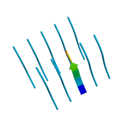 | |
1YJP
 | | Structure of GNNQQNY from yeast prion Sup35 | | 分子名称: | Eukaryotic peptide chain release factor GTP-binding subunit | | 著者 | Nelson, R, Sawaya, M.R, Balbirnie, M, Madsen, A.O, Riekel, C, Grothe, R, Eisenberg, D. | | 登録日 | 2005-01-15 | | 公開日 | 2005-06-14 | | 最終更新日 | 2024-02-14 | | 実験手法 | X-RAY DIFFRACTION (1.8 Å) | | 主引用文献 | Structure of the cross-beta spine of amyloid-like fibrils.
Nature, 435, 2005
|
|
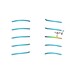
3Q9G
 | | VQIVY segment from Alzheimer's tau displayed on 42-membered macrocycle scaffold | | 分子名称: | ACETIC ACID, Cyclic pseudo-peptide VQIV(4BF)(ORN)(HAO)KL(ORN), GLYCEROL | | 著者 | Liu, C, Sawaya, M.R, Eisenberg, D, Nowick, J.S, Cheng, P, Zheng, J. | | 登録日 | 2011-01-07 | | 公開日 | 2011-06-08 | | 最終更新日 | 2023-11-15 | | 実験手法 | X-RAY DIFFRACTION (2.05 Å) | | 主引用文献 | Characteristics of Amyloid-Related Oligomers Revealed by Crystal Structures of Macrocyclic beta-Sheet Mimics.
J.Am.Chem.Soc., 133, 2011
|
|
3NVF
 | |
3NVG
 | |
1LU4
 | | 1.1 ANGSTROM RESOLUTION CRYSTAL STRUCTURE OF A SECRETED MYCOBACTERIUM TUBERCULOSIS DISULFIDE OXIDOREDUCTASE HOMOLOGOUS TO E. COLI DSBE: IMPLICATIONS FOR FUNCTIONS | | 分子名称: | SOLUBLE SECRETED ANTIGEN MPT53 | | 著者 | Goulding, C.W, Apostol, M.I, Gleiter, S, Parseghian, A, Bardwell, J, Gennaro, M, Eisenberg, D, TB Structural Genomics Consortium (TBSGC) | | 登録日 | 2002-05-21 | | 公開日 | 2003-10-14 | | 最終更新日 | 2024-02-14 | | 実験手法 | X-RAY DIFFRACTION (1.12 Å) | | 主引用文献 | Gram-positive DsbE Proteins Function Differently from Gram-negative DsbE Homologs: A STRUCTURE TO FUNCTION ANALYSIS OF DsbE FROM MYCOBACTERIUM TUBERCULOSIS.
J.Biol.Chem., 279, 2004
|
|
1F1G
 | | Crystal structure of yeast cuznsod exposed to nitric oxide | | 分子名称: | COPPER (II) ION, COPPER-ZINC SUPEROXIDE DISMUTASE, PHOSPHATE ION, ... | | 著者 | Hart, P.J, Ogihara, N.L, Liu, H, Nersissian, A.M, Valentine, J.S, Eisenberg, D. | | 登録日 | 2000-05-18 | | 公開日 | 2002-12-12 | | 最終更新日 | 2017-10-04 | | 実験手法 | X-RAY DIFFRACTION (1.35 Å) | | 主引用文献 | A structure-based mechanism for copper-zinc superoxide dismutase.
Biochemistry, 38, 1999
|
|
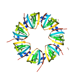
1HTQ
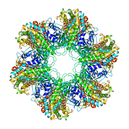 | | Multicopy crystallographic structure of a relaxed glutamine synthetase from Mycobacterium tuberculosis | | 分子名称: | ADENOSINE MONOPHOSPHATE, CITRIC ACID, MANGANESE (II) ION, ... | | 著者 | Gill, H.S, Pfluegl, G.M, Eisenberg, D, TB Structural Genomics Consortium (TBSGC) | | 登録日 | 2001-01-01 | | 公開日 | 2002-07-24 | | 最終更新日 | 2023-08-09 | | 実験手法 | X-RAY DIFFRACTION (2.4 Å) | | 主引用文献 | Multicopy crystallographic refinement of a relaxed glutamine synthetase from Mycobacterium tuberculosis highlights flexible loops in the enzymatic mechanism and its regulation.
Biochemistry, 41, 2002
|
|
3NVH
 | |
3OVL
 | |
1JRI
 | |
1EWF
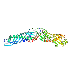 | | THE 1.7 ANGSTROM CRYSTAL STRUCTURE OF BPI | | 分子名称: | 1,2-DIACYL-SN-GLYCERO-3-PHOSPHOCHOLINE, BACTERICIDAL/PERMEABILITY-INCREASING PROTEIN | | 著者 | Kleiger, G, Beamer, L.J, Grothe, R, Mallick, P, Eisenberg, D. | | 登録日 | 2000-04-25 | | 公開日 | 2000-06-21 | | 最終更新日 | 2021-11-03 | | 実験手法 | X-RAY DIFFRACTION (1.7 Å) | | 主引用文献 | The 1.7 A crystal structure of BPI: a study of how two dissimilar amino acid sequences can adopt the same fold.
J.Mol.Biol., 299, 2000
|
|
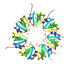
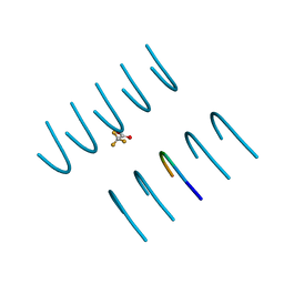
1G6U
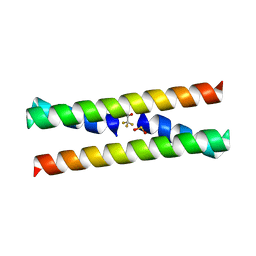 | | CRYSTAL STRUCTURE OF A DOMAIN SWAPPED DIMER | | 分子名称: | DOMAIN SWAPPED DIMER, SULFATE ION, trifluoroacetic acid | | 著者 | Ogihara, N.L, Ghirlanda, G, Bryson, J.W, Gingery, M, DeGrado, W.F, Eisenberg, D. | | 登録日 | 2000-11-07 | | 公開日 | 2001-02-21 | | 最終更新日 | 2024-02-07 | | 実験手法 | X-RAY DIFFRACTION (1.48 Å) | | 主引用文献 | Design of three-dimensional domain-swapped dimers and fibrous oligomers.
Proc.Natl.Acad.Sci.USA, 98, 2001
|
|
1JS0
 | | Crystal Structure of 3D Domain-swapped RNase A Minor Trimer | | 分子名称: | RIBONUCLEASE A, SULFATE ION | | 著者 | Liu, Y, Gotte, G, Libonati, M, Eisenberg, D. | | 登録日 | 2001-08-15 | | 公開日 | 2002-03-13 | | 最終更新日 | 2023-08-16 | | 実験手法 | X-RAY DIFFRACTION (2.2 Å) | | 主引用文献 | Structures of the two 3D domain-swapped RNase A trimers.
Protein Sci., 11, 2002
|
|


