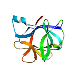
Loading
PDBj
MenuPDBj@FacebookPDBj@TwitterPDBj@YouTubewwPDB FoundationwwPDB
RCSB PDBPDBeBMRBAdv. SearchSearch help
PDB: 5 results

1I1B
DownloadVisualize
BU of 1i1b by Molmil
CRYSTAL STRUCTURE OF RECOMBINANT HUMAN INTERLEUKIN-1BETA AT 2.0 ANGSTROMS RESOLUTION
Descriptor: INTERLEUKIN-1 BETA
Authors:Finzel, B.C, Watenpaugh, K.D, Einspahr, H.M.
Deposit date:1989-12-05
Release date:1990-01-15
Last modified:2024-02-07
Method:X-RAY DIFFRACTION (2 Å)
Cite:Crystal structure of recombinant human interleukin-1 beta at 2.0 A resolution.
J.Mol.Biol., 209, 1989
2AYW
DownloadVisualize
BU of 2ayw by Molmil
Crystal Structure of the complex formed between trypsin and a designed synthetic highly potent inhibitor in the presence of benzamidine at 0.97 A resolution
Descriptor: 2-(N-MORPHOLINO)-ETHANESULFONIC ACID, 2-[2-({[4-(DIAMINOMETHYL)PHENYL]AMINO}CARBONYL)-6-METHOXYPYRIDIN-3-YL]-5-{[(1-FORMYL-2,2-DIMETHYLPROPYL)AMINO]CARBONYL}BENZOIC ACID, BENZAMIDINE, ...
Authors:Sherawat, M, Kaur, P, Perbandt, M, Betzel, C, Slusarchyk, W.A, Bisacchi, G.S, Chang, C, Jacobson, B.L, Einspahr, H.M, Singh, T.P.
Deposit date:2005-09-09
Release date:2006-01-17
Last modified:2023-10-25
Method:X-RAY DIFFRACTION (0.97 Å)
Cite:Structure of the complex of trypsin with a highly potent synthetic inhibitor at 0.97 A resolution.
Acta Crystallogr.,Sect.D, 63, 2007
2IRT
DownloadVisualize
BU of 2irt by Molmil
INITIAL CRYSTALLOGRAPHIC ANALYSES OF A RECOMBINANT INTERLEUKIN-1 RECEPTOR ANTAGONIST PROTEIN
Descriptor: INTERLEUKIN-1 RECEPTOR ANTAGONIST
Authors:Finzel, B.C, Clancy, L.L, Einspahr, H.M.
Deposit date:1994-07-13
Release date:1994-10-15
Last modified:2024-02-21
Method:X-RAY DIFFRACTION (3.2 Å)
Cite:Initial crystallographic analysis of a recombinant human interleukin-1 receptor antagonist protein.
Acta Crystallogr.,Sect.D, 50, 1994
1PPA
DownloadVisualize
BU of 1ppa by Molmil
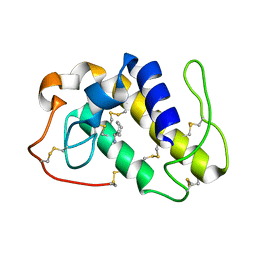
THE CRYSTAL STRUCTURE OF A LYSINE 49 PHOSPHOLIPASE A2 FROM THE VENOM OF THE COTTONMOUTH SNAKE AT 2.0 ANGSTROMS RESOLUTION
Descriptor: ANILINE, PHOSPHOLIPASE A2
Authors:Holland, D.R, Clancy, L.L, Muchmore, S.W, Rydel, T.J, Einspahr, H.M, Finzel, B.C, Heinrikson, R.L, Watenpaugh, K.D.
Deposit date:1991-10-29
Release date:1994-01-31
Last modified:2011-07-13
Method:X-RAY DIFFRACTION (2 Å)
Cite:The crystal structure of a lysine 49 phospholipase A2 from the venom of the cottonmouth snake at 2.0-A resolution.
J.Biol.Chem., 265, 1990
1RDH
DownloadVisualize
BU of 1rdh by Molmil
CRYSTALLOGRAPHIC ANALYSES OF AN ACTIVE HIV-1 RIBONUCLEASE H DOMAIN SHOW STRUCTURAL FEATURES THAT DISTINGUISH IT FROM THE INACTIVE FORM
Descriptor: HIV-1 REVERSE TRANSCRIPTASE (RIBONUCLEASE H DOMAIN)
Authors:Finzel, B.C, Chattopadhyay, D, Einspahr, H.M.
Deposit date:1993-03-05
Release date:1994-05-31
Last modified:2024-02-14
Method:X-RAY DIFFRACTION (2.8 Å)
Cite:Crystallographic analyses of an active HIV-1 ribonuclease H domain show structural features that distinguish it from the inactive form.
Acta Crystallogr.,Sect.D, 49, 1993

218853

PDB entries from 2024-04-24

PDB statisticsPDBj update infoContact PDBjnumon