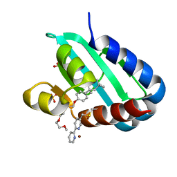
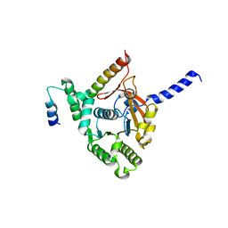
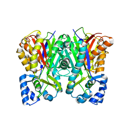
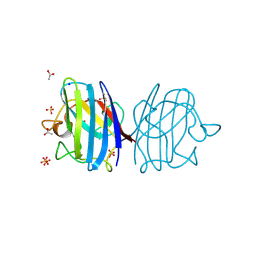
1ACM
 
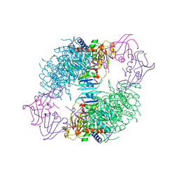 | | ARGININE 54 IN THE ACTIVE SITE OF ESCHERICHIA COLI ASPARTATE TRANSCARBAMOYLASE IS CRITICAL FOR CATALYSIS: A SITE-SPECIFIC MUTAGENESIS, NMR AND X-RAY CRYSTALLOGRAPHY STUDY | | 分子名称: | ASPARTATE CARBAMOYLTRANSFERASE REGULATORY CHAIN, ASPARTATE CARBAMOYLTRANSFERASE, CATALYTIC CHAIN, ... | | 著者 | Stevens, R.C, Kantrowitz, E.R, Lipscomb, W.N. | | 登録日 | 1992-07-08 | | 公開日 | 1992-07-15 | | 最終更新日 | 2024-02-07 | | 実験手法 | X-RAY DIFFRACTION (2.8 Å) | | 主引用文献 | Arginine 54 in the active site of Escherichia coli aspartate transcarbamoylase is critical for catalysis: a site-specific mutagenesis, NMR, and X-ray crystallographic study.
Protein Sci., 1, 1992
|
|
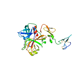
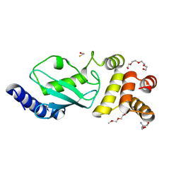
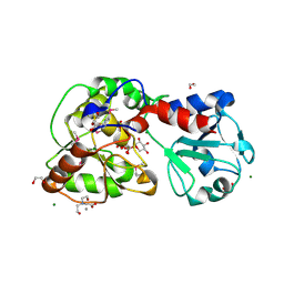
1A73
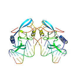 | | INTRON-ENCODED ENDONUCLEASE I-PPOI COMPLEXED WITH DNA | | 分子名称: | DNA (5'-D(*TP*TP*GP*AP*CP*TP*CP*TP*CP*TP*TP*AP*A)-3'), DNA (5'-D(P*GP*AP*GP*AP*GP*TP*CP*A)-3'), INTRON 3 (I-PPO) ENCODED ENDONUCLEASE, ... | | 著者 | Flick, K.E, Monnat Junior, R.J, Stoddard, B.L. | | 登録日 | 1998-03-19 | | 公開日 | 1998-10-14 | | 最終更新日 | 2024-02-07 | | 実験手法 | X-RAY DIFFRACTION (1.8 Å) | | 主引用文献 | DNA binding and cleavage by the nuclear intron-encoded homing endonuclease I-PpoI.
Nature, 394, 1998
|
|
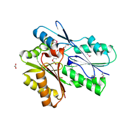
3F1T
 | | Crystal structure of the Q9I3C8_PSEAE protein from Pseudomonas aeruginosa. Northeast Structural Genomics Consortium target PaR319a. | | 分子名称: | MAGNESIUM ION, uncharacterized protein Q9I3C8_PSEAE | | 著者 | Vorobiev, S.M, Chen, Y, Abashidze, M, Seetharaman, J, Wang, D, Foote, E.L, Ciccosanti, C, Mao, L, Xiao, R, Acton, T.B, Montelione, G.T, Tong, L, Hunt, J.F, Northeast Structural Genomics Consortium (NESG) | | 登録日 | 2008-10-28 | | 公開日 | 2008-11-18 | | 最終更新日 | 2023-12-27 | | 実験手法 | X-RAY DIFFRACTION (2.2 Å) | | 主引用文献 | Crystal structure of the Q9I3C8_PSEAE protein from Pseudomonas aeruginosa.
To be Published
|
|
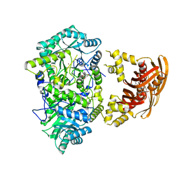
3F3C
 | | Crystal structure of LeuT bound to 4-Fluoro-L-Phenylalanine and sodium | | 分子名称: | 4-FLUORO-L-PHENYLALANINE, SODIUM ION, Transporter, ... | | 著者 | Singh, S.K, Piscitelli, C.L, Yamashita, A, Gouaux, E. | | 登録日 | 2008-10-30 | | 公開日 | 2008-12-23 | | 最終更新日 | 2023-09-06 | | 実験手法 | X-RAY DIFFRACTION (2.1 Å) | | 主引用文献 | A competitive inhibitor traps LeuT in an open-to-out conformation.
Science, 322, 2008
|
|
8AF3
 | | Sterol carrier protein Artifical metalloenzyme incorporating Q111C mutation coupled to 2,2'-bipyridine | | 分子名称: | COPPER (II) ION, Enoyl-CoA hydratase 2, FRAGMENT OF TRITON X-100, ... | | 著者 | Richardson, J.M, Klemencic, E, Jarvis, A.G. | | 登録日 | 2022-07-15 | | 公開日 | 2023-08-16 | | 最終更新日 | 2024-07-10 | | 実験手法 | X-RAY DIFFRACTION (1.52 Å) | | 主引用文献 | Using BpyAla to generate copper artificial metalloenzymes: a catalytic and structural study.
Catalysis Science And Technology, 14, 2024
|
|
3F4F
 | | Crystal structure of dUT1p, a dUTPase from Saccharomyces cerevisiae | | 分子名称: | 1,2-ETHANEDIOL, 2'-DEOXYURIDINE 5'-MONOPHOSPHATE, DI(HYDROXYETHYL)ETHER, ... | | 著者 | Singer, A.U, Evdokimova, E, Kudritska, M, Edwards, A.M, Yakunin, A.F, Savchenko, A. | | 登録日 | 2008-10-31 | | 公開日 | 2008-11-11 | | 最終更新日 | 2023-09-06 | | 実験手法 | X-RAY DIFFRACTION (2 Å) | | 主引用文献 | Structure and activity of the Saccharomyces cerevisiae dUTP pyrophosphatase DUT1, an essential housekeeping enzyme.
Biochem.J., 437, 2011
|
|
3F6U
 | | Crystal structure of human Activated Protein C (APC) complexed with PPACK | | 分子名称: | CALCIUM ION, D-phenylalanyl-N-[(2S,3S)-6-{[amino(iminio)methyl]amino}-1-chloro-2-hydroxyhexan-3-yl]-L-prolinamide, SODIUM ION, ... | | 著者 | Schmidt, A.E, Padmanabhan, K, Underwood, M.C, Bode, W, Mather, T, Bajaj, S.P. | | 登録日 | 2008-11-06 | | 公開日 | 2008-11-25 | | 最終更新日 | 2024-03-13 | | 実験手法 | X-RAY DIFFRACTION (2.8 Å) | | 主引用文献 | Thermodynamic linkage between the S1 site, the Na+ site, and the Ca2+ site in the protease domain of human activated protein C (APC).
J.Biol.Chem., 277, 2002
|
|
3F7K
 | | X-ray Crystal Structure of an Alvinella pompejana Cu,Zn Superoxide Dismutase- Hydrogen Peroxide Complex | | 分子名称: | COPPER (I) ION, COPPER (II) ION, Copper,Zinc Superoxide Dismutase, ... | | 著者 | Shin, D.S, DiDonato, M, Barondeau, D.P, Getzoff, E.D, Tainer, J.A. | | 登録日 | 2008-11-09 | | 公開日 | 2009-02-10 | | 最終更新日 | 2023-12-27 | | 実験手法 | X-RAY DIFFRACTION (1.35 Å) | | 主引用文献 | Superoxide Dismutase from the Eukaryotic Thermophile Alvinella pompejana: Structures, Stability, Mechanism, and Insights into Amyotrophic Lateral Sclerosis.
J.Mol.Biol., 385, 2009
|
|
8AH3
 | |
3F8M
 | | Crystal Structure of PhnF from Mycobacterium smegmatis | | 分子名称: | GLYCEROL, GntR-family protein transcriptional regulator | | 著者 | Busby, J.N, Gebhard, S, Cook, G.M, Lott, S.J, Baker, E.N, Money, V.A. | | 登録日 | 2008-11-12 | | 公開日 | 2009-11-17 | | 最終更新日 | 2023-11-01 | | 実験手法 | X-RAY DIFFRACTION (1.8 Å) | | 主引用文献 | Crystal structure of PhnF, a GntR-family transcription regulator in Mycobacterium smegmatis
To be Published
|
|
6KZA
 | | Crystal structure of the complex of the interaction domains of E. coli DnaB helicase and DnaC helicase loader | | 分子名称: | DNA replication protein DnaC, Replicative DNA helicase | | 著者 | Nagata, K, Okada, A, Ohtsuka, J, Ohkuri, T, Akama, Y, Sakiyama, Y, Miyazaki, E, Horita, S, Katayama, T, Ueda, T, Tanokura, M. | | 登録日 | 2019-09-23 | | 公開日 | 2019-11-20 | | 最終更新日 | 2024-03-27 | | 実験手法 | X-RAY DIFFRACTION (3.1 Å) | | 主引用文献 | Crystal structure of the complex of the interaction domains of Escherichia coli DnaB helicase and DnaC helicase loader: structural basis implying a distortion-accumulation mechanism for the DnaB ring opening caused by DnaC binding.
J.Biochem., 167, 2020
|
|
3F92
 | | Crystal structure of ubiquitin-conjugating enzyme E2-25kDa (Huntington Interacting Protein 2) M172A mutant crystallized at pH 8.5 | | 分子名称: | 2-AMINO-2-HYDROXYMETHYL-PROPANE-1,3-DIOL, BETA-MERCAPTOETHANOL, CALCIUM ION, ... | | 著者 | Wilson, R.C, Hughes, R.C, Flatt, J.W, Meehan, E.J, Ng, J.D, Twigg, P.D. | | 登録日 | 2008-11-13 | | 公開日 | 2008-11-25 | | 最終更新日 | 2023-09-06 | | 実験手法 | X-RAY DIFFRACTION (2.23 Å) | | 主引用文献 | Structure of full-length ubiquitin-conjugating enzyme E2-25K (huntingtin-interacting protein 2).
Acta Crystallogr.,Sect.F, 65, 2009
|
|
3FB8
 | | KcsA Potassium channel in the open-conductive state with 20 A opening at T112 in the presence of Rb+ ion | | 分子名称: | RUBIDIUM ION, Voltage-gated potassium channel, antibody fab fragment heavy chain, ... | | 著者 | Cuello, L.G, Jogini, V, Cortes, D.M, Perozo, E. | | 登録日 | 2008-11-18 | | 公開日 | 2010-05-19 | | 最終更新日 | 2023-09-06 | | 実験手法 | X-RAY DIFFRACTION (3.4 Å) | | 主引用文献 | KcsA Potassium channel in the open-conductive state
with 20 A opening at T112 in the presence of Rb+ ion
TO BE PUBLISHED
|
|
3FBK
 | | Crystal structure of the C2 domain of the human regulator of G-protein signaling 3 isoform 6 (RGP3), Northeast Structural Genomics Consortium Target HR5550A | | 分子名称: | Regulator of G-protein signaling 3, SULFATE ION | | 著者 | Forouhar, F, Lew, S, Seetharaman, J, Mao, L, Xiao, R, Ciccosanti, C, Foote, E.L, Shastry, R, Everett, J.K, Nair, R, Acton, T.B, Rost, B, Montelione, G.T, Hunt, J.F, Tong, L, Northeast Structural Genomics Consortium (NESG) | | 登録日 | 2008-11-19 | | 公開日 | 2008-12-02 | | 最終更新日 | 2023-12-27 | | 実験手法 | X-RAY DIFFRACTION (2 Å) | | 主引用文献 | Crystal structure of the C2 domain of the human regulator of G-protein signaling 3 isoform 6 (RGP3), Northeast Structural Genomics Consortium Target HR5550A
To be Published
|
|
6L78
 | |
8AVQ
 | | AO75L in Complex with UDP-Xylose | | 分子名称: | 1,2-ETHANEDIOL, AO75L, BICINE, ... | | 著者 | Laugeri, M.E, Speciale, I, Gimeno, A, Lin, S, Poveda, A, Lowary, T, Van Etten, J.L, Barbero, J.J, De Castro, C, Tonetti, M, Rojas, A.L. | | 登録日 | 2022-08-26 | | 公開日 | 2023-09-06 | | 最終更新日 | 2023-11-15 | | 実験手法 | X-RAY DIFFRACTION (2 Å) | | 主引用文献 | AO75L in Complex with UDP-Xylose
To Be Published
|
|
3FMP
 | | Crystal structure of the nucleoporin Nup214 in complex with the DEAD-box helicase Ddx19 | | 分子名称: | ADENOSINE-5'-DIPHOSPHATE, ATP-dependent RNA helicase DDX19B, Nuclear pore complex protein Nup214 | | 著者 | Napetschnig, J, Debler, E.W, Blobel, G, Hoelz, A. | | 登録日 | 2008-12-22 | | 公開日 | 2009-05-19 | | 最終更新日 | 2023-09-06 | | 実験手法 | X-RAY DIFFRACTION (3.19 Å) | | 主引用文献 | Structural and functional analysis of the interaction between the nucleoporin Nup214 and the DEAD-box helicase Ddx19.
Proc.Natl.Acad.Sci.USA, 106, 2009
|
|
3FO0
 | |
3F7L
 | | X-ray Crystal Structure of Alvinella pompejana Cu,Zn Superoxide Dismutase | | 分子名称: | ACETIC ACID, COPPER (I) ION, COPPER (II) ION, ... | | 著者 | Shin, D.S, DiDonato, M, Barondeau, D.P, Getzoff, E.D, Tainer, J.A. | | 登録日 | 2008-11-09 | | 公開日 | 2009-02-10 | | 最終更新日 | 2023-09-06 | | 実験手法 | X-RAY DIFFRACTION (0.99 Å) | | 主引用文献 | Superoxide Dismutase from the Eukaryotic Thermophile Alvinella pompejana: Structures, Stability, Mechanism, and Insights into Amyotrophic Lateral Sclerosis.
J.Mol.Biol., 385, 2009
|
|
3F7V
 | | KcsA Potassium channel in the open-inactivated state with 23 A opening at T112 | | 分子名称: | POTASSIUM ION, Voltage-gated potassium channel, antibody fab fragment Heavy chain, ... | | 著者 | Cuello, L.G, Jogini, V, Cortes, D.M, Perozo, E. | | 登録日 | 2008-11-10 | | 公開日 | 2010-05-19 | | 最終更新日 | 2023-12-27 | | 実験手法 | X-RAY DIFFRACTION (3.2 Å) | | 主引用文献 | KcsA Potassium channel in the open-inactivated state with 23 A opening at T112
TO BE PUBLISHED
|
|
3FDJ
 | | The structure of a DegV family protein from Eubacterium eligens. | | 分子名称: | 1,2-ETHANEDIOL, ACETIC ACID, DegV family protein, ... | | 著者 | Cuff, M.E, Hendricks, R, Freeman, L, Joachimiak, A, Midwest Center for Structural Genomics (MCSG) | | 登録日 | 2008-11-25 | | 公開日 | 2009-02-03 | | 最終更新日 | 2023-12-27 | | 実験手法 | X-RAY DIFFRACTION (1.8 Å) | | 主引用文献 | The structure of a DegV family protein from Eubacterium eligens.
TO BE PUBLISHED
|
|
8AK4
 | | Structure of the C-terminally truncated NAD+-dependent DNA ligase from the poly-extremophile Deinococcus radiodurans | | 分子名称: | DNA ligase, MANGANESE (II) ION, ZINC ION | | 著者 | Fernandes, A, Williamson, A.K, Matias, P.M, Moe, E. | | 登録日 | 2022-07-29 | | 公開日 | 2023-09-27 | | 実験手法 | X-RAY DIFFRACTION (3.36 Å) | | 主引用文献 | Structure/function studies of the NAD + -dependent DNA ligase from the poly-extremophile Deinococcus radiodurans reveal importance of the BRCT domain for DNA binding.
Extremophiles, 27, 2023
|
|
3FIG
 | | Crystal Structure of Leucine-bound LeuA from Mycobacterium tuberculosis | | 分子名称: | 2-isopropylmalate synthase, GLYCEROL, LEUCINE, ... | | 著者 | Koon, N, Squire, C.J, Baker, E.N. | | 登録日 | 2008-12-11 | | 公開日 | 2008-12-23 | | 最終更新日 | 2023-11-01 | | 実験手法 | X-RAY DIFFRACTION (2.3 Å) | | 主引用文献 | Crystal structure of LeuA from Mycobacterium tuberculosis, a key enzyme in leucine biosynthesis.
Proc.Natl.Acad.Sci.USA, 101, 2004
|
|
8AWT
 | | IAPP S20G lag-phase fibril polymorph 2PF-P | | 分子名称: | Islet amyloid polypeptide | | 著者 | Wilkinson, M, Xu, Y, Gallardo, R, Radford, S.E, Ranson, N.A. | | 登録日 | 2022-08-30 | | 公開日 | 2024-01-10 | | 実験手法 | ELECTRON MICROSCOPY (3 Å) | | 主引用文献 | Structural evolution of fibril polymorphs during amyloid assembly.
Cell, 186, 2023
|
|
8AZ3
 | | IAPP S20G growth-phase fibril polymorph 4PF-CU | | 分子名称: | Islet amyloid polypeptide | | 著者 | Wilkinson, M, Xu, Y, Gallardo, R, Radford, S.E, Ranson, N.A. | | 登録日 | 2022-09-05 | | 公開日 | 2024-01-10 | | 実験手法 | ELECTRON MICROSCOPY (3.4 Å) | | 主引用文献 | Structural evolution of fibril polymorphs during amyloid assembly.
Cell, 186, 2023
|
|


