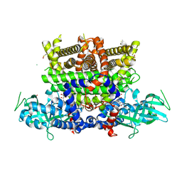
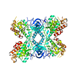
8GH2
 
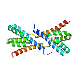
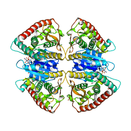
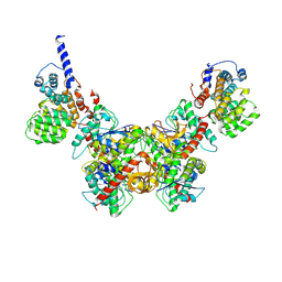 | | Structure of Trypanosoma (MDH)4-(Pex5)2, close conformation | | 分子名称: | Peroxisome targeting signal 1 receptor, malate dehydrogenase | | 著者 | Sonani, R.R, Artur, B, Jemiola-Rzeminska, M, Lipinski, O, Patel, S.N, Sood, T, Dubin, G. | | 登録日 | 2023-03-09 | | 公開日 | 2024-03-27 | | 実験手法 | ELECTRON MICROSCOPY (3.66 Å) | | 主引用文献 | Structure of Trypanosoma (MDH)4-(Pex5)2, close conformation
To Be Published
|
|
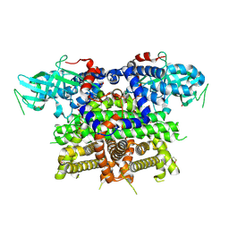
8GI0
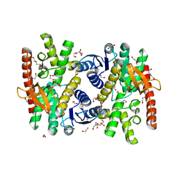
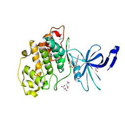
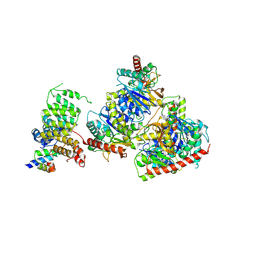 | | Structure of Trypanosoma docking complex | | 分子名称: | Peroxisomal membrane protein PEX14, Peroxisome targeting signal 1 receptor, malate dehydrogenase | | 著者 | Sonani, R.R, Artur, B, Jemiola-Rzeminska, M, Lipinski, O, Patel, S.N, Sood, T, Dubin, G. | | 登録日 | 2023-03-13 | | 公開日 | 2024-03-27 | | 実験手法 | ELECTRON MICROSCOPY (3.5 Å) | | 主引用文献 | Structure of Trypanosoma docking complex
To Be Published
|
|
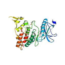
8GGH
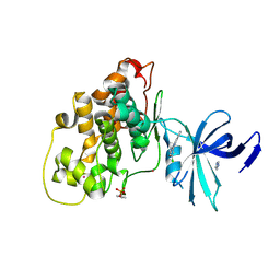
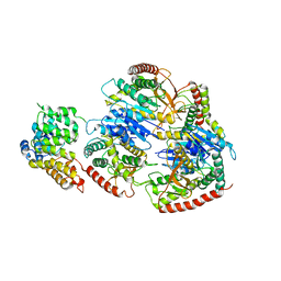 | | Structure of Trypanosoma (MDH)4-PEX5, distal conformation | | 分子名称: | Peroxisome targeting signal 1 receptor, malate dehydrogenase | | 著者 | Sonani, R.R, Artur, B, Jemiola-Rzeminska, M, Lipinski, O, Patel, S.N, Sood, T, Dubin, G. | | 登録日 | 2023-03-08 | | 公開日 | 2024-03-27 | | 実験手法 | ELECTRON MICROSCOPY (3.29 Å) | | 主引用文献 | Structure of Trypanosoma (MDH)4-PEX5, distal conformation
To Be Published
|
|
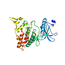
8GGD
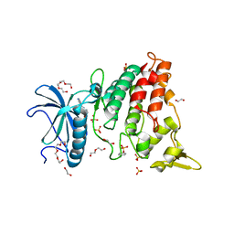
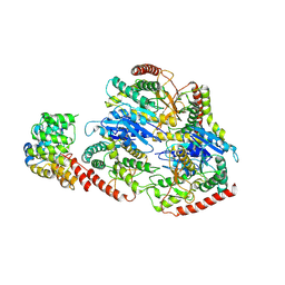 | | Structure of Trypanosoma (MDH)4-Pex5, close conformation | | 分子名称: | Peroxisome targeting signal 1 receptor, malate dehydrogenase | | 著者 | Sonani, R.R, Artur, B, Jemiola-Rzeminska, M, Lipinski, O, Patel, S.N, Sood, T, Dubin, G. | | 登録日 | 2023-03-08 | | 公開日 | 2024-03-27 | | 実験手法 | ELECTRON MICROSCOPY (3.33 Å) | | 主引用文献 | Structure of Trypanosoma (MDH)4-Pex5, close conformation
To Be Published
|
|
7Z35
 | |
5C3T
 | |
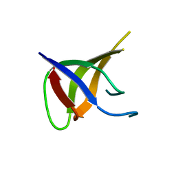
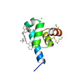
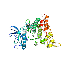
5EPW
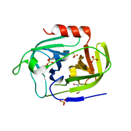
 | | C-Terminal Domain Of Human Coronavirus Nl63 Nucleocapsid Protein | | 分子名称: | Nucleoprotein | | 著者 | Szelazek, B, Kabala, W, Kus, K, Zdzalik, M, Golik, P, Florek, D, Burmistrz, M, Pyrc, K, Dubin, G. | | 登録日 | 2015-11-12 | | 公開日 | 2017-02-22 | | 最終更新日 | 2024-05-08 | | 実験手法 | X-RAY DIFFRACTION (1.5 Å) | | 主引用文献 | Structural Characterization of Human Coronavirus NL63 N Protein.
J. Virol., 91, 2017
|
|
4GR6
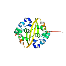
 | | Crystal structure of AtRbcX2 from Arabidopsis thaliana | | 分子名称: | 1,2-ETHANEDIOL, AtRbcX2 | | 著者 | Grudnik, P, Golik, P, Kolesinski, P, Dubin, G, Szczepaniak, A. | | 登録日 | 2012-08-24 | | 公開日 | 2013-01-30 | | 最終更新日 | 2023-09-13 | | 実験手法 | X-RAY DIFFRACTION (2 Å) | | 主引用文献 | Insights into eukaryotic Rubisco assembly - Crystal structures of RbcX chaperones from Arabidopsis thaliana.
Biochim.Biophys.Acta, 1830, 2013
|
|
4GR2
 | | Structure of AtRbcX1 from Arabidopsis thaliana. | | 分子名称: | AtRbcX1 | | 著者 | Golik, P, Grudnik, P, Kolesinski, P, Dubin, G, Szczepaniak, A. | | 登録日 | 2012-08-24 | | 公開日 | 2013-01-30 | | 最終更新日 | 2023-09-13 | | 実験手法 | X-RAY DIFFRACTION (2 Å) | | 主引用文献 | Insights into eukaryotic Rubisco assembly - Crystal structures of RbcX chaperones from Arabidopsis thaliana.
Biochim.Biophys.Acta, 1830, 2013
|
|
6SF7
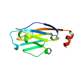
 | | Atomic resolution structure of SplF protease from Staphylococcus aureus | | 分子名称: | 1,2-ETHANEDIOL, DI(HYDROXYETHYL)ETHER, SULFATE ION, ... | | 著者 | Golik, P, Stach, N, Karim, A, Dubin, G. | | 登録日 | 2019-08-01 | | 公開日 | 2021-03-03 | | 最終更新日 | 2024-05-15 | | 実験手法 | X-RAY DIFFRACTION (1.7 Å) | | 主引用文献 | Structural Determinants of Substrate Specificity of SplF Protease from Staphylococcus aureus .
Int J Mol Sci, 22, 2021
|
|
7NRZ
 | | Crystal structure of malate dehydrogenase from Trypanosoma cruzi | | 分子名称: | 1,2-ETHANEDIOL, CHLORIDE ION, DI(HYDROXYETHYL)ETHER, ... | | 著者 | Sonani, R.R, Kurpiewska, K, Lewinski, K, Dubin, G. | | 登録日 | 2021-03-04 | | 公開日 | 2022-02-16 | | 最終更新日 | 2024-01-31 | | 実験手法 | X-RAY DIFFRACTION (2.6 Å) | | 主引用文献 | Distinct sequence and structural feature of trypanosoma malate dehydrogenase.
Biochem.Biophys.Res.Commun., 557, 2021
|
|
7OUN
 | |
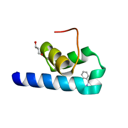
6SPT
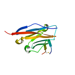
 | | High resolution crystal structure of N-terminal domain of PEX14 from Trypanosoma brucei in complex with the fist compound with sub-micromolar trypanocidal activity | | 分子名称: | 5-[(4-methoxynaphthalen-1-yl)methyl]-1-[2-[(2-methyl-1-oxidanyl-propan-2-yl)amino]ethyl]-~{N}-(naphthalen-1-ylmethyl)-6,7-dihydro-4~{H}-pyrazolo[4,3-c]pyridine-3-carboxamide, BETA-MERCAPTOETHANOL, CHLORIDE ION, ... | | 著者 | Napolitano, V, Dawidowski, M, Kalel, V.C, Fino, R, Emmanouilidis, L, Lenhart, D, Ostertag, M, Kaiser, M, Kolonko, M, Schilebs, W, Maser, P, Tetko, I, Hadian, K, Plettenburg, O, Erdmann, R, Sattler, M, Popowicz, G.M, Dubin, G. | | 登録日 | 2019-09-02 | | 公開日 | 2020-01-01 | | 最終更新日 | 2020-02-05 | | 実験手法 | X-RAY DIFFRACTION (1.2 Å) | | 主引用文献 | Structure-Activity Relationship in Pyrazolo[4,3-c]pyridines, First Inhibitors of PEX14-PEX5 Protein-Protein Interaction with Trypanocidal Activity.
J.Med.Chem., 63, 2020
|
|
7Q86
 | |
7Q84
 | | Crystal structure of human peroxisomal acyl-Co-A oxidase 1a, apo-form | | 分子名称: | 1,2-ETHANEDIOL, DI(HYDROXYETHYL)ETHER, GLYCEROL, ... | | 著者 | Sonani, R.R, Blat, A, Dubin, G. | | 登録日 | 2021-11-10 | | 公開日 | 2022-03-02 | | 最終更新日 | 2024-01-31 | | 実験手法 | X-RAY DIFFRACTION (2 Å) | | 主引用文献 | Crystal structures of apo- and FAD-bound human peroxisomal acyl-CoA oxidase provide mechanistic basis explaining clinical observations.
Int.J.Biol.Macromol., 205, 2022
|
|
7QRC
 | | X-ray structure of Trypanosoma cruzi PEX14 in complex with a PEX5-PEX14 PPI inhibitor | | 分子名称: | GLYCEROL, Peroxin-14, ~{N}-(5-ethyl-6-oxidanylidene-benzo[b][1,4]benzothiazepin-2-yl)-2-(4-fluorophenyl)ethanamide | | 著者 | Napolitano, V, Popowicz, G.M, Dawidowski, M, Dubin, G. | | 登録日 | 2022-01-10 | | 公開日 | 2022-11-23 | | 最終更新日 | 2024-01-31 | | 実験手法 | X-RAY DIFFRACTION (2.18 Å) | | 主引用文献 | Structure-based design, synthesis and evaluation of a novel family of PEX5-PEX14 interaction inhibitors against Trypanosoma.
Eur.J.Med.Chem., 243, 2022
|
|
7QOZ
 | |
7Z1G
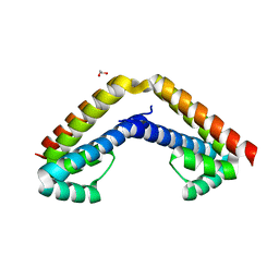
 | | Crystal structure of nonphosphorylated (Tyr216) GSK3b in complex with CX-4945 | | 分子名称: | 5-[(3-chlorophenyl)amino]benzo[c][2,6]naphthyridine-8-carboxylic acid, Glycogen synthase kinase-3 beta, IMIDAZOLE, ... | | 著者 | Grygier, P, Pustelny, K, Dubin, G, Czarna, A. | | 登録日 | 2022-02-24 | | 公開日 | 2023-03-08 | | 最終更新日 | 2024-02-07 | | 実験手法 | X-RAY DIFFRACTION (2.85 Å) | | 主引用文献 | Crystal structure of nonphosphorylated (Tyr216) GSK3b kinase in complex with CX-4945
To Be Published
|
|
7Z1F
 | | Crystal structure of GSK3b in complex with CX-4945 | | 分子名称: | 5-[(3-chlorophenyl)amino]benzo[c][2,6]naphthyridine-8-carboxylic acid, Glycogen synthase kinase-3 beta, IMIDAZOLE, ... | | 著者 | Grygier, P, Pustelny, K, Dubin, G, Czarna, A. | | 登録日 | 2022-02-24 | | 公開日 | 2023-03-08 | | 最終更新日 | 2024-02-07 | | 実験手法 | X-RAY DIFFRACTION (3 Å) | | 主引用文献 | Crystal structure of phosphorylated (Tyr216) GSK3b kinase in complex with CX-4945
To Be Published
|
|
7Z5N
 | | Crystal structure of DYRK1A in complex with CX-4945 | | 分子名称: | 1,2-ETHANEDIOL, 5-[(3-chlorophenyl)amino]benzo[c][2,6]naphthyridine-8-carboxylic acid, DI(HYDROXYETHYL)ETHER, ... | | 著者 | Pustelny, K, Grygier, P, Golik, P, Dubin, G, Czarna, A. | | 登録日 | 2022-03-09 | | 公開日 | 2023-03-22 | | 最終更新日 | 2024-02-07 | | 実験手法 | X-RAY DIFFRACTION (2.77 Å) | | 主引用文献 | Crystal structure DYRK1A in complex with CX-4945
To Be Published
|
|
8C3G
 | | Crystal structure of DYRK1A in complex with AZ191 | | 分子名称: | 1,2-ETHANEDIOL, Dual specificity tyrosine-phosphorylation-regulated kinase 1A, N-[2-methoxy-4-(4-methylpiperazin-1-yl)phenyl]-4-(1-methylpyrrolo[2,3-c]pyridin-3-yl)pyrimidin-2-amine, ... | | 著者 | Grygier, P, Pustelny, K, Dubin, G, Czarna, A. | | 登録日 | 2022-12-23 | | 公開日 | 2024-01-10 | | 実験手法 | X-RAY DIFFRACTION (2.08 Å) | | 主引用文献 | Structural perspective on the design of selective DYRK1B inhibitors
To Be Published
|
|
8C3Q
 | |
8C3R
 | |
8C2Z
 | | Crystal structure of DYRK1B in complex with AZ191 | | 分子名称: | Dual specificity tyrosine-phosphorylation-regulated kinase 1B, MANGANESE (II) ION, N-[2-methoxy-4-(4-methylpiperazin-1-yl)phenyl]-4-(1-methylpyrrolo[2,3-c]pyridin-3-yl)pyrimidin-2-amine | | 著者 | Grygier, P, Pustelny, K, Dubin, G, Czarna, A. | | 登録日 | 2022-12-23 | | 公開日 | 2024-01-17 | | 実験手法 | X-RAY DIFFRACTION (1.91 Å) | | 主引用文献 | Structural perspective on the design of selective DYRK1B inhibitors
To Be Published
|
|
7R2V
 | | Structure of nsp14 from SARS-CoV-2 in complex with SAH | | 分子名称: | 2-AMINO-2-HYDROXYMETHYL-PROPANE-1,3-DIOL, DI(HYDROXYETHYL)ETHER, Proofreading exoribonuclease nsp14, ... | | 著者 | Czarna, A, Plewka, J, Kresik, L, Matsuda, A, Abdulkarim, K, Robinson, C, OByrne, S, Cunningham, F, Georgiou, I, Pachota, M, Popowicz, G.M, Wyatt, P.G, Dubin, G, Pyrc, K. | | 登録日 | 2022-02-06 | | 公開日 | 2022-03-09 | | 最終更新日 | 2024-01-31 | | 実験手法 | X-RAY DIFFRACTION (2.53 Å) | | 主引用文献 | Refolding of lid subdomain of SARS-CoV-2 nsp14 upon nsp10 interaction releases exonuclease activity.
Structure, 30, 2022
|
|


