5I5D
 
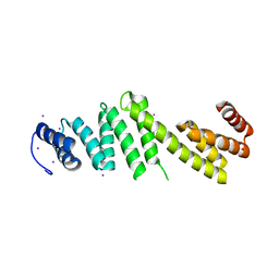
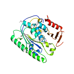 | | Salmonella global domain 245 | | 分子名称: | Inner membrane protein YejM | | 著者 | Dong, C, Dong, H. | | 登録日 | 2016-02-15 | | 公開日 | 2017-04-19 | | 実験手法 | X-RAY DIFFRACTION (1.64 Å) | | 主引用文献 | Structural insights into cardiolipin transfer from the Inner membrane to the outer membrane by PbgA in Gram-negative bacteria.
Sci Rep, 6, 2016
|
|
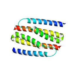
5I5F
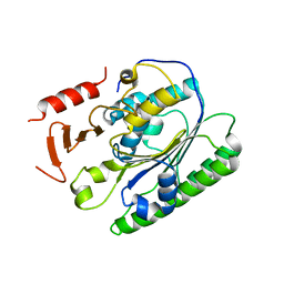 | | Salmonella global domain 191 | | 分子名称: | Inner membrane protein YejM | | 著者 | Dong, C, Dong, H. | | 登録日 | 2016-02-15 | | 公開日 | 2016-08-17 | | 最終更新日 | 2024-05-08 | | 実験手法 | X-RAY DIFFRACTION (1.84 Å) | | 主引用文献 | Structural insights into cardiolipin transfer from the Inner membrane to the outer membrane by PbgA in Gram-negative bacteria.
Sci Rep, 6, 2016
|
|
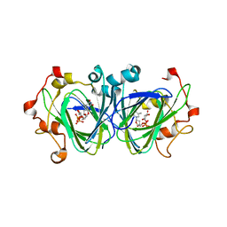
6ZY3
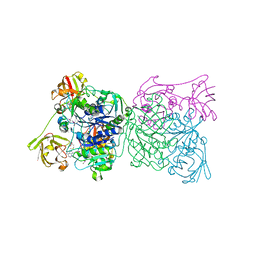
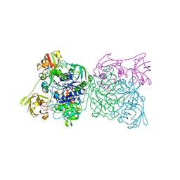
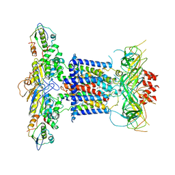 | | Cryo-EM structure of MlaFEDB in complex with phospholipid | | 分子名称: | 1,2-dioleoyl-sn-glycero-3-phosphoethanolamine, ABC transporter maintaining OM lipid asymmetry, cytoplasmic STAS component, ... | | 著者 | Dong, C.J, Dong, H.H. | | 登録日 | 2020-07-30 | | 公開日 | 2020-11-25 | | 最終更新日 | 2024-05-01 | | 実験手法 | ELECTRON MICROSCOPY (3.3 Å) | | 主引用文献 | Structural insights into outer membrane asymmetry maintenance in Gram-negative bacteria by MlaFEDB.
Nat.Struct.Mol.Biol., 28, 2021
|
|
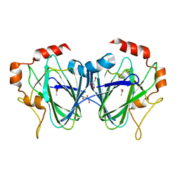
6ZY2
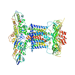 | | Cryo-EM structure of apo MlaFEDB | | 分子名称: | ABC transporter maintaining OM lipid asymmetry, cytoplasmic STAS component, Toluene tolerance protein Ttg2A, ... | | 著者 | Dong, C.J, Dong, H.H. | | 登録日 | 2020-07-30 | | 公開日 | 2020-11-25 | | 最終更新日 | 2024-05-01 | | 実験手法 | ELECTRON MICROSCOPY (3.6 Å) | | 主引用文献 | Structural insights into outer membrane asymmetry maintenance in Gram-negative bacteria by MlaFEDB.
Nat.Struct.Mol.Biol., 28, 2021
|
|
6ZY4
 | | Cryo-EM structure of MlaFEDB in complex with ADP | | 分子名称: | ABC transporter maintaining OM lipid asymmetry, cytoplasmic STAS component, ADENOSINE-5'-DIPHOSPHATE, ... | | 著者 | Dong, C.J, Dong, H.H. | | 登録日 | 2020-07-30 | | 公開日 | 2020-11-25 | | 最終更新日 | 2024-05-01 | | 実験手法 | ELECTRON MICROSCOPY (4.1 Å) | | 主引用文献 | Structural insights into outer membrane asymmetry maintenance in Gram-negative bacteria by MlaFEDB.
Nat.Struct.Mol.Biol., 28, 2021
|
|
7YNX
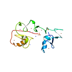 | | Crystal structure of Pirh2 bound to poly-Ala peptide | | 分子名称: | RING finger and CHY zinc finger domain-containing protein 1, SODIUM ION, SULFATE ION, ... | | 著者 | Dong, C, Yan, X, Li, Y. | | 登録日 | 2022-08-01 | | 公開日 | 2023-04-26 | | 最終更新日 | 2024-05-29 | | 実験手法 | X-RAY DIFFRACTION (2.3 Å) | | 主引用文献 | Recognition of an Ala-rich C-degron by the E3 ligase Pirh2.
Nat Commun, 14, 2023
|
|
6L1G
 | |
5WP3
 | | Crystal Structure of EED in complex with EB22 | | 分子名称: | EB22, Polycomb protein EED, UNKNOWN ATOM OR ION | | 著者 | Dong, C, Tempel, W, Zhu, L, Moody, J.D, Baker, D, Bountra, C, Arrowsmith, C.H, Edwards, A.M, Min, J, Structural Genomics Consortium (SGC) | | 登録日 | 2017-08-03 | | 公開日 | 2017-09-13 | | 最終更新日 | 2023-10-04 | | 実験手法 | X-RAY DIFFRACTION (2.55 Å) | | 主引用文献 | First critical repressive H3K27me3 marks in embryonic stem cells identified using designed protein inhibitor.
Proc. Natl. Acad. Sci. U.S.A., 114, 2017
|
|
6ZY9
 | | Cryo-EM structure of MlaFEDB in complex with AMP-PNP | | 分子名称: | ABC transporter maintaining OM lipid asymmetry, cytoplasmic STAS component, MAGNESIUM ION, ... | | 著者 | Dong, C.J, Dong, H.H. | | 登録日 | 2020-07-30 | | 公開日 | 2020-11-25 | | 最終更新日 | 2024-05-01 | | 実験手法 | ELECTRON MICROSCOPY (3.3 Å) | | 主引用文献 | Structural insights into outer membrane asymmetry maintenance in Gram-negative bacteria by MlaFEDB.
Nat.Struct.Mol.Biol., 28, 2021
|
|
5L75
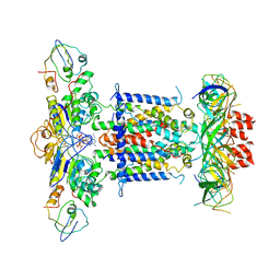
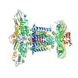
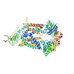 | | A protein structure | | 分子名称: | FIG000906: Predicted Permease, FIG000988: Predicted permease, Lipopolysaccharide ABC transporter, ... | | 著者 | Dong, C, Dong, H, Zhang, Z, Paterson, N, Tang, X. | | 登録日 | 2016-06-01 | | 公開日 | 2017-08-16 | | 最終更新日 | 2024-05-08 | | 実験手法 | X-RAY DIFFRACTION (3.7 Å) | | 主引用文献 | Structural and functional insights into the lipopolysaccharide ABC transporter LptB2FG.
Nat Commun, 8, 2017
|
|
1RQR
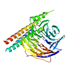
 | | Crystal structure and mechanism of a bacterial fluorinating enzyme, product complex | | 分子名称: | 5'-FLUORO-5'-DEOXYADENOSINE, 5'-fluoro-5'-deoxyadenosine synthase, METHIONINE | | 著者 | Dong, C, Huang, F, Deng, H, Schaffrath, C, Spencer, J.B, O'Hagan, D, Naismith, J.H. | | 登録日 | 2003-12-07 | | 公開日 | 2004-03-02 | | 最終更新日 | 2022-06-15 | | 実験手法 | X-RAY DIFFRACTION (2.67 Å) | | 主引用文献 | Crystal structure and mechanism of a bacterial fluorinating enzyme
Nature, 427, 2004
|
|
1RQP
 | | Crystal structure and mechanism of a bacterial fluorinating enzyme | | 分子名称: | 5'-fluoro-5'-deoxyadenosine synthase, S-ADENOSYLMETHIONINE | | 著者 | Dong, C, Huang, F, Deng, H, Schaffrath, C, Spencer, J.B, O'Hagan, D, Naismith, J.H. | | 登録日 | 2003-12-06 | | 公開日 | 2004-03-02 | | 最終更新日 | 2024-02-14 | | 実験手法 | X-RAY DIFFRACTION (1.8 Å) | | 主引用文献 | Crystal structure and mechanism of a bacterial fluorinating enzyme
Nature, 427, 2004
|
|
5I5H
 | | Ecoli global domain 245-586 | | 分子名称: | Inner membrane protein YejM | | 著者 | Dong, C, Dong, H. | | 登録日 | 2016-02-15 | | 公開日 | 2016-08-17 | | 最終更新日 | 2024-01-10 | | 実験手法 | X-RAY DIFFRACTION (1.65 Å) | | 主引用文献 | Structural insights into cardiolipin transfer from the Inner membrane to the outer membrane by PbgA in Gram-negative bacteria.
Sci Rep, 6, 2016
|
|
5OR1
 | | BamA structure of Salmonella enterica | | 分子名称: | Outer membrane protein assembly factor BamA | | 著者 | Dong, C, Gu, Y. | | 登録日 | 2017-08-14 | | 公開日 | 2018-02-14 | | 最終更新日 | 2024-05-08 | | 実験手法 | X-RAY DIFFRACTION (2.92 Å) | | 主引用文献 | BamA beta 16C strand and periplasmic turns are critical for outer membrane protein insertion and assembly.
Biochem. J., 474, 2017
|
|
6WAV
 | | Crystal structure of PHF1 in complex with H3K36me3 substitution | | 分子名称: | Histone H3.1, PHD finger protein 1, SULFATE ION, ... | | 著者 | Dong, C, Bountra, C, Edwards, A.M, Arrowsmith, C.H, Min, J.R, Structural Genomics Consortium (SGC) | | 登録日 | 2020-03-26 | | 公開日 | 2020-08-26 | | 最終更新日 | 2023-10-18 | | 実験手法 | X-RAY DIFFRACTION (1.7 Å) | | 主引用文献 | Structural basis for histone variant H3tK27me3 recognition by PHF1 and PHF19.
Elife, 9, 2020
|
|
8V1P
 | | CRYSTAL STRUCTURE OF GID4 IN COMPLEX WITH UBF9092 | | 分子名称: | Glucose-induced degradation protein 4 homolog, N,N~2~-bis[(4-methoxyphenyl)methyl]glycinamide | | 著者 | Dong, C, Dong, A, Calabrese, M, Wang, F, Owen, D, Arrowsmith, C.H, Edwards, A.M, Min, J, Structural Genomics Consortium (SGC) | | 登録日 | 2023-11-21 | | 公開日 | 2023-12-06 | | 実験手法 | X-RAY DIFFRACTION (2.21 Å) | | 主引用文献 | CRYSTAL STRUCTURE OF GID4 IN COMPLEX WITH UBF9092
To be published
|
|
6WAT
 | | complex structure of PHF1 | | 分子名称: | Histone H3.1t peptide, PHD finger protein 1, UNKNOWN ATOM OR ION | | 著者 | Dong, C, Bountra, C, Edwards, A.M, Arrowsmith, C.H, Min, J.R, Structural Genomics Consortium (SGC) | | 登録日 | 2020-03-26 | | 公開日 | 2020-08-26 | | 最終更新日 | 2023-10-18 | | 実験手法 | X-RAY DIFFRACTION (1.8 Å) | | 主引用文献 | Structural basis for histone variant H3tK27me3 recognition by PHF1 and PHF19.
Elife, 9, 2020
|
|
6WAU
 | | Complex structure of PHF19 | | 分子名称: | Histone H3.1t peptide, PHD finger protein 19, UNKNOWN ATOM OR ION | | 著者 | Dong, C, Bountra, C, Edwards, A.M, Arrowsmith, C.H, Min, J.R, Structural Genomics Consortium (SGC) | | 登録日 | 2020-03-26 | | 公開日 | 2020-08-26 | | 最終更新日 | 2023-10-18 | | 実験手法 | X-RAY DIFFRACTION (1.75 Å) | | 主引用文献 | Structural basis for histone variant H3tK27me3 recognition by PHF1 and PHF19.
Elife, 9, 2020
|
|
3Q5M
 | | Crystal structure of Escherichia coli BamD | | 分子名称: | IODIDE ION, UPF0169 lipoprotein yfiO | | 著者 | Dong, C, Hou, H, Yang, X, Dong, Y, Shen, Y. | | 登録日 | 2010-12-28 | | 公開日 | 2011-12-28 | | 最終更新日 | 2024-03-20 | | 実験手法 | X-RAY DIFFRACTION (2.604 Å) | | 主引用文献 | Structure of Escherichia coli BamD and its functional implications in outer membrane protein assembly
Acta Crystallogr.,Sect.D, 68, 2012
|
|
4NSR
 | |
1NZC
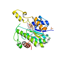
 | | The high resolution structures of RmlC from Streptococcus suis in complex with dTDP-D-xylose | | 分子名称: | NICKEL (II) ION, THYMIDINE-5'-DIPHOSPHO-BETA-D-XYLOSE, dTDP-6-deoxy-D-xylo-4-hexulose 3,5-epimerase | | 著者 | Dong, C, Major, L.L, Allen, A, Blankenfeldt, W, Maskell, D, Naismith, J.H. | | 登録日 | 2003-02-17 | | 公開日 | 2003-06-24 | | 最終更新日 | 2023-08-16 | | 実験手法 | X-RAY DIFFRACTION (1.8 Å) | | 主引用文献 | High-Resolution Structures of RmlC from Streptococcus suis in Complex with Substrate Analogs Locate the Active Site of This Class of Enzyme
Structure, 11, 2003
|
|
1PM7
 | |
6V8W
 | | CDYL2 chromodomain in complex with a synthetic peptide | | 分子名称: | Chromodomain Y-like protein 2, IVA-PHE-ALA-PHE-5T3-SER-NH2, UNKNOWN ATOM OR ION | | 著者 | Dong, C, Tempel, W, James, L.I, Lamb, K.N, Bountra, C, Edwards, A.M, Arrowsmith, C.H, Min, J, Structural Genomics Consortium, Structural Genomics Consortium (SGC) | | 登録日 | 2019-12-12 | | 公開日 | 2020-01-15 | | 最終更新日 | 2023-11-15 | | 実験手法 | X-RAY DIFFRACTION (2.8 Å) | | 主引用文献 | Structural Basis for the Binding Selectivity of Human CDY Chromodomains.
Cell Chem Biol, 27, 2020
|
|
1NXM
 | | The high resolution structures of RmlC from Streptococcus suis | | 分子名称: | dTDP-6-deoxy-D-xylo-4-hexulose 3,5-epimerase | | 著者 | Dong, C, Major, L.L, Allen, A, Blankenfeldt, W, Maskell, D, Naismith, J.H. | | 登録日 | 2003-02-11 | | 公開日 | 2003-06-24 | | 最終更新日 | 2024-02-14 | | 実験手法 | X-RAY DIFFRACTION (1.3 Å) | | 主引用文献 | High-Resolution Structures of RmlC from Streptococcus suis in Complex with Substrate Analogs Locate the Active Site of This Class of Enzyme
Structure, 11, 2003
|
|
1NYW
 | | The high resolution structures of RmlC from Streptoccus suis in complex with dTDP-D-glucose | | 分子名称: | 2'DEOXY-THYMIDINE-5'-DIPHOSPHO-ALPHA-D-GLUCOSE, dTDP-6-deoxy-D-xylo-4-hexulose 3,5-epimerase | | 著者 | Dong, C, Major, L.L, Allen, A, Blankenfeldt, W, Maskell, D, Naismith, J.H. | | 登録日 | 2003-02-14 | | 公開日 | 2003-06-24 | | 最終更新日 | 2024-02-14 | | 実験手法 | X-RAY DIFFRACTION (1.6 Å) | | 主引用文献 | High-Resolution Structures of RmlC from Streptococcus suis in Complex with Substrate Analogs Locate the Active Site of This Class of Enzyme
Structure, 11, 2003
|
|


