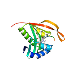
8CH7
 
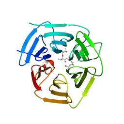
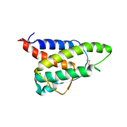 | | RDC-refined Interleukin-4 (wild type) pH 5.6 | | 分子名称: | Interleukin-4 | | 著者 | Vaz, D.C, Rodrigues, J.R, Loureiro-Ferreira, N, Mueller, T, Sebald, W, Redfield, C, Brito, R.M.M. | | 登録日 | 2023-02-07 | | 公開日 | 2023-10-18 | | 最終更新日 | 2024-01-17 | | 実験手法 | SOLUTION NMR | | 主引用文献 | Lessons on protein structure from interleukin-4: All disulfides are not created equal.
Proteins, 92, 2024
|
|
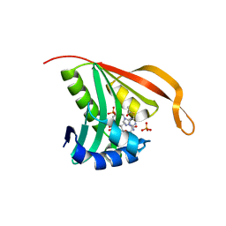
8CGF
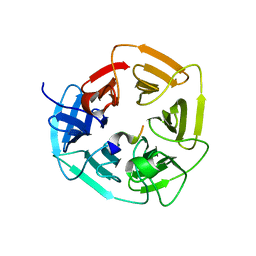
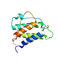 | | Interleukin-4 (wild type) pH 2.4 | | 分子名称: | Interleukin-4 | | 著者 | Vaz, D.C, Rodrigues, J.R, Loureiro-Ferreira, N, Mueller, T, Sebald, W, Redfield, C, Brito, R.M.M. | | 登録日 | 2023-02-04 | | 公開日 | 2023-10-18 | | 最終更新日 | 2024-01-17 | | 実験手法 | SOLUTION NMR | | 主引用文献 | Lessons on protein structure from interleukin-4: All disulfides are not created equal.
Proteins, 92, 2024
|
|
8C12
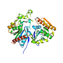
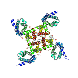
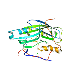
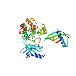 | | Identification of an intermediate activation state of PAK5 reveals a novel mechanism of kinase inhibition. | | 分子名称: | PAK5-Af17, Serine/threonine-protein kinase PAK 5 | | 著者 | Martin, H.L, Turner, A.L, Trinh, C.H, Bayliss, R.W, Tomlinson, D.C. | | 登録日 | 2022-12-19 | | 公開日 | 2023-10-25 | | 実験手法 | X-RAY DIFFRACTION (1.549 Å) | | 主引用文献 | Affimer-mediated locking of p21-activated kinase 5 in an intermediate activation state results in kinase inhibition.
Cell Rep, 42, 2023
|
|
2HMG
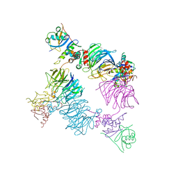
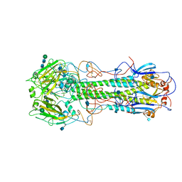 | | REFINEMENT OF THE INFLUENZA VIRUS HEMAGGLUTININ BY SIMULATED ANNEALING | | 分子名称: | 2-acetamido-2-deoxy-beta-D-glucopyranose, HEMAGGLUTININ (HA1 CHAIN), HEMAGGLUTININ (HA2 CHAIN), ... | | 著者 | Weis, W.I, Bruenger, A.T, Skehel, J.J, Wiley, D.C. | | 登録日 | 1989-09-11 | | 公開日 | 1991-01-15 | | 最終更新日 | 2020-07-29 | | 実験手法 | X-RAY DIFFRACTION (3 Å) | | 主引用文献 | Refinement of the influenza virus hemagglutinin by simulated annealing.
J.Mol.Biol., 212, 1990
|
|
8DFC
 | |
8DFD
 | |
8DBX
 | |
8DBY
 | |
8EBM
 | |
8EBN
 | |
8EBL
 | |
7Z2X
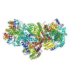
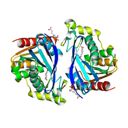
 | | Wild-type ferulic acid esterase from Lactobacillus buchneri | | 分子名称: | Ferulic acid esterase | | 著者 | Mogodiniyai, K.K, Reichenbach, T, Kalyani, D.C, Keskitalo, M.M, Divne, C. | | 登録日 | 2022-03-01 | | 公開日 | 2022-11-30 | | 最終更新日 | 2024-05-01 | | 実験手法 | X-RAY DIFFRACTION (1.5 Å) | | 主引用文献 | Crystal structure of the feruloyl esterase from Lentilactobacillus buchneri reveals a novel homodimeric state.
Front Microbiol, 13, 2022
|
|
7Z2U
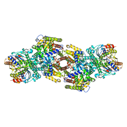
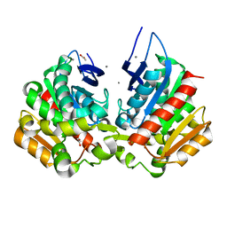
 | | Wild-type ferulic acid esterase from Lactobacillus buchneri in complex with ferulate | | 分子名称: | 3-(4-HYDROXY-3-METHOXYPHENYL)-2-PROPENOIC ACID, CALCIUM ION, Ferulic acid esterase | | 著者 | Mogodiniyai, K.K, Reichenbach, T, Kalyani, D.C, Keskitalo, M.M, Divne, C. | | 登録日 | 2022-02-28 | | 公開日 | 2022-11-30 | | 最終更新日 | 2024-05-01 | | 実験手法 | X-RAY DIFFRACTION (1.9 Å) | | 主引用文献 | Crystal structure of the feruloyl esterase from Lentilactobacillus buchneri reveals a novel homodimeric state.
Front Microbiol, 13, 2022
|
|
7Z2V
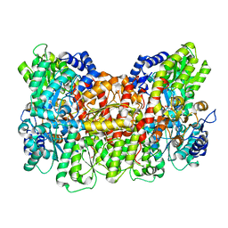
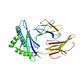
 | | Ferulic acid esterase variant S114A from Lactobacillus buchneri | | 分子名称: | ACETATE ION, CALCIUM ION, Ferulic acid esterase | | 著者 | Mogodiniyai, K.K, Reichenbach, T, Kalyani, D.C, Keskitalo, M.M, Divne, C. | | 登録日 | 2022-02-28 | | 公開日 | 2022-11-30 | | 最終更新日 | 2024-05-01 | | 実験手法 | X-RAY DIFFRACTION (1.45 Å) | | 主引用文献 | Crystal structure of the feruloyl esterase from Lentilactobacillus buchneri reveals a novel homodimeric state.
Front Microbiol, 13, 2022
|
|
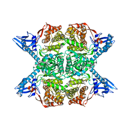
2HLA
 | |
5JM8
 | |
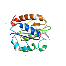
2CNM
 | | RimI - Ribosomal S18 N-alpha-protein acetyltransferase in complex with a bisubstrate inhibitor (Cterm-Arg-Arg-Phe-Tyr-Arg-Ala-N-alpha- acetyl-S-CoA). | | 分子名称: | 30S RIBOSOMAL PROTEIN S18, COENZYME A, MODIFICATION OF 30S RIBOSOMAL SUBUNIT PROTEIN S18 | | 著者 | Vetting, M.W, Yu, M, Bareich, D.C, Blanchard, J.S. | | 登録日 | 2006-05-22 | | 公開日 | 2007-05-22 | | 最終更新日 | 2019-10-23 | | 実験手法 | X-RAY DIFFRACTION (2.6 Å) | | 主引用文献 | Crystal Structure of Rimi from Salmonella Typhimurium Lt2, the Gnat Responsible for N{Alpha}- Acetylation of Ribosomal Protein S18.
Protein Sci., 17, 2008
|
|
2CNS
 | | RimI - Ribosomal S18 N-alpha-protein acetyltransferase in complex with acetylCoA. | | 分子名称: | ACETYL COENZYME *A, MODIFICATION OF 30S RIBOSOMAL SUBUNIT PROTEIN S18, PHOSPHATE ION | | 著者 | Vetting, M.W, Bareich, D.C, Yu, M, Blanchard, J.S. | | 登録日 | 2006-05-23 | | 公開日 | 2007-06-19 | | 最終更新日 | 2024-05-08 | | 実験手法 | X-RAY DIFFRACTION (2.5 Å) | | 主引用文献 | Crystal Structure of Rimi from Salmonella Typhimurium Lt2, the Gnat Responsible for N{Alpha}- Acetylation of Ribosomal Protein S18.
Protein Sci., 17, 2008
|
|
5K3Q
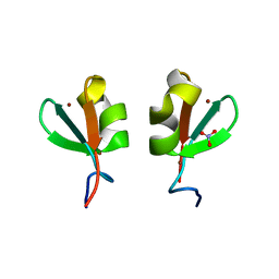 | | Rhesus macaques Trim5alpha Bbox2 domain | | 分子名称: | NITRATE ION, Tripartite motif-containing protein 5,Tripartite motif-containing protein 5, ZINC ION | | 著者 | Keown, J.K, Goldstone, D.C. | | 登録日 | 2016-05-19 | | 公開日 | 2016-07-20 | | 最終更新日 | 2024-03-06 | | 実験手法 | X-RAY DIFFRACTION (1.8 Å) | | 主引用文献 | Crystal structure of the Trim5 alpha Bbox2 domain from rhesus macaques describes a plastic oligomerisation interface.
J.Struct.Biol., 195, 2016
|
|
2CSE
 | | Features of Reovirus Outer-Capsid Protein mu1 Revealed by Electron and Image Reconstruction of the virion at 7.0-A Resolution | | 分子名称: | Minor core protein lambda 3, Sigma 2 protein, guanylyltransferase, ... | | 著者 | Zhang, X, Ji, Y, Zhang, L, Harrison, S.C, Marinescu, D.C, Nibert, M.L, Baker, T.S. | | 登録日 | 2005-05-21 | | 公開日 | 2005-10-18 | | 最終更新日 | 2024-02-14 | | 実験手法 | ELECTRON MICROSCOPY (7 Å) | | 主引用文献 | Features of reovirus outer capsid protein mu1 revealed by electron cryomicroscopy and image reconstruction of the virion at 7.0 Angstrom resolution.
Structure, 13, 2005
|
|
2BOG
 | | Catalytic domain of endo-1,4-glucanase Cel6A mutant Y73S from Thermobifida fusca in complex with methyl cellobiosyl-4-thio-beta- cellobioside | | 分子名称: | ENDOGLUCANASE E-2, beta-D-glucopyranose-(1-4)-beta-D-glucopyranose-(1-4)-4-thio-beta-D-glucopyranose-(1-4)-methyl beta-D-glucopyranoside | | 著者 | Larsson, A.M, Bergfors, T, Dultz, E, Irwin, D.C, Roos, A, Driguez, H, Wilson, D.B, Jones, T.A. | | 登録日 | 2005-04-10 | | 公開日 | 2005-10-05 | | 最終更新日 | 2023-12-13 | | 実験手法 | X-RAY DIFFRACTION (1.04 Å) | | 主引用文献 | Crystal Structure of Thermobifida Fusca Endoglucanase Cel6A in Complex with Substrate and Inhibitor: The Role of Tyrosine Y73 in Substrate Ring Distortion.
Biochemistry, 44, 2005
|
|
5K9B
 | |
5KMD
 | | Structure of CavAb in complex with amlodipine | | 分子名称: | 1,2-DIMYRISTOYL-RAC-GLYCERO-3-PHOSPHOCHOLINE, CALCIUM ION, Ion transport protein, ... | | 著者 | Tang, L, Gamal EL-Din, T.M, Swanson, T.M, Pryde, D.C, Scheuer, T, Zheng, N, Catterall, W.A. | | 登録日 | 2016-06-26 | | 公開日 | 2016-08-31 | | 最終更新日 | 2023-09-27 | | 実験手法 | X-RAY DIFFRACTION (3.2 Å) | | 主引用文献 | Structural basis for inhibition of a voltage-gated Ca(2+) channel by Ca(2+) antagonist drugs.
Nature, 537, 2016
|
|
2BOD
 | | Catalytic domain of endo-1,4-glucanase Cel6A from Thermobifida fusca in complex with methyl cellobiosyl-4-thio-beta-cellobioside | | 分子名称: | ENDOGLUCANASE E-2, beta-D-glucopyranose-(1-4)-beta-D-glucopyranose-(1-4)-4-thio-beta-D-glucopyranose-(1-4)-methyl beta-D-glucopyranoside | | 著者 | Larsson, A.M, Bergfors, T, Dultz, E, Irwin, D.C, Roos, A, Driguez, H, Wilson, D.B, Jones, T.A. | | 登録日 | 2005-04-10 | | 公開日 | 2005-10-05 | | 最終更新日 | 2023-12-13 | | 実験手法 | X-RAY DIFFRACTION (1.5 Å) | | 主引用文献 | Crystal Structure of Thermobifida Fusca Endoglucanase Cel6A in Complex with Substrate and Inhibitor: The Role of Tyrosine Y73 in Substrate Ring Distortion.
Biochemistry, 44, 2005
|
|
2C3A
 | | Structure of unliganded HSV gD reveals a mechanism for receptor- mediated activation of virus entry | | 分子名称: | 2-acetamido-2-deoxy-beta-D-glucopyranose-(1-4)-2-acetamido-2-deoxy-beta-D-glucopyranose, CHLORIDE ION, GLYCOPROTEIN D, ... | | 著者 | Krummenacher, C, Supekar, V.M, Whitbeck, J.C, Lazear, E, Connolly, S.A, Eisenberg, R.J, Cohen, G.H, Wiley, D.C, Carfi, A. | | 登録日 | 2005-10-05 | | 公開日 | 2005-12-21 | | 最終更新日 | 2023-12-13 | | 実験手法 | X-RAY DIFFRACTION (2.5 Å) | | 主引用文献 | Structure of Unliganded Hsv Gd Reveals a Mechanism for Receptor-Mediated Activation of Virus Entry.
Embo J., 24, 2005
|
|


