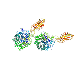
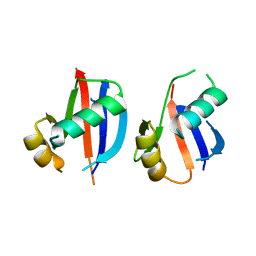
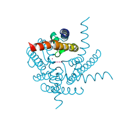
6JLI
 
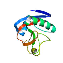 | | Crystal structure of CTLD7 domain of human PLA2R | | 分子名称: | 2-acetamido-2-deoxy-beta-D-glucopyranose, Secretory phospholipase A2 receptor | | 著者 | Yu, B, Hu, Z, Kong, D, Cheng, C, He, Y. | | 登録日 | 2019-03-06 | | 公開日 | 2019-07-17 | | 最終更新日 | 2023-11-22 | | 実験手法 | X-RAY DIFFRACTION (1.778 Å) | | 主引用文献 | Crystal structure of the CTLD7 domain of human M-type phospholipase A2 receptor.
J.Struct.Biol., 207, 2019
|
|
7QPZ
 | |
7QQ1
 | |
1K3R
 | | Crystal Structure of the Methyltransferase with a Knot from Methanobacterium thermoautotrophicum | | 分子名称: | conserved protein MT0001 | | 著者 | Zarembinski, T.I, Kim, Y, Peterson, K, Christendat, D, Dharamsi, A, Arrowsmith, C.H, Edwards, A.M, Joachimiak, A, Midwest Center for Structural Genomics (MCSG) | | 登録日 | 2001-10-03 | | 公開日 | 2002-05-15 | | 最終更新日 | 2024-02-07 | | 実験手法 | X-RAY DIFFRACTION (2.3 Å) | | 主引用文献 | Deep trefoil knot implicated in RNA binding found in an archaebacterial protein.
Proteins, 50, 2003
|
|
1K52
 | | Monomeric Protein L B1 Domain with a K54G mutation | | 分子名称: | Protein L, ZINC ION | | 著者 | O'Neill, J.W, Kim, D.E, Johnsen, K, Baker, D, Zhang, K.Y.J. | | 登録日 | 2001-10-09 | | 公開日 | 2001-12-05 | | 最終更新日 | 2023-08-16 | | 実験手法 | X-RAY DIFFRACTION (1.8 Å) | | 主引用文献 | Single-site mutations induce 3D domain swapping in the B1 domain of protein L from Peptostreptococcus magnus.
Structure, 9, 2001
|
|
7QQY
 | |
6JHS
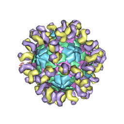 | | The cryo-EM structure of HAV bound to a neutralizing antibody-F7 | | 分子名称: | FAB Heavy Chain, FAB Light Chain, VP1, ... | | 著者 | Cao, L, Liu, P, Yang, P, Gao, Q, Li, H, Sun, Y, Zhu, L, Lin, J, Su, D, Rao, Z, Wang, X. | | 登録日 | 2019-02-19 | | 公開日 | 2020-03-18 | | 実験手法 | ELECTRON MICROSCOPY (3.05 Å) | | 主引用文献 | Structural basis for neutralization of hepatitis A virus informs a rational design of highly potent inhibitors.
Plos Biol., 17, 2019
|
|
8D2W
 | | Zebrafish MFSD2A isoform B in inward open ligand 2B conformation | | 分子名称: | DODECYL-BETA-D-MALTOSIDE, FAB heavy chain, FAB light chain, ... | | 著者 | Nguyen, C, Lei, H.T, Lai, L.T.F, Gallentino, M.J, Mu, X, Matthies, D, Gonen, T. | | 登録日 | 2022-05-30 | | 公開日 | 2023-05-10 | | 最終更新日 | 2023-05-24 | | 実験手法 | ELECTRON MICROSCOPY (3.4 Å) | | 主引用文献 | Lipid flipping in the omega-3 fatty-acid transporter.
Nat Commun, 14, 2023
|
|
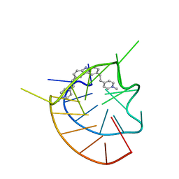
1K6G
 | | Solution Structure of Conserved AGNN Tetraloops: Insights into Rnt1p RNA Processing | | 分子名称: | RNA (5'-R(P*GP*GP*CP*GP*UP*CP*AP*UP*GP*AP*GP*UP*CP*CP*AP*UP*GP*GP*CP*GP*CP*C)-3') | | 著者 | Lebars, I, Lamontagne, B, Yoshizawa, S, Abou Elela, S, Fourmy, D. | | 登録日 | 2001-10-16 | | 公開日 | 2001-12-19 | | 最終更新日 | 2024-05-22 | | 実験手法 | SOLUTION NMR | | 主引用文献 | Solution structure of conserved AGNN tetraloops: insights into Rnt1p RNA processing.
EMBO J., 20, 2001
|
|
7QCG
 | | Structure of SARS-CoV-2 Papain-like Protease bound to N-(2-pyrrolidyl)-3,4,5-trihydroxybenzoylhydrazone | | 分子名称: | 3,4,5-tris(oxidanyl)-N-[(E)-1H-pyrrol-2-ylmethylideneamino]benzamide, CHLORIDE ION, GLYCEROL, ... | | 著者 | Ewert, W, Gunther, S, Reinke, P, Falke, S, Lieske, J, Miglioli, F, Carcelli, M, Srinivasan, V, Betzel, C, Han, H, Lorenzen, K, Guenther, C, Niebling, S, Garcia-Alai, M, Hinrichs, W, Rogolino, D, Meents, A. | | 登録日 | 2021-11-23 | | 公開日 | 2022-03-23 | | 最終更新日 | 2024-01-31 | | 実験手法 | X-RAY DIFFRACTION (1.75 Å) | | 主引用文献 | Hydrazones and Thiosemicarbazones Targeting Protein-Protein-Interactions of SARS-CoV-2 Papain-like Protease.
Front Chem, 10, 2022
|
|
6JJ0
 | |
1K72
 | | The X-ray Crystal Structure Of Cel9G Complexed With cellotriose | | 分子名称: | CALCIUM ION, Endoglucanase 9G, GLYCEROL, ... | | 著者 | Mandelman, D, Belaich, A, Belaich, J.P, Aghajari, N, Driguez, H, Haser, R. | | 登録日 | 2001-10-18 | | 公開日 | 2003-07-15 | | 最終更新日 | 2023-08-16 | | 実験手法 | X-RAY DIFFRACTION (1.8 Å) | | 主引用文献 | X-Ray Crystal Structure of the Multidomain Endoglucanase Cel9G from Clostridium
cellulolyticum Complexed with Natural and Synthetic Cello-Oligosaccharides
J.BACTERIOL., 185, 2003
|
|
8D2T
 | | Zebrafish MFSD2A isoform B in inward open ligand-free conformation | | 分子名称: | DODECYL-BETA-D-MALTOSIDE, FAB heavy chain, FAB light chain, ... | | 著者 | Nguyen, C, Lei, H.T, Lai, L.T.F, Gallentino, M.J, Mu, X, Matthies, D, Gonen, T. | | 登録日 | 2022-05-30 | | 公開日 | 2023-05-10 | | 最終更新日 | 2023-05-24 | | 実験手法 | ELECTRON MICROSCOPY (3.4 Å) | | 主引用文献 | Lipid flipping in the omega-3 fatty-acid transporter.
Nat Commun, 14, 2023
|
|
8D2X
 | | Zebrafish MFSD2A isoform B in inward open ligand 3C conformation | | 分子名称: | DODECYL-BETA-D-MALTOSIDE, FAB heavy chain, FAB light chain, ... | | 著者 | Nguyen, C, Lei, H.T, Lai, L.T.F, Gallentino, M.J, Mu, X, Matthies, D, Gonen, T. | | 登録日 | 2022-05-30 | | 公開日 | 2023-05-10 | | 最終更新日 | 2023-05-24 | | 実験手法 | ELECTRON MICROSCOPY (3.4 Å) | | 主引用文献 | Lipid flipping in the omega-3 fatty-acid transporter.
Nat Commun, 14, 2023
|
|
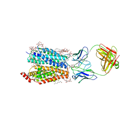
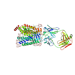
6JKH
 | | The NAD+-bound form of human NSDHL | | 分子名称: | NICOTINAMIDE-ADENINE-DINUCLEOTIDE, Sterol-4-alpha-carboxylate 3-dehydrogenase, decarboxylating | | 著者 | Kim, D, Lee, S.J, Lee, B. | | 登録日 | 2019-02-28 | | 公開日 | 2020-03-04 | | 最終更新日 | 2023-11-22 | | 実験手法 | X-RAY DIFFRACTION (3 Å) | | 主引用文献 | Crystal structures of human NSDHL and development of its novel inhibitor with the potential to suppress EGFR activity.
Cell.Mol.Life Sci., 78, 2021
|
|
8CTN
 | | Structure of a K+ selective NaK mutant (NaK2K, Laue diffraction, no electric field) | | 分子名称: | (4S)-2-METHYL-2,4-PENTANEDIOL, POTASSIUM ION, Potassium channel protein, ... | | 著者 | Lee, B, White, K.I, Socolich, M.A, Klureza, M.A, Henning, R, Srajer, V, Ranganathan, R, Hekstra, D. | | 登録日 | 2022-05-16 | | 公開日 | 2023-06-14 | | 最終更新日 | 2024-04-03 | | 実験手法 | X-RAY DIFFRACTION (2.01 Å) | | 主引用文献 | Direct visualization of electric field-stimulated ion conduction in a potassium channel
To Be Published
|
|
6JM4
 | |
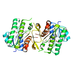
6JKV
 | | PppA, a key regulatory component of T6SS in Pseudomonas aeruginosa | | 分子名称: | MANGANESE (II) ION, PppA | | 著者 | Wang, T, Liu, L, Wu, Y, Li, D. | | 登録日 | 2019-03-02 | | 公開日 | 2019-06-26 | | 最終更新日 | 2023-11-22 | | 実験手法 | X-RAY DIFFRACTION (2.1 Å) | | 主引用文献 | Crystal structure of PppA from Pseudomonas aeruginosa, a key regulatory component of type VI secretion systems.
Biochem.Biophys.Res.Commun., 516, 2019
|
|
8CTT
 | | Crystal structure of a K+ selective NaK mutant (NaK2K) at 100K | | 分子名称: | (4S)-2-METHYL-2,4-PENTANEDIOL, POTASSIUM ION, Potassium channel protein | | 著者 | Lee, B, White, K.I, Socolich, M.A, Klureza, M.A, Henning, R, Srajer, V, Ranganathan, R, Hekstra, D. | | 登録日 | 2022-05-16 | | 公開日 | 2023-06-14 | | 最終更新日 | 2023-10-25 | | 実験手法 | X-RAY DIFFRACTION (2.05 Å) | | 主引用文献 | Direct visualization of electric field-stimulated ion conduction in a potassium channel
To Be Published
|
|
1K5O
 | | CPI-17(35-120) deletion mutant | | 分子名称: | CPI-17 | | 著者 | Ohki, S, Eto, M, Kariya, E, Hayano, T, Hayashi, Y, Yazawa, M, Brautigan, D, Kainosho, M. | | 登録日 | 2001-10-11 | | 公開日 | 2002-10-11 | | 最終更新日 | 2024-05-29 | | 実験手法 | SOLUTION NMR | | 主引用文献 | Solution NMR Structure of the Myosin Phosphatase Inhibitor Protein CPI-17 Shows Phosphorylation-induced Conformational Changes Responsible for Activation
J.Mol.Biol., 314, 2001
|
|
8CTW
 | | Crystal structure of a K+ selective NaK mutant (NaK2K) -Na+,Tl+ complex | | 分子名称: | (4S)-2-METHYL-2,4-PENTANEDIOL, Potassium channel protein, SODIUM ION, ... | | 著者 | Lee, B, White, K.I, Socolich, M.A, Klureza, M.A, Henning, R, Srajer, V, Ranganathan, R, Hekstra, D. | | 登録日 | 2022-05-16 | | 公開日 | 2023-06-14 | | 最終更新日 | 2023-10-25 | | 実験手法 | X-RAY DIFFRACTION (2 Å) | | 主引用文献 | Direct visualization of electric field-stimulated ion conduction in a potassium channel
To Be Published
|
|
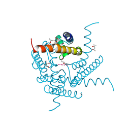
1K5W
 | | THREE-DIMENSIONAL STRUCTURE OF THE SYNAPTOTAGMIN 1 C2B-DOMAIN: SYNAPTOTAGMIN 1 AS A PHOSPHOLIPID BINDING MACHINE | | 分子名称: | CALCIUM ION, Synaptotagmin I | | 著者 | Fernandez, I, Arac, D, Ubach, J, Gerber, S.H, Shin, O, Gao, Y, Anderson, R.G.W, Sudhof, T.C, Rizo, J. | | 登録日 | 2001-10-12 | | 公開日 | 2002-01-23 | | 最終更新日 | 2024-05-22 | | 実験手法 | SOLUTION NMR | | 主引用文献 | Three-dimensional structure of the synaptotagmin 1 C2B-domain: synaptotagmin 1 as a phospholipid binding machine.
Neuron, 32, 2001
|
|
8CTX
 | | Crystal structure of a K+ selective NaK mutant (NaK2K) -K+,Tl+ complex | | 分子名称: | POTASSIUM ION, Potassium channel protein, THALLIUM (I) ION | | 著者 | Lee, B, White, K.I, Socolich, M.A, Klureza, M.A, Henning, R, Srajer, V, Ranganathan, R, Hekstra, D. | | 登録日 | 2022-05-16 | | 公開日 | 2023-06-14 | | 最終更新日 | 2023-10-25 | | 実験手法 | X-RAY DIFFRACTION (1.99 Å) | | 主引用文献 | Direct visualization of electric field-stimulated ion conduction in a potassium channel
To Be Published
|
|
8DE4
 | |
8CTV
 | | Crystal structure of a K+ selective NaK mutant (NaK2K) -Tl+ complex | | 分子名称: | (4S)-2-METHYL-2,4-PENTANEDIOL, Potassium channel protein, SODIUM ION, ... | | 著者 | Lee, B, White, K.I, Socolich, M.A, Klureza, M.A, Henning, R, Srajer, V, Ranganathan, R, Hekstra, D. | | 登録日 | 2022-05-16 | | 公開日 | 2023-06-14 | | 最終更新日 | 2024-04-03 | | 実験手法 | X-RAY DIFFRACTION (2.1 Å) | | 主引用文献 | Direct visualization of electric field-stimulated ion conduction in a potassium channel
To Be Published
|
|


