9CK0
 
 | |
8WML
 | |
8ZWJ
 | |
8ZWK
 | |
8X2V
 | |
9B34
 | |
9CA2
 | |
6OED
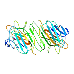
 | | CRYSTAL STRUCTURE OF THE RV144 C1-C2 SPECIFIC ANTIBODY CH55 FAB | | 分子名称: | CH55 Fab heavy chain, CH55 Fab light chain | | 著者 | Yan, F, Van, V, Tolbert, W.D, Pazgier, M. | | 登録日 | 2019-03-27 | | 公開日 | 2020-07-15 | | 最終更新日 | 2023-10-11 | | 実験手法 | X-RAY DIFFRACTION (2.461 Å) | | 主引用文献 | Recognition Patterns of the C1/C2 Epitopes Involved in Fc-Mediated Response in HIV-1 Natural Infection and the RV114 Vaccine Trial.
Mbio, 11, 2020
|
|
6M8H
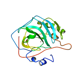
 | | Crystal Structure of the R208Q mutant of G(i) subunit alpha-1 | | 分子名称: | 5'-GUANOSINE-DIPHOSPHATE-MONOTHIOPHOSPHATE, Guanine nucleotide-binding protein G(i) subunit alpha-1, MAGNESIUM ION | | 著者 | Mascarenhas, R, Goossens, J, Leverson, B, Kothawala, S, Ballicora, M, Olsen, K, de freitas, D, Liu, D. | | 登録日 | 2018-08-21 | | 公開日 | 2019-08-21 | | 最終更新日 | 2024-03-13 | | 実験手法 | X-RAY DIFFRACTION (2.07 Å) | | 主引用文献 | FUNCTIONAL CONSEQUENCES OF ONCOGENIC MUTATIONS IN THE SWITCH II REGION OF Galphai1 and Galphas PROTEINS
To Be Published
|
|
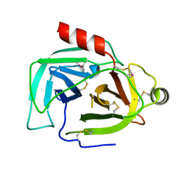
6U1V
 | | Crystal structure of acyl-ACP/acyl-CoA dehydrogenase from allylmalonyl-CoA and FK506 biosynthesis, TcsD | | 分子名称: | Acyl-CoA dehydrogenase domain-containing protein, DIHYDROFLAVINE-ADENINE DINUCLEOTIDE | | 著者 | Blake-Hedges, J.M, Pereira, J.H, Barajas, J.F, Adams, P.D, Keasling, J.D. | | 登録日 | 2019-08-16 | | 公開日 | 2019-12-18 | | 最終更新日 | 2023-10-11 | | 実験手法 | X-RAY DIFFRACTION (1.75 Å) | | 主引用文献 | Structural Mechanism of Regioselectivity in an Unusual Bacterial Acyl-CoA Dehydrogenase.
J.Am.Chem.Soc., 142, 2020
|
|
9BFL
 | |
9BKG
 | |
8WAG
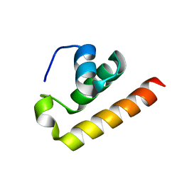
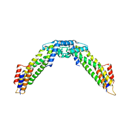 | | Crystal structure of the C-terminal fragment (residues 716-982) of Arabidopsis thaliana CHUP1 | | 分子名称: | Protein CHUP1, chloroplastic | | 著者 | Shimada, A, Nakamura, Y, Takano, A, Kohda, D. | | 登録日 | 2023-09-07 | | 公開日 | 2024-01-17 | | 最終更新日 | 2024-04-10 | | 実験手法 | X-RAY DIFFRACTION (3.003 Å) | | 主引用文献 | CHLOROPLAST UNUSUAL POSITIONING 1 is a plant-specific actin polymerization factor regulating chloroplast movement.
Plant Cell, 36, 2024
|
|
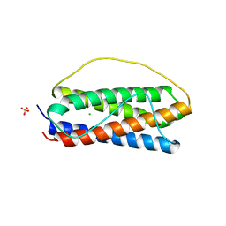
8WAF
 | | Crystal structure of the C-terminal fragment (residues 756-982 with the C864S mutation) of Arabidopsis thaliana CHUP1 | | 分子名称: | Protein CHUP1, chloroplastic | | 著者 | Shimada, A, Takano, A, Nakamura, Y, Kohda, D. | | 登録日 | 2023-09-07 | | 公開日 | 2024-01-17 | | 最終更新日 | 2024-04-10 | | 実験手法 | X-RAY DIFFRACTION (2.8 Å) | | 主引用文献 | CHLOROPLAST UNUSUAL POSITIONING 1 is a plant-specific actin polymerization factor regulating chloroplast movement.
Plant Cell, 36, 2024
|
|
8X43
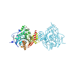
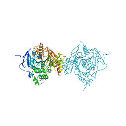
 | | human KCNQ2-CaM-Ebio1-S1 complex in the presence of PIP2 | | 分子名称: | Calmodulin-1, N-(4-azanyl-1,2-dihydroacenaphthylen-5-yl)-4-fluoranyl-benzamide, Potassium voltage-gated channel subfamily KQT member 2 | | 著者 | Ma, D, Guo, J. | | 登録日 | 2023-11-15 | | 公開日 | 2024-01-17 | | 最終更新日 | 2024-07-17 | | 実験手法 | ELECTRON MICROSCOPY (3 Å) | | 主引用文献 | A small-molecule activation mechanism that directly opens the KCNQ2 channel.
Nat.Chem.Biol., 20, 2024
|
|
6ONO
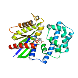
 | | Complex structure of WhiB1 and region 4 of SigA in C2221 space group | | 分子名称: | DI(HYDROXYETHYL)ETHER, IRON/SULFUR CLUSTER, RNA polymerase sigma factor SigA, ... | | 著者 | Wan, T, Li, S.R, Beltran, D.G, Schacht, A, Becker, D.C, Zhang, L.M. | | 登録日 | 2019-04-22 | | 公開日 | 2019-11-27 | | 最終更新日 | 2020-01-22 | | 実験手法 | X-RAY DIFFRACTION (1.85 Å) | | 主引用文献 | Structural basis of non-canonical transcriptional regulation by the sigma A-bound iron-sulfur protein WhiB1 in M. tuberculosis.
Nucleic Acids Res., 48, 2020
|
|
9BKL
 | |
9BKP
 | |
7A9W
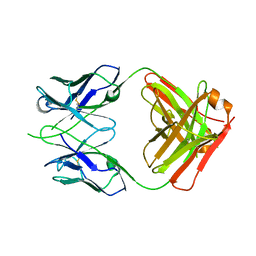
 | | Structure of yeast Rmd9p in complex with 20nt target RNA | | 分子名称: | CHLORIDE ION, Protein RMD9, mitochondrial, ... | | 著者 | Hillen, H.S, Markov, D.A, Ireneusz, W.D, Hofmann, K.B, Cowan, A.T, Jones, J.L, Temiakov, D, Cramer, P, Anikin, M. | | 登録日 | 2020-09-02 | | 公開日 | 2021-04-07 | | 最終更新日 | 2021-05-05 | | 実験手法 | X-RAY DIFFRACTION (2.55 Å) | | 主引用文献 | The pentatricopeptide repeat protein Rmd9 recognizes the dodecameric element in the 3'-UTRs of yeast mitochondrial mRNAs.
Proc.Natl.Acad.Sci.USA, 118, 2021
|
|
7A9X
 | | Structure of yeast Rmd9p in complex with 16nt target RNA | | 分子名称: | CHLORIDE ION, Protein RMD9, mitochondrial, ... | | 著者 | Hillen, H.S, Markov, D.A, Ireneusz, W.D, Hofmann, K.B, Cowan, A.T, Jones, J.L, Temiakov, D, Cramer, P, Anikin, M. | | 登録日 | 2020-09-02 | | 公開日 | 2021-04-07 | | 最終更新日 | 2024-01-31 | | 実験手法 | X-RAY DIFFRACTION (2.45 Å) | | 主引用文献 | The pentatricopeptide repeat protein Rmd9 recognizes the dodecameric element in the 3'-UTRs of yeast mitochondrial mRNAs.
Proc.Natl.Acad.Sci.USA, 118, 2021
|
|
6OJC
 | |
8ZEY
 | |
6O4O
 | | The structure of human interleukin 11 | | 分子名称: | CHLORIDE ION, Interleukin-11, SULFATE ION | | 著者 | Metcalfe, R.D, Griffin, M.D.W. | | 登録日 | 2019-02-28 | | 公開日 | 2020-05-06 | | 最終更新日 | 2023-10-11 | | 実験手法 | X-RAY DIFFRACTION (1.62 Å) | | 主引用文献 | The structure of the extracellular domains of human interleukin 11 alpha receptor reveals mechanisms of cytokine engagement.
J.Biol.Chem., 295, 2020
|
|
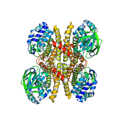
6WVQ
 | | Crystal Structure of Recombinant Human Acetylcholinesterase Inhibited by GP | | 分子名称: | (1S)-2,2-dimethylcyclopentyl (R)-methylphosphinate, 2-(2-(2-(2-(2-(2-ETHOXYETHOXY)ETHOXY)ETHOXY)ETHOXY)ETHOXY)ETHANOL, 2-acetamido-2-deoxy-beta-D-glucopyranose, ... | | 著者 | McGuire, J.R, Bester, S.M, Pegan, S.D, Height, J.J. | | 登録日 | 2020-05-06 | | 公開日 | 2021-02-17 | | 最終更新日 | 2023-10-18 | | 実験手法 | X-RAY DIFFRACTION (2.289 Å) | | 主引用文献 | Structural and Biochemical Insights into the Inhibition of Human Acetylcholinesterase by G-Series Nerve Agents and Subsequent Reactivation by HI-6.
Chem.Res.Toxicol., 34, 2021
|
|
6WVC
 | | Crystal Structure of Recombinant Human Acetylcholinesterase Inhibited by GD | | 分子名称: | (1S)-2,2-dimethylcyclopentyl (R)-methylphosphinate, 2-(2-(2-(2-(2-(2-ETHOXYETHOXY)ETHOXY)ETHOXY)ETHOXY)ETHOXY)ETHANOL, 2-acetamido-2-deoxy-beta-D-glucopyranose, ... | | 著者 | McGuire, J.R, Bester, S.M, Pegan, S.D, Height, J.J. | | 登録日 | 2020-05-05 | | 公開日 | 2021-02-17 | | 最終更新日 | 2023-10-18 | | 実験手法 | X-RAY DIFFRACTION (2.599 Å) | | 主引用文献 | Structural and Biochemical Insights into the Inhibition of Human Acetylcholinesterase by G-Series Nerve Agents and Subsequent Reactivation by HI-6.
Chem.Res.Toxicol., 34, 2021
|
|


