Loading
PDBj
MenuPDBj@FacebookPDBj@TwitterPDBj@YouTubewwPDB FoundationwwPDB
RCSB PDBPDBeBMRBAdv. SearchSearch help
PDB: 787 results

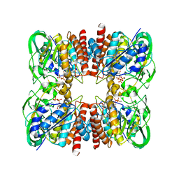
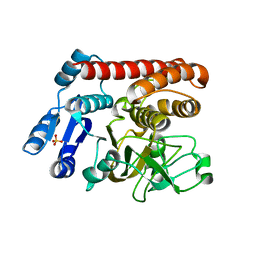
8J5V
DownloadVisualize
BU of 8j5v by Molmil
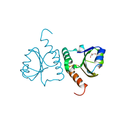
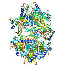
Crystal structure of estZF172 as a novel biocatalyst for the efficient biosynthesis of a chiral intermediate of pregabalin
Descriptor: Carboxylesterase
Authors:Chi, C.B, Liang, Z.D, Huo, B.Q, Hu, C.X, Sun, Q.Y.
Deposit date:2023-04-24
Release date:2024-05-01
Method:X-RAY DIFFRACTION (2.05 Å)
Cite:Crystal structure of estZF172 as a novel biocatalyst for the efficient biosynthesis of a chiral intermediate of pregabalin
To Be Published
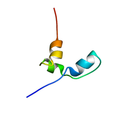
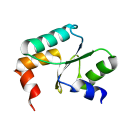
7OSR
DownloadVisualize
BU of 7osr by Molmil
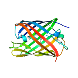
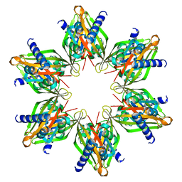
Structure and folding of a 600-million-year-old nuclear coactivator binding domain suggest conservation of dynamic properties
Descriptor: Nuclear co-activator binding domain
Authors:Chi, C.
Deposit date:2021-06-09
Release date:2022-04-20
Last modified:2024-06-19
Method:SOLUTION NMR
Cite:The dynamic properties of a nuclear coactivator binding domain are evolutionarily conserved.
Commun Biol, 5, 2022
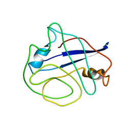
7OSW
DownloadVisualize
BU of 7osw by Molmil
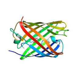
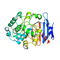
Structure and folding of a 600-million-year-old nuclear coactivator binding domain suggest conservation of dynamic properties
Descriptor: NCBD
Authors:Chi, C.
Deposit date:2021-06-09
Release date:2022-04-20
Last modified:2024-06-19
Method:SOLUTION NMR
Cite:The dynamic properties of a nuclear coactivator binding domain are evolutionarily conserved.
Commun Biol, 5, 2022
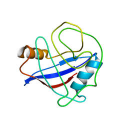
2N0T
DownloadVisualize
BU of 2n0t by Molmil
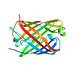
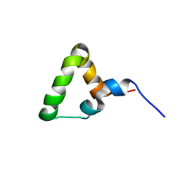
Structural ensemble of the enzyme cyclophilin reveals an orchestrated mode of action at atomic resolution
Descriptor: Peptidyl-prolyl cis-trans isomerase A
Authors:Chi, C.N, Voegeli, B, Bibow, S, Strotz, D, Orts, J, Guntert, P, Riek, R.
Deposit date:2015-03-13
Release date:2015-08-26
Last modified:2024-05-15
Method:SOLUTION NMR
Cite:A Structural Ensemble for the Enzyme Cyclophilin Reveals an Orchestrated Mode of Action at Atomic Resolution.
Angew.Chem.Int.Ed.Engl., 54, 2015
2MZU
DownloadVisualize
BU of 2mzu by Molmil
Extending the eNOE data set of large proteins by evaluation of NOEs with unresolved diagonals
Descriptor: Peptidyl-prolyl cis-trans isomerase A
Authors:Chi, C.N, Strotz, D, Riek, R, Voegeli, B.
Deposit date:2015-02-24
Release date:2015-04-22
Last modified:2024-05-15
Method:SOLUTION NMR
Cite:Extending the eNOE data set of large proteins by evaluation of NOEs with unresolved diagonals.
J.Biomol.Nmr, 62, 2015
7YUA
DownloadVisualize
BU of 7yua by Molmil
Structural Insight into a Metal-Dependent Mutase MtdL Revealing an Arginine Residue Covalently Mediated Interconversion between Nucleotide-Based furanose and pyranose
Descriptor: GUANOSINE-5'-DIPHOSPHATE, MAGNESIUM ION, SULFATE ION, ...
Authors:Chi, C.B, Ma, M.
Deposit date:2022-08-16
Release date:2023-07-26
Last modified:2024-10-09
Method:X-RAY DIFFRACTION (2.5 Å)
Cite:Structural Insight into a Metal-Dependent Mutase Revealing an Arginine Residue-Covalently Mediated Interconversion between Nucleotide-Based Pyranose and Furanose.
Acs Catalysis, 13, 2023
7YV0
DownloadVisualize
BU of 7yv0 by Molmil
Structural Insight into a Metal-Dependent Mutase MtdL Revealing an Arginine Residue Covalently Mediated Interconversion between Nucleotide-Based furanose and pyranose
Descriptor: SULFATE ION, Transglycosylse
Authors:Chi, C.B, Ma, M.
Deposit date:2022-08-18
Release date:2023-07-26
Last modified:2024-10-16
Method:X-RAY DIFFRACTION (2.34 Å)
Cite:Structural Insight into a Metal-Dependent Mutase Revealing an Arginine Residue-Covalently Mediated Interconversion between Nucleotide-Based Pyranose and Furanose.
Acs Catalysis, 13, 2023
7DMN
DownloadVisualize
BU of 7dmn by Molmil
Crystal structure of two pericyclases catalyzing [4+2] cycloaddition
Descriptor: Diels-Alderase fsa2, GLYCEROL
Authors:Chi, C.B, Wang, Z.D, Liu, T, Zhang, Z.Y, Ma, M.
Deposit date:2020-12-04
Release date:2021-10-06
Last modified:2024-04-03
Method:X-RAY DIFFRACTION (2 Å)
Cite:Crystal Structures of Fsa2 and Phm7 Catalyzing [4 + 2] Cycloaddition Reactions with Reverse Stereoselectivities in Equisetin and Phomasetin Biosynthesis.
Acs Omega, 6, 2021
7E22
DownloadVisualize
BU of 7e22 by Molmil
Crystal structure of two pericyclases catalyzing [4+2] cycloaddition
Descriptor: Diels-Alderase fsa2, Equisetin
Authors:Chi, C.B, Wang, Z.D.
Deposit date:2021-02-04
Release date:2022-02-09
Last modified:2023-11-29
Method:X-RAY DIFFRACTION (2.63 Å)
Cite:Crystal structure of two pericyclases catalyzing [4+2] cycloaddition
To Be Published
5Y4U
DownloadVisualize
BU of 5y4u by Molmil
Crystal structure of Grx domain of Grx3 from Saccharomyces cerevisiae
Descriptor: Monothiol glutaredoxin-3
Authors:Chi, C.B, Tang, Y.J, Zhang, J.H, Dai, Y.N, Abdalla, M, Chen, Y.X, Zhou, C.Z.
Deposit date:2017-08-05
Release date:2018-08-15
Last modified:2023-11-22
Method:X-RAY DIFFRACTION (1.7 Å)
Cite:Structural and Biochemical Insights into the Multiple Functions of Yeast Grx3.
J.Mol.Biol., 430, 2018
5Y4T
DownloadVisualize
BU of 5y4t by Molmil
Crystal structure of Trx domain of Grx3 from Saccharomyces cerevisiae
Descriptor: GLYCEROL, Glutaredoxin
Authors:Chi, C.B, Tang, Y.J, Zhang, J.H, Dai, Y.N, Abdalla, M, Chen, Y.X, Zhou, C.Z.
Deposit date:2017-08-05
Release date:2018-08-15
Last modified:2023-11-22
Method:X-RAY DIFFRACTION (1.4 Å)
Cite:Structural and Biochemical Insights into the Multiple Functions of Yeast Grx3.
J.Mol.Biol., 430, 2018
5OQA
DownloadVisualize
BU of 5oqa by Molmil
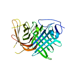
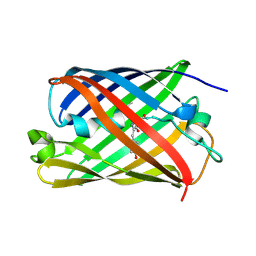
XFEL structure of the off state of a reversibly photoswitching fluorescent protein determined using the GDVN injection method
Descriptor: Green to red photoconvertible GFP-like protein EosFP
Authors:Hutchison, C.D.M, Cordon-Preciado, V, Morgan, R.M.L, van Thor, J.J.
Deposit date:2017-08-11
Release date:2017-12-06
Last modified:2023-11-15
Method:X-RAY DIFFRACTION (1.53 Å)
Cite:X-ray Free Electron Laser Determination of Crystal Structures of Dark and Light States of a Reversibly Photoswitching Fluorescent Protein at Room Temperature.
Int J Mol Sci, 18, 2017
5OOZ
DownloadVisualize
BU of 5ooz by Molmil
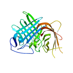
XFEL structure of the on state of a reversibly photoswitching fluorescent protein determined using the grease injection method
Descriptor: Green to red photoconvertible GFP-like protein EosFP
Authors:Hutchison, C.D.M, Cordon-Preciado, V, Morgan, R.M.L, van Thor, J.J.
Deposit date:2017-08-09
Release date:2017-12-06
Last modified:2024-10-23
Method:X-RAY DIFFRACTION (1.92 Å)
Cite:X-ray Free Electron Laser Determination of Crystal Structures of Dark and Light States of a Reversibly Photoswitching Fluorescent Protein at Room Temperature.
Int J Mol Sci, 18, 2017
5OQE
DownloadVisualize
BU of 5oqe by Molmil
XFEL structure of the on state of a reversibly photoswitching fluorescent protein determined using the droplet injection method
Descriptor: Green to red photoconvertible GFP-like protein EosFP
Authors:Hutchison, C.D.M, Cordon-Preciado, V, Morgan, R.M.L, van Thor, J.J.
Deposit date:2017-08-11
Release date:2017-12-06
Last modified:2024-10-16
Method:X-RAY DIFFRACTION (1.91 Å)
Cite:X-ray Free Electron Laser Determination of Crystal Structures of Dark and Light States of a Reversibly Photoswitching Fluorescent Protein at Room Temperature.
Int J Mol Sci, 18, 2017
5OQ9
DownloadVisualize
BU of 5oq9 by Molmil
XFEL structure of the off state of a reversibly photoswitching fluorescent protein determined using the droplet on demand injection method
Descriptor: Green to red photoconvertible GFP-like protein EosFP
Authors:Hutchison, C.D.M, Cordon-Preciado, V, Morgan, R.M.L, van Thor, J.J.
Deposit date:2017-08-11
Release date:2017-12-06
Last modified:2023-11-15
Method:X-RAY DIFFRACTION (1.51 Å)
Cite:X-ray Free Electron Laser Determination of Crystal Structures of Dark and Light States of a Reversibly Photoswitching Fluorescent Protein at Room Temperature.
Int J Mol Sci, 18, 2017
2PSV
DownloadVisualize
BU of 2psv by Molmil
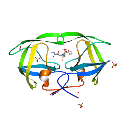
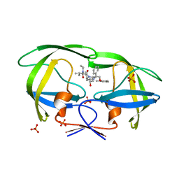
Crystal Structure of wild type HIV-1 protease in complex with CARB-KB45
Descriptor: ACETATE ION, N-{(1S,2R)-1-BENZYL-3-[(CYCLOPROPYLMETHYL)(2-FURYLSULFONYL)AMINO]-2-HYDROXYPROPYL}-N'-METHYLSUCCINAMIDE, PHOSPHATE ION, ...
Authors:Schiffer, C.A, Nalam, M.N.L.
Deposit date:2007-05-07
Release date:2007-06-05
Last modified:2023-08-30
Method:X-RAY DIFFRACTION (1.75 Å)
Cite:Design of Mutation-resistant HIV Protease Inhibitors with the Substrate Envelope Hypothesis.
Chem.Biol.Drug Des., 69, 2007
2Q54
DownloadVisualize
BU of 2q54 by Molmil
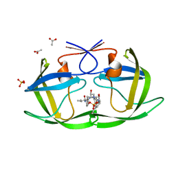
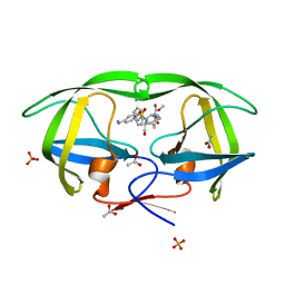
Crystal structure of KB73 bound to HIV-1 protease
Descriptor: ACETATE ION, N~2~-ACETYL-N-[(1S,3S,4S)-4-({[(5S)-3-(3-ACETYLPHENYL)-2-OXO-1,3-OXAZOLIDIN-5-YL]CARBONYL}AMINO)-1-BENZYL-3-HYDROXY-5-PHENYLPENTYL]-L-VALINAMIDE, PHOSPHATE ION, ...
Authors:Schiffer, C.A, Nalam, M.N.L.
Deposit date:2007-05-31
Release date:2007-09-04
Last modified:2024-03-13
Method:X-RAY DIFFRACTION (1.85 Å)
Cite:Design and Synthesis of HIV-1 Protease Inhibitors Incorporating Oxazolidinones as P2/P2' Ligands in Pseudosymmetric Dipeptide Isosteres.
J.Med.Chem., 50, 2007
2Q55
DownloadVisualize
BU of 2q55 by Molmil
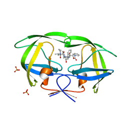
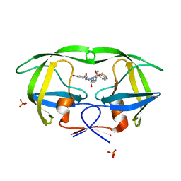
Crystal structure of KK44 bound to HIV-1 protease
Descriptor: (5S)-N-[(1S,2S,4S)-1-BENZYL-2-HYDROXY-4-{[(2S)-3-METHYL-2-(2-OXOTETRAHYDROPYRIMIDIN-1(2H)-YL)BUTANOYL]AMINO}-5-PHENYLPENTYL]-2-OXO-3-PHENYL-1,3-OXAZOLIDINE-5-CARBOXAMIDE, PHOSPHATE ION, Protease
Authors:Schiffer, C.A, Nalam, M.N.L.
Deposit date:2007-05-31
Release date:2007-09-04
Last modified:2024-03-13
Method:X-RAY DIFFRACTION (1.9 Å)
Cite:Design and Synthesis of HIV-1 Protease Inhibitors Incorporating Oxazolidinones as P2/P2' Ligands in Pseudosymmetric Dipeptide Isosteres.
J.Med.Chem., 50, 2007
2Q5K
DownloadVisualize
BU of 2q5k by Molmil
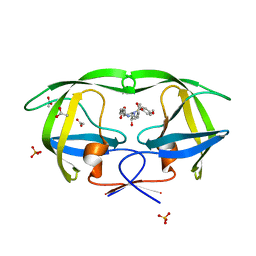
Crystal structure of lopinavir bound to wild type HIV-1 protease
Descriptor: N-{1-BENZYL-4-[2-(2,6-DIMETHYL-PHENOXY)-ACETYLAMINO]-3-HYDROXY-5-PHENYL-PENTYL}-3-METHYL-2-(2-OXO-TETRAHYDRO-PYRIMIDIN-1-YL)-BUTYRAMIDE, PHOSPHATE ION, Protease
Authors:Schiffer, C.A, Nalam, M.N.L.
Deposit date:2007-06-01
Release date:2007-09-04
Last modified:2024-03-13
Method:X-RAY DIFFRACTION (1.95 Å)
Cite:Design and Synthesis of HIV-1 Protease Inhibitors Incorporating Oxazolidinones as P2/P2' Ligands in Pseudosymmetric Dipeptide Isosteres.
J.Med.Chem., 50, 2007
2Q3K
DownloadVisualize
BU of 2q3k by Molmil
Crystal Structure of Lysine Sulfonamide Inhibitor Reveals the Displacement of the Conserved Flap Water Molecule in HIV-1 Protease
Descriptor: ACETATE ION, N-{(5S)-5-[{[4-(AMINOMETHYL)PHENYL]SULFONYL}(ISOBUTYL)AMINO]-6-HYDROXYHEXYL}-NALPHA-(METHOXYCARBONYL)-BETA-PHENYL-L-PHENYLALANINAMIDE, PHOSPHATE ION, ...
Authors:Schiffer, C.A, Nalam, M.N.L.
Deposit date:2007-05-30
Release date:2007-08-21
Last modified:2023-08-30
Method:X-RAY DIFFRACTION (2 Å)
Cite:Crystal structure of lysine sulfonamide inhibitor reveals the displacement of the conserved flap water molecule in human immunodeficiency virus type 1 protease.
J.Virol., 81, 2007
2QI6
DownloadVisualize
BU of 2qi6 by Molmil
Crystal structure of protease inhibitor, MIT-2-KB98 in complex with wild type HIV-1 protease
Descriptor: N-{(1S,2R)-3-[(1,3-BENZOTHIAZOL-6-YLSULFONYL)(ISOBUTYL)AMINO]-1-BENZYL-2-HYDROXYPROPYL}-3-HYDROXYBENZAMIDE, PHOSPHATE ION, Protease
Authors:Schiffer, C.A, Nalam, M.N.L.
Deposit date:2007-07-03
Release date:2008-04-22
Last modified:2023-08-30
Method:X-RAY DIFFRACTION (1.85 Å)
Cite:HIV-1 protease inhibitors from inverse design in the substrate envelope exhibit subnanomolar binding to drug-resistant variants.
J.Am.Chem.Soc., 130, 2008
2QI7
DownloadVisualize
BU of 2qi7 by Molmil
Crystal structure of protease inhibitor, MIT-2-AD86 in complex with wild type HIV-1 protease
Descriptor: ACETATE ION, N-[(1S,2R)-1-BENZYL-2-HYDROXY-3-{[(4-METHOXYPHENYL)SULFONYL][(2S)-2-METHYLBUTYL]AMINO}PROPYL]-4-OXOHEXANAMIDE, PHOSPHATE ION, ...
Authors:Schiffer, C.A, Nalam, M.N.L.
Deposit date:2007-07-03
Release date:2008-04-22
Last modified:2023-08-30
Method:X-RAY DIFFRACTION (1.85 Å)
Cite:HIV-1 protease inhibitors from inverse design in the substrate envelope exhibit subnanomolar binding to drug-resistant variants.
J.Am.Chem.Soc., 130, 2008
8T17
DownloadVisualize
BU of 8t17 by Molmil
Cryo-EM structure of tetradecameric hub domain of CaMKII beta
Descriptor: Venus-tagged CaMKII Beta Association Domain
Authors:Chien, C.-T, Chiu, W, Khan, S.
Deposit date:2023-06-02
Release date:2024-06-12
Last modified:2024-07-10
Method:ELECTRON MICROSCOPY (2.6 Å)
Cite:Hub stability in the calcium calmodulin-dependent protein kinase II.
Commun Biol, 7, 2024
8T18
DownloadVisualize
BU of 8t18 by Molmil
Cryo-EM structure of dodecameric hub domain of CaMKII beta
Descriptor: Venus-tagged CaMKII Alpha Association Domain
Authors:Chien, C.-T, Chiu, W, Khan, S.
Deposit date:2023-06-02
Release date:2024-06-12
Last modified:2024-07-10
Method:ELECTRON MICROSCOPY (2.6 Å)
Cite:Hub stability in the calcium calmodulin-dependent protein kinase II.
Commun Biol, 7, 2024
8T15
DownloadVisualize
BU of 8t15 by Molmil
Cryo-EM structure of dodecameric hub domain of CaMKII alpha
Descriptor: Venus-tagged CaMKII Alpha Association Domain
Authors:Chien, C.-T, Chiu, W, Khan, S.
Deposit date:2023-06-01
Release date:2024-06-12
Last modified:2024-07-10
Method:ELECTRON MICROSCOPY (2.7 Å)
Cite:Hub stability in the calcium calmodulin-dependent protein kinase II.
Commun Biol, 7, 2024

226707

数据于2024-10-30公开中

PDB statisticsPDBj update infoContact PDBjnumon