8JOV
 
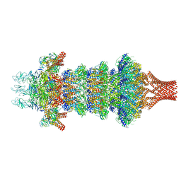 | | Portal-tail complex of phage GP4 | | 分子名称: | Portal protein, Putative tail fiber protein, Virion associated protein, ... | | 著者 | Liu, H, Chen, W. | | 登録日 | 2023-06-08 | | 公開日 | 2023-11-01 | | 実験手法 | ELECTRON MICROSCOPY (3.8 Å) | | 主引用文献 | Asymmetric Structure of Podophage GP4 Reveals a Novel Architecture of Three Types of Tail Fibers.
J.Mol.Biol., 435, 2023
|
|
6K2H
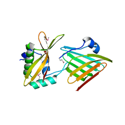
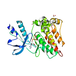
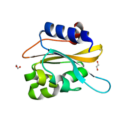 | | structural characterization of mutated NreA protein in nitrate binding site from staphylococcus aureus. | | 分子名称: | 1,2-ETHANEDIOL, NreA | | 著者 | Sangare, L, Chen, W, Wang, C, Chen, X, Wu, M, Zhang, X, Zang, J. | | 登録日 | 2019-05-14 | | 公開日 | 2020-03-11 | | 最終更新日 | 2023-11-22 | | 実験手法 | X-RAY DIFFRACTION (1.8 Å) | | 主引用文献 | Structural insights into the conformational change of Staphylococcus aureus NreA at C-terminus.
Biotechnol.Lett., 42, 2020
|
|
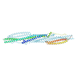
8JOU
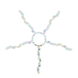 | | Fiber I and fiber-tail-adaptor of phage GP4 | | 分子名称: | Virion-associated phage protein, rope protein of phage GP4 | | 著者 | Liu, H, Chen, W. | | 登録日 | 2023-06-08 | | 公開日 | 2024-01-03 | | 実験手法 | ELECTRON MICROSCOPY (4.1 Å) | | 主引用文献 | Asymmetric Structure of Podophage GP4 Reveals a Novel Architecture of Three Types of Tail Fibers.
J.Mol.Biol., 435, 2023
|
|
8HUD
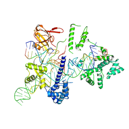 | | Cryo-EM structure of the EvCas9-sgRNA-target DNA ternary complex | | 分子名称: | CRISPR-associated endonuclease Cas9, Non-target DNA strand, Target DNA strand, ... | | 著者 | Tang, N, Wu, Z, Gao, Y, Chen, W, Su, M, Wang, Z, Ji, Q. | | 登録日 | 2022-12-23 | | 公開日 | 2023-12-27 | | 実験手法 | ELECTRON MICROSCOPY (3.43 Å) | | 主引用文献 | Cryo-EM structure of the EvCas9-sgRNA-target DNA ternary complex
To Be Published
|
|
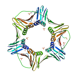
4DNW
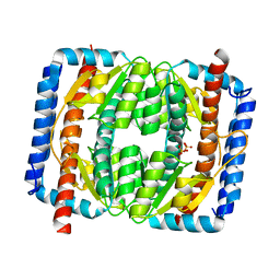
 | | Crystal structure of UVB-resistance protein UVR8 | | 分子名称: | AT5g63860/MGI19_6 | | 著者 | Wu, D, Hu, Q, Yan, Z, Chen, W, Yan, C, Wang, J, Shi, Y. | | 登録日 | 2012-02-09 | | 公開日 | 2012-03-07 | | 最終更新日 | 2023-11-08 | | 実験手法 | X-RAY DIFFRACTION (1.773 Å) | | 主引用文献 | Structural basis of ultraviolet-B perception by UVR8.
Nature, 484, 2012
|
|
4DNU
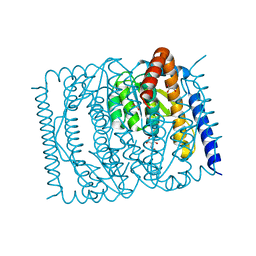
 | | Crystal structure of the W285A mutant of UVB-resistance protein UVR8 | | 分子名称: | AT5g63860/MGI19_6 | | 著者 | Wu, D, Hu, Q, Yan, Z, Chen, W, Yan, C, Zhang, J, Wang, J, Shi, Y. | | 登録日 | 2012-02-09 | | 公開日 | 2012-03-07 | | 最終更新日 | 2013-07-17 | | 実験手法 | X-RAY DIFFRACTION (1.764 Å) | | 主引用文献 | Structural basis of ultraviolet-B perception by UVR8.
Nature, 484, 2012
|
|
4DNV
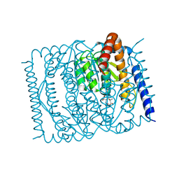
 | | Crystal structure of the W285F mutant of UVB-resistance protein UVR8 | | 分子名称: | AT5g63860/MGI19_6 | | 著者 | Wu, D, Hu, Q, Yan, Z, Chen, W, Yan, C, Zhang, J, Wang, J, Shi, Y. | | 登録日 | 2012-02-09 | | 公開日 | 2012-03-07 | | 最終更新日 | 2023-11-08 | | 実験手法 | X-RAY DIFFRACTION (1.999 Å) | | 主引用文献 | Structural basis of ultraviolet-B perception by UVR8.
Nature, 484, 2012
|
|
5YCO
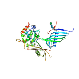
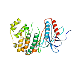
 | | Complex structure of PCNA with UHRF2 | | 分子名称: | E3 ubiquitin-protein ligase UHRF2, GLYCEROL, Proliferating cell nuclear antigen, ... | | 著者 | Wu, M, Chen, W, Hang, T, Wang, C, Zhang, X, Zang, J. | | 登録日 | 2017-09-07 | | 公開日 | 2017-11-15 | | 最終更新日 | 2023-11-22 | | 実験手法 | X-RAY DIFFRACTION (2.199 Å) | | 主引用文献 | Structure insights into the molecular mechanism of the interaction between UHRF2 and PCNA.
Biochem. Biophys. Res. Commun., 494, 2017
|
|
6M0X
 | | Crystal structure of Streptococcus thermophilus Cas9 in complex with AGGA PAM | | 分子名称: | BARIUM ION, CRISPR-associated endonuclease Cas9 1, DNA (28-MER), ... | | 著者 | Zhang, Y, Zhang, H, Xu, X, Wang, Y, Chen, W, Wang, Y, Wu, Z, Tang, N, Wang, Y, Zhao, S, Gan, J, Ji, Q. | | 登録日 | 2020-02-23 | | 公開日 | 2020-09-09 | | 最終更新日 | 2023-11-29 | | 実験手法 | X-RAY DIFFRACTION (2.561 Å) | | 主引用文献 | Catalytic-state structure and engineering of Streptococcus thermophilus Cas9
Nat Catal, 2020
|
|
6M0W
 | | Crystal structure of Streptococcus thermophilus Cas9 in complex with the AGAA PAM | | 分子名称: | CRISPR-associated endonuclease Cas9 1, DNA (28-MER), DNA (5'-D(*AP*AP*AP*GP*AP*AP*GP*C)-3'), ... | | 著者 | Zhang, Y, Zhang, H, Xu, X, Wang, Y, Chen, W, Wang, Y, Wu, Z, Tang, N, Wang, Y, Zhao, S, Gan, J, Ji, Q. | | 登録日 | 2020-02-23 | | 公開日 | 2020-09-09 | | 最終更新日 | 2023-11-29 | | 実験手法 | X-RAY DIFFRACTION (2.76 Å) | | 主引用文献 | Catalytic-state structure and engineering of Streptococcus thermophilus Cas9
Nat Catal, 2020
|
|
6M0V
 | | Crsytal structure of streptococcus thermophilus Cas9 in complex with the GGAA PAM | | 分子名称: | BARIUM ION, CRISPR-associated endonuclease Cas9 1, DNA (28-MER), ... | | 著者 | Zhang, Y, Zhang, H, Xu, X, Wang, Y, Chen, W, Wang, Y, Wu, Z, Tang, N, Wang, Y, Zhao, S, Gan, J, Ji, Q. | | 登録日 | 2020-02-22 | | 公開日 | 2020-09-09 | | 最終更新日 | 2023-11-29 | | 実験手法 | X-RAY DIFFRACTION (3 Å) | | 主引用文献 | Catalytic-state structure and engineering of Streptococcus thermophilus Cas9
Nat Catal, 2020
|
|
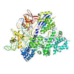
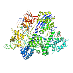
3R1K
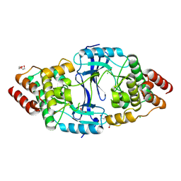
 | | Crystal structure of acetyltransferase Eis from Mycobacterium tuberculosis H37Rv in complex with CoA and an acetamide moiety | | 分子名称: | ACETAMIDE, COENZYME A, Enhanced intracellular survival protein | | 著者 | Biswas, T, Chen, W, Garneau-Tsodikova, S, Tsodikov, O.V. | | 登録日 | 2011-03-10 | | 公開日 | 2011-06-01 | | 最終更新日 | 2024-02-21 | | 実験手法 | X-RAY DIFFRACTION (1.95 Å) | | 主引用文献 | Unusual regioversatility of acetyltransferase Eis, a cause of drug resistance in XDR-TB.
Proc.Natl.Acad.Sci.USA, 108, 2011
|
|
7EN6
 | |
7EN5
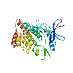
 | | The crystal structure of Escherichia coli MurR in complex with N-acetylglucosamine-6-phosphate | | 分子名称: | 2-METHOXYETHANOL, 2-acetamido-2-deoxy-6-O-phosphono-beta-D-glucopyranose, GLYCEROL, ... | | 著者 | Zhang, Y, Chen, W, Ji, Q. | | 登録日 | 2021-04-16 | | 公開日 | 2022-04-20 | | 最終更新日 | 2023-11-29 | | 実験手法 | X-RAY DIFFRACTION (1.25 Å) | | 主引用文献 | Molecular basis for cell-wall recycling regulation by transcriptional repressor MurR in Escherichia coli.
Nucleic Acids Res., 50, 2022
|
|
7EN7
 | | The crystal structure of Escherichia coli MurR in complex with N-acetylmuramic-acid-6-phosphate | | 分子名称: | (2R)-2-[(2R,3R,4R,5S,6R)-3-acetamido-2,5-bis(oxidanyl)-6-(phosphonooxymethyl)oxan-4-yl]oxypropanoic acid, HTH-type transcriptional regulator MurR | | 著者 | Zhang, Y, Chen, W, Ji, Q. | | 登録日 | 2021-04-16 | | 公開日 | 2022-04-20 | | 最終更新日 | 2023-11-29 | | 実験手法 | X-RAY DIFFRACTION (1.22 Å) | | 主引用文献 | Molecular basis for cell-wall recycling regulation by transcriptional repressor MurR in Escherichia coli.
Nucleic Acids Res., 50, 2022
|
|
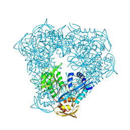
5NJT
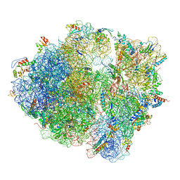 | | Structure of the Bacillus subtilis hibernating 100S ribosome reveals the basis for 70S dimerization. | | 分子名称: | 16S ribosomal RNA, 23S ribosomal RNA, 30S ribosomal protein S10, ... | | 著者 | Beckert, B, Abdelshahid, M, Schaefer, H, Steinchen, W, Arenz, S, Berninghausen, O, Beckmann, R, Bange, G, Turgay, K, Wilson, D.N. | | 登録日 | 2017-03-29 | | 公開日 | 2017-06-14 | | 最終更新日 | 2024-05-15 | | 実験手法 | ELECTRON MICROSCOPY (3.8 Å) | | 主引用文献 | Structure of the Bacillus subtilis hibernating 100S ribosome reveals the basis for 70S dimerization.
EMBO J., 36, 2017
|
|
6CP9
 | | Contact-dependent growth inhibition toxin - immunity protein complex from Klebsiella pneumoniae 342 | | 分子名称: | CdiA, CdiI | | 著者 | Michalska, K, Stols, L, Eschenfeldt, W, Hayes, C.S, Goulding, C.W, Joachimiak, A, Midwest Center for Structural Genomics (MCSG), Structure-Function Analysis of Polymorphic CDI Toxin-Immunity Protein Complexes (UC4CDI) | | 登録日 | 2018-03-13 | | 公開日 | 2019-03-13 | | 最終更新日 | 2020-01-01 | | 実験手法 | X-RAY DIFFRACTION (2.55 Å) | | 主引用文献 | Convergent Evolution of the Barnase/EndoU/Colicin/RelE (BECR) Fold in Antibacterial tRNase Toxins.
Structure, 27, 2019
|
|
6SAZ
 | | Cleaved human fetuin-b in complex with crayfish astacin | | 分子名称: | 2-acetamido-2-deoxy-beta-D-glucopyranose, 2-acetamido-2-deoxy-beta-D-glucopyranose-(1-4)-2-acetamido-2-deoxy-beta-D-glucopyranose, Astacin, ... | | 著者 | Gomis-Ruth, F.X, Guevara, T, Cuppari, A, Korschgen, H, Schmitz, C, Kuske, M, Yiallouros, I, Floehr, J, Jahnen-Dechent, W, Stocker, W. | | 登録日 | 2019-07-18 | | 公開日 | 2019-10-23 | | 最終更新日 | 2024-01-24 | | 実験手法 | X-RAY DIFFRACTION (3 Å) | | 主引用文献 | The C-terminal region of human plasma fetuin-B is dispensable for the raised-elephant-trunk mechanism of inhibition of astacin metallopeptidases.
Sci Rep, 9, 2019
|
|
6ZZR
 | | The Crystal Structure of human LDHA from Biortus. | | 分子名称: | 1,2-ETHANEDIOL, DI(HYDROXYETHYL)ETHER, L-lactate dehydrogenase A chain | | 著者 | Wang, F, Lin, D, Cheng, W, Bao, X, Zhu, B, Shang, H. | | 登録日 | 2020-08-05 | | 公開日 | 2020-08-19 | | 最終更新日 | 2024-04-24 | | 実験手法 | X-RAY DIFFRACTION (2.65 Å) | | 主引用文献 | The Crystal Structure of human LDHA from Biortus
To Be Published
|
|
8XFY
 | | The Crystal Structure of RSK2 from Biortus. | | 分子名称: | 2,6-bis(fluoranyl)-4-[4-(4-morpholin-4-ylphenyl)pyridin-3-yl]phenol, Ribosomal protein S6 kinase alpha-3 | | 著者 | Wang, F, Cheng, W, Lv, Z, Lin, D, Pan, W. | | 登録日 | 2023-12-14 | | 公開日 | 2024-03-06 | | 実験手法 | X-RAY DIFFRACTION (3.2 Å) | | 主引用文献 | The Crystal Structure of RSK2 from Biortus.
To Be Published
|
|
8YVV
 | | The Crystal Structure of BTK from Biortus | | 分子名称: | 1,2-ETHANEDIOL, CHLORIDE ION, DI(HYDROXYETHYL)ETHER, ... | | 著者 | Wang, F, Cheng, W, Yuan, Z, Lin, D, Pan, W. | | 登録日 | 2024-03-29 | | 公開日 | 2024-07-03 | | 実験手法 | X-RAY DIFFRACTION (2.25 Å) | | 主引用文献 | The Crystal Structure of BTK from Biortus.
To Be Published
|
|
8WSW
 | | The Crystal Structure of LIMK2a from Biortus | | 分子名称: | 1,2-ETHANEDIOL, LIM domain kinase 2, ~{N}-[5-[2-[2,6-bis(chloranyl)phenyl]-5-[bis(fluoranyl)methyl]pyrazol-3-yl]-1,3-thiazol-2-yl]-2-methyl-propanamide | | 著者 | Wang, F, Cheng, W, Yuan, Z, Lin, D, Pan, W. | | 登録日 | 2023-10-17 | | 公開日 | 2023-11-15 | | 実験手法 | X-RAY DIFFRACTION (2.5 Å) | | 主引用文献 | The Crystal Structure of LIMK2a from Biortus.
To Be Published
|
|
8ZKD
 | | The Crystal Structure of the RON from Biortus. | | 分子名称: | 1,2-ETHANEDIOL, MAGNESIUM ION, Macrophage-stimulating protein receptor beta chain, ... | | 著者 | Wang, F, Cheng, W, Yuan, Z, Qi, J, Pan, W. | | 登録日 | 2024-05-16 | | 公開日 | 2024-06-26 | | 実験手法 | X-RAY DIFFRACTION (2.05 Å) | | 主引用文献 | The Crystal Structure of the RON from Biortus.
To Be Published
|
|
8X23
 | | The Crystal Structure of MAPK13 from Biortus. | | 分子名称: | 1,2-ETHANEDIOL, GLYCEROL, Mitogen-activated protein kinase 13 | | 著者 | Wang, F, Cheng, W, Yuan, Z, Lin, D, Pan, W. | | 登録日 | 2023-11-09 | | 公開日 | 2023-12-27 | | 実験手法 | X-RAY DIFFRACTION (1.5 Å) | | 主引用文献 | The Crystal Structure of MAPK13 from Biortus.
To Be Published
|
|
8X2T
 | |


