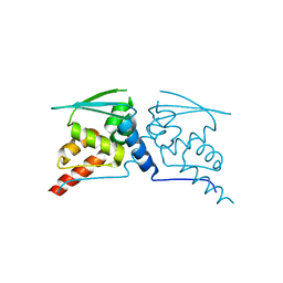
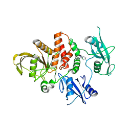
6FPG
 
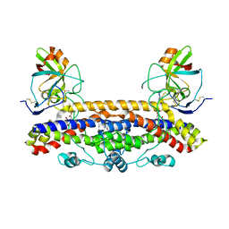 | | Structure of the Ustilago maydis chorismate mutase 1 in complex with a Zea mays kiwellin | | 分子名称: | CITRIC ACID, Chromosome 16, whole genome shotgun sequence, ... | | 著者 | Altegoer, F, Steinchen, W, Bange, G. | | 登録日 | 2018-02-09 | | 公開日 | 2019-01-16 | | 最終更新日 | 2024-01-17 | | 実験手法 | X-RAY DIFFRACTION (1.8 Å) | | 主引用文献 | A kiwellin disarms the metabolic activity of a secreted fungal virulence factor.
Nature, 565, 2019
|
|
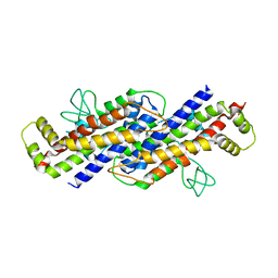
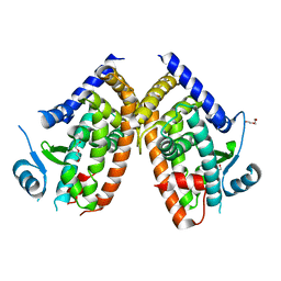
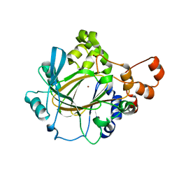
6K7P
 | | Crystal structure of human AFF4-THD domain | | 分子名称: | AF4/FMR2 family member 4 | | 著者 | Tang, D, Xue, Y, Li, S, Cheng, W, Duan, J, Wang, J, Qi, S. | | 登録日 | 2019-06-08 | | 公開日 | 2020-03-11 | | 最終更新日 | 2024-03-27 | | 実験手法 | X-RAY DIFFRACTION (2.4 Å) | | 主引用文献 | Structural and functional insight into the effect of AFF4 dimerization on activation of HIV-1 proviral transcription.
Cell Discov, 6, 2020
|
|
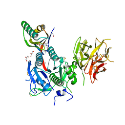
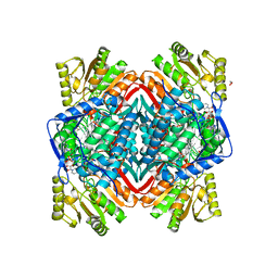
7C8U
 | | The crystal structure of COVID-19 main protease in complex with GC376 | | 分子名称: | (1S,2S)-2-({N-[(benzyloxy)carbonyl]-L-leucyl}amino)-1-hydroxy-3-[(3S)-2-oxopyrrolidin-3-yl]propane-1-sulfonic acid, 3C-like proteinase | | 著者 | Luan, X, Shang, W, Wang, Y, Yin, W, Jiang, Y, Feng, S, Wang, Y, Liu, M, Zhou, R, Zhang, Z, Wang, F, Cheng, W, Gao, M, Wang, H, Wu, W, Tian, R, Tian, Z, Jin, Y, Jiang, H.W, Zhang, L, Xu, H.E, Zhang, S. | | 登録日 | 2020-06-03 | | 公開日 | 2020-06-24 | | 最終更新日 | 2023-11-29 | | 実験手法 | X-RAY DIFFRACTION (2.35 Å) | | 主引用文献 | The crystal structure of COVID-19 main protease in complex with GC376
To Be Published
|
|
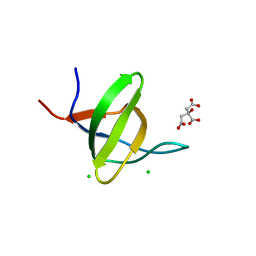
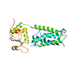
6FPF
 | |
8TPU
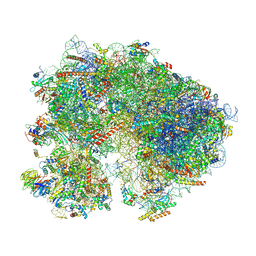 | | Subtomogram averaged consensus structure of the malarial 80S ribosome in Plasmodium falciparum-infected human erythrocytes | | 分子名称: | 18S ribosomal RNA, 28S ribosomal RNA, 40S ribosomal protein S10, ... | | 著者 | Anton, L, Cheng, W, Zhu, X, Ho, C.M. | | 登録日 | 2023-08-05 | | 公開日 | 2024-08-14 | | 実験手法 | ELECTRON MICROSCOPY (4.1 Å) | | 主引用文献 | Divergent translational landscape reflects adaptation to biased codon usage in malaria parasites
To Be Published
|
|
6VEK
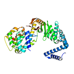 | | Contact-dependent growth inhibition toxin-immunity protein complex from from E. coli 3006, full-length | | 分子名称: | contact-dependent immunity protein CdiI, contact-dependent toxin CdiA | | 著者 | Michalska, K, Stols, L, Eschenfeldt, W, Hayes, C.S, Goulding, C.W, Joachimiak, A, Midwest Center for Structural Genomics (MCSG), Structure-Function Analysis of Polymorphic CDI Toxin-Immunity Protein Complexes (UC4CDI), Center for Structural Genomics of Infectious Diseases (CSGID) | | 登録日 | 2020-01-02 | | 公開日 | 2021-01-27 | | 最終更新日 | 2023-10-11 | | 実験手法 | X-RAY DIFFRACTION (2.25 Å) | | 主引用文献 | Contact-dependent growth inhibition toxin-immunity protein complex from from E. coli 3006, full-length
To Be Published
|
|
8WF7
 | | The Crystal Structure of integrase from Biortus | | 分子名称: | ACETATE ION, Integrase, SULFATE ION | | 著者 | Wang, F, Cheng, W, Yuan, Z, Qi, J, Li, J. | | 登録日 | 2023-09-19 | | 公開日 | 2023-10-04 | | 実験手法 | X-RAY DIFFRACTION (1.55 Å) | | 主引用文献 | The Crystal Structure of integrase from Biortus
To Be Published
|
|
8WD2
 | | The Crystal Structure of p53 from Biortus. | | 分子名称: | 1,2-ETHANEDIOL, Cellular tumor antigen p53, PHOSPHATE ION, ... | | 著者 | Wang, F, Cheng, W, Yuan, Z, Qi, J, Lu, Y. | | 登録日 | 2023-09-14 | | 公開日 | 2023-10-04 | | 実験手法 | X-RAY DIFFRACTION (1.85 Å) | | 主引用文献 | The Crystal Structure of p53 from Biortus.
To Be Published
|
|
8WFG
 | |
8WGF
 | | The Crystal Structure of JNK3 from Biortus. | | 分子名称: | MAGNESIUM ION, Mitogen-activated protein kinase 10, PHOSPHOAMINOPHOSPHONIC ACID-ADENYLATE ESTER | | 著者 | Wang, F, Cheng, W, Lv, Z, Ju, C, Wang, J. | | 登録日 | 2023-09-21 | | 公開日 | 2023-11-22 | | 実験手法 | X-RAY DIFFRACTION (1.85 Å) | | 主引用文献 | The Crystal Structure of JNK3 from Biortus.
To Be Published
|
|
8WGQ
 | | The Crystal Structure of L-asparaginase from Biortus. | | 分子名称: | 1,2-ETHANEDIOL, GLYCEROL, L-asparaginase | | 著者 | Wang, F, Cheng, W, Lv, Z, Ju, C, Wang, J. | | 登録日 | 2023-09-22 | | 公開日 | 2023-11-22 | | 実験手法 | X-RAY DIFFRACTION (2.75 Å) | | 主引用文献 | The Crystal Structure of L-asparaginase from Biortus.
To Be Published
|
|
8WFE
 | | The Crystal Structure of PPARg from Biortus. | | 分子名称: | 1,2-ETHANEDIOL, DI(HYDROXYETHYL)ETHER, Peroxisome proliferator-activated receptor gamma | | 著者 | Wang, F, Cheng, W, Lv, Z, Guo, S, Lin, D. | | 登録日 | 2023-09-19 | | 公開日 | 2023-11-22 | | 実験手法 | X-RAY DIFFRACTION (2.2 Å) | | 主引用文献 | The Crystal Structure of PPARg from Biortus.
To Be Published
|
|
8WFR
 | | The Crystal Structure of PCSK9 from Biortus. | | 分子名称: | 1,2-ETHANEDIOL, 4-(2-HYDROXYETHYL)-1-PIPERAZINE ETHANESULFONIC ACID, GLYCEROL, ... | | 著者 | Wang, F, Cheng, W, Lv, Z, Meng, Q, Lu, Y. | | 登録日 | 2023-09-20 | | 公開日 | 2023-11-22 | | 実験手法 | X-RAY DIFFRACTION (1.95 Å) | | 主引用文献 | The Crystal Structure of PCSK9 from Biortus.
To Be Published
|
|
8WF4
 | | The Crystal Structure of RSK1 from Biortus. | | 分子名称: | 1,2-ETHANEDIOL, Ribosomal protein S6 kinase alpha-1 | | 著者 | Wang, F, Cheng, W, Lv, Z, Qi, J, Li, J. | | 登録日 | 2023-09-19 | | 公開日 | 2023-11-22 | | 実験手法 | X-RAY DIFFRACTION (2.65 Å) | | 主引用文献 | The Crystal Structure of RSK1 from Biortus.
To Be Published
|
|
8WFF
 | | The Crystal Structure of LYN from Biortus. | | 分子名称: | CHLORIDE ION, CITRATE ANION, Tyrosine-protein kinase Lyn | | 著者 | Wang, F, Cheng, W, Yuan, Z, Qi, J, Lu, Y. | | 登録日 | 2023-09-19 | | 公開日 | 2023-11-22 | | 実験手法 | X-RAY DIFFRACTION (1.3 Å) | | 主引用文献 | The Crystal Structure of LYN from Biortus.
To Be Published
|
|
8WFY
 | | The Crystal Structure of SHP2 from Biortus. | | 分子名称: | 6-(4-azanyl-4-methyl-piperidin-1-yl)-3-[2,3-bis(chloranyl)phenyl]pyrazin-2-amine, Tyrosine-protein phosphatase non-receptor type 11 | | 著者 | Wang, F, Cheng, W, Yuan, Z, Qi, J, Li, J. | | 登録日 | 2023-09-20 | | 公開日 | 2023-11-22 | | 実験手法 | X-RAY DIFFRACTION (2.6 Å) | | 主引用文献 | The Crystal Structure of SHP2 from Biortus.
To Be Published
|
|
8WD3
 | | The Crystal Structure of JMJD2A(M1-L359) from Biortus. | | 分子名称: | Lysine-specific demethylase 4A, NICKEL (II) ION, ZINC ION | | 著者 | Wang, F, Cheng, W, Lv, Z, Ju, C, Bao, C. | | 登録日 | 2023-09-14 | | 公開日 | 2023-11-22 | | 実験手法 | X-RAY DIFFRACTION (3.3 Å) | | 主引用文献 | The Crystal Structure of JMJD2A(M1-L359) from Biortus.
To Be Published
|
|
8WFQ
 | | The Crystal Structure of RALDH1 from Biortus. | | 分子名称: | 1,2-ETHANEDIOL, 1,4-DIHYDRONICOTINAMIDE ADENINE DINUCLEOTIDE, Aldehyde dehydrogenase 1A1 | | 著者 | Wang, F, Cheng, W, Lv, Z, Qi, J, Shen, Z. | | 登録日 | 2023-09-20 | | 公開日 | 2023-11-22 | | 実験手法 | X-RAY DIFFRACTION (3.5 Å) | | 主引用文献 | The Crystal Structure of RALDH1 from Biortus.
To Be Published
|
|
4NV5
 | | C50A mutant of Synechococcus VKOR, C2 crystal form (dehydrated) | | 分子名称: | UBIQUINONE-10, VKORC1/thioredoxin domain protein | | 著者 | Liu, S, Cheng, W, Fowle Grider, R, Shen, G, Li, W. | | 登録日 | 2013-12-04 | | 公開日 | 2014-02-12 | | 実験手法 | X-RAY DIFFRACTION (2.79 Å) | | 主引用文献 | Structures of an intramembrane vitamin K epoxide reductase homolog reveal control mechanisms for electron transfer.
Nat Commun, 5, 2014
|
|
4NV6
 | | C212A mutant of Synechococcus VKOR | | 分子名称: | UBIQUINONE-10, VKORC1/thioredoxin domain protein | | 著者 | Liu, S, Cheng, W, Fowle Grider, R, Shen, G, Li, W. | | 登録日 | 2013-12-04 | | 公開日 | 2014-02-12 | | 実験手法 | X-RAY DIFFRACTION (4.19 Å) | | 主引用文献 | Structures of an intramembrane vitamin K epoxide reductase homolog reveal control mechanisms for electron transfer.
Nat Commun, 5, 2014
|
|
4NV2
 | | C50A mutant of Synechococcus VKOR, C2221 crystal form | | 分子名称: | UBIQUINONE-10, VKORC1/thioredoxin domain protein | | 著者 | Liu, S, Cheng, W, Fowle Grider, R, Shen, G, Li, W. | | 登録日 | 2013-12-04 | | 公開日 | 2014-02-12 | | 実験手法 | X-RAY DIFFRACTION (3.61 Å) | | 主引用文献 | Structures of an intramembrane vitamin K epoxide reductase homolog reveal control mechanisms for electron transfer.
Nat Commun, 5, 2014
|
|
3CDK
 | | Crystal structure of the co-expressed succinyl-CoA transferase A and B complex from Bacillus subtilis | | 分子名称: | Succinyl-CoA:3-ketoacid-coenzyme A transferase subunit A, Succinyl-CoA:3-ketoacid-coenzyme A transferase subunit B | | 著者 | Kim, Y, Zhou, M, Stols, L, Eschenfeldt, W, Donnelly, M, Joachimiak, A, Midwest Center for Structural Genomics (MCSG) | | 登録日 | 2008-02-27 | | 公開日 | 2008-03-18 | | 最終更新日 | 2023-08-30 | | 実験手法 | X-RAY DIFFRACTION (2.59 Å) | | 主引用文献 | Crystal structure of the co-expressed succinyl-CoA transferase A and B complex from Bacillus subtilis.
To be Published
|
|
8H4R
 | | The Crystal Structure of CDK3 and CyclinE1 Complex with Dinaciclib from Biortus | | 分子名称: | 2-(N-MORPHOLINO)-ETHANESULFONIC ACID, 3-[({3-ethyl-5-[(2S)-2-(2-hydroxyethyl)piperidin-1-yl]pyrazolo[1,5-a]pyrimidin-7-yl}amino)methyl]-1-hydroxypyridinium, G1/S-specific cyclin-E1, ... | | 著者 | Gui, W, Wang, F, Cheng, W, Gao, J, Huang, Y, Ouyang, Z. | | 登録日 | 2022-10-11 | | 公開日 | 2023-10-11 | | 実験手法 | X-RAY DIFFRACTION (2.75 Å) | | 主引用文献 | The Crystal Structure of CDK3 and CyclinE1 Complex with Dinaciclib from Biortus
To Be Published
|
|
6A82
 | |
8GYF
 | | Crystal structure of a bright green fluorescent protein (StayGold) with single mutation (K192Y) in jellyfish Cytaeis uchidae from Biortus | | 分子名称: | 1,2-ETHANEDIOL, staygold(K192Y) | | 著者 | Wu, J, Wang, F, Gui, W, Cheng, W, Yang, Y. | | 登録日 | 2022-09-22 | | 公開日 | 2023-10-04 | | 最終更新日 | 2023-11-15 | | 実験手法 | X-RAY DIFFRACTION (2 Å) | | 主引用文献 | Crystal structure of a bright green fluorescent protein (StayGold) with single mutation (K192Y) in jellyfish Cytaeis uchidae from Biortus
To Be Published
|
|


